báo cáo khoa học: " A rare case of giant leiomyosarcoma in a filarial scrotum: a case report" ppt
Bạn đang xem bản rút gọn của tài liệu. Xem và tải ngay bản đầy đủ của tài liệu tại đây (2.27 MB, 5 trang )
WORLD JOURNAL OF
SURGICAL ONCOLOGY
A rare case of giant leiomyosarcoma in a filarial
scrotum: a case report
Talikoti et al.
Talikoti et al. World Journal of Surgical Oncology 2011, 9:20
(10 February 2011)
CAS E REP O R T Open Access
A rare case of giant leiomyosarcoma in a filarial
scrotum: a case report
Majid Ahmed Talikoti
*
, SV S Deo, Nootan K Shukla, Ashwin A Kallianpur, Mamraj Gupta
Abstract
Giant leiomyosarcoma of scrotum is a rare tumour. A case of scrotum leiomyosarcoma is presented in a 67 year
old patient with scrotal filariasis which was managed successfully with total scrotectomy with bilateral
orchidectomy, degloved penis reconstructed with rotation advancement sup ra pubic fasciocutaneous flap. We
made a literature search proving the rarity of this lesion type. Only 36 cases have been described and the first case
in a filarial scrotum
Introduction
Leiomyosarcoma of the scro tum is a rare tumour. Scro -
tal leiomyosarcomas (LMS) are slow growing tumours
that present as firm rubbery nontender irregular mass
[1]. They may arise from paratesticular or scrotal skin.
Over 95% of all paratesticular leiomyosarcomas are
located in the spermatic cord or epididymis; their loca-
tion in the scrota l skin is exceptional. To date approxi-
mate 36 scrotal LMS have been reported in literatur e.
We report a rare case of giant primary scrotal LMS aris-
ing in a filarial scrotum. There is no report of such giant
LMS and none in the background of scrotal
elephantiasis.
Case Presentation
A 67 years old patient of high socioeconomic status
reported to the outpatient clinic wit h complain of
rapidly ulcerated mass in the scrotum. He had history of
filariasis and scrotal swelling of more than 40 years
duration. His Eastern Cooperative Oncology G roup
(ECOG) performance Status was 3, capable of only lim-
ited self ca re, confined to bed or chair more than 50%
of waking hours, due to large painful swelling of the
scrotum [2].
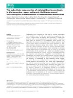
On local examination, huge filarial scrotum with skin
changes and buried penis, 30 × 30 cm ulcerated firm
diffuse mass of the scrotal skin encroaching over the
root of penile skin [figures 1, 2]. The tumour was
mobile not fixed to the testes. Metastatic work up
included CT chest and abdomen and was negative.
Wedge biopsy was compatible with Leiomyosarcoma.
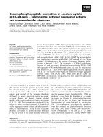
After an informed written consent patient underwent a
radical resection of tumour including total scrotectomy
and bilateral orchidectomy, degloving of buried penis
with reconstruction by rotation adva ncement supra
pubic fasciocutaneous flap [figures 3, 4, 5, 6]. Patient
recovery was uneventful. Patients ECOG performance
scale improved from 3 to 1, he became ambulatory with
his ability to perform routine plus outdoor activity on
his own.
Post operative histopathology revealed 28 × 25 × 15
cm fleshy tumour with an over lying ulcer. Testes were
not involved by the tumour. All resected margins were
negative. Microscopically a malignant mesenchymal neo-
plasm was identified with smooth muscle differentiated
intermingling bundle of cells with eosinophilic abundant
mitotic figure 15 mitosis per 10 high power. Tumour
was positive for actin, vimentin and desmin and negative
for S 100. Based o n histopathology findings and Immu-
nohistochemistry markers diagnosis of scrotal leiomyo-
sarcoma was made.
Patient re ceived postoperative external beam radiation
60G/30fractions. H e is on regular follow up for past 19
months and is disease free.
Discussion
Soft tissue Sarcomas are 1% of all malignancies. Leio-
myosarcomas consti tutes 10 to 20% of soft tissue sarco-
mas. They arise most often from uterus, gastrointestinal
tract and retroperitoneal region [1]. Subcutaneous LMS
are 1 to 2% of all superficial soft tissue malignancies [3].
* Correspondence:
Department of Surgical Oncology, BRAIRCH, AIIMS, New Delhi, 110029, India
Talikoti et al. World Journal of Surgical Oncology 2011, 9:20
/>WORLD JOURNAL OF
SURGICAL ONCOLOGY
© 2011 Talikoti et al; licensee BioMed Central Ltd. This is an Open Access article distributed under the terms of the Creative Commons
Attribution License (http://creativecommo ns.org/licenses/by/2.0), which permits unrestricted use, distr ibution, and reproduction in
any medium, provided the original work is properly cited.
Among genitourinary sarcomas in adults, leiomyosarco -
mas ar e the most common type and arises in the blad-
der, kidney, or prostate. Its origin in scrotum is
exceptional with only 36 cases have been reported in lit-
erature. LMS are malignant mesenchymal line neoplasm
with size of the tumor varying from 2 to 9 cm, with an
average of 5 cm. Biopsy is done to confirm the diag-
noses of LMS. A confirmative diagnosis of LMS is based
on histological examination. They show spindle cells
with cigar shaped nuclei arranged in interweaving fasci-
cles [1]. On Immunoh istochemistry t hey are positive for
actin, desmin and CD 34 [4]. The mode of spread is pri-
marily haematogenous to lung, liver, and bone. Prog-
noses of LMS depend on tumour size, depth, grade and
evidence of distant metastases.
Scrotal elephantiasis is caused by acquired filarial
infestation with Wuchereria bancrofti. Occasionally it
has been attributed to radiotherapy, neoplasm and lym-
phadenectomy. It is emotionally distressing and physi-
cally disabling. With problems of hygiene, urinary
incontinence, unesthetic appearance, loss of libido and
immobility are severely debilitating symptom [5].
Filariasis in advance stages may evolve into scrotal lym-
phedema and scrotal elephantiasis. In 1948 Stewart and
Treves first described the associat ion of chronic l ymphe-
dema with lymphangiosarcoma [6]. Lymphangiosarcomas
are common malignancies in those with chronic f ilarial
infections [7]. Sarcomas have been seen after filarial
infection and chronic lymphedema [8]. Scrotal leiomyo-
sarcoma have a potential of distant metastases. Here in
we report the first case of large Leiomyosarcoma of a
filarial scrotum. Scrotal Leiomyosarcoma in our case was
a high grade, large size, stage III having a potential of dis-
tant metastases.
The paucity of literature in this area often makes
treatment decisions difficult. Simple excision proved to
Figure 1 Large 30x30 cm tumour with overlying ulcer over the
right scrotum.
Figure 2 Large 30x30 cm tumour with overlying ulcer over the
right scrotum.
Figure 3 Radical resection of tumour.
Figure 4 Radical resection of tumour.
Talikoti et al. World Journal of Surgical Oncology 2011, 9:20
/>Page 2 of 4
be inadequate treatment for sarcomas in the paratesticu-
lar region. In the Princess Margaret Hospital report,
wide excision revealed microscopic residual disease in
27% of completely excised cases [9]. The primary treat-
ment of LMS of scrotum is complete resection with his-
tological negative margins. The difficult y in achieving an
oncological safe margin reflects the tumo ur biology. An
aggressive initial resection is required at the time of the
first operation [10]. Many surgical methods have been
described for scrotal and penile reconstruction. Like
pedicle groin flap based on superficial circumflex iliac
artery to cover the penis and bilateral superior medial
thigh flap for scrotal reconstruction [11]. We preferred
a single staged, simpler reconstruction by using rotation
advancement flap of the supra pubic area over the two
staged pedicle groin flap to reconstruct the penis. The
scrotal defect was primarily closed. For most patients,
Figure 5 Radical resection of tumour.
Figure 6 Reconstruction of degloved penis done with suprapubic fasciocutaneous flap.
Talikoti et al. World Journal of Surgical Oncology 2011, 9:20
/>Page 3 of 4
local control is improved with preoperative or post-
operative radiotherapy. Hensl ey and colleagues were t he
first to report the activity of the gemcitabine-docetaxel
combination in patients with lei omyosarcomas [12]. The
role of chemotherapy for high-risk patients remains con-
troversial, but chemotherapy is used at several major
centres’ for high-risk patients. In our case we treated
our patient with wide local e xcision and postoperative
60 Gy radiotherapy.
Conclusions
• Scrotal leiomyosarcoma is a rare clinical entity and
scrotal leiomyosarcoma in a filarial lymphadenomatous
scrotum; this is the first case to be reported.
• Aggressive surgical resection including tumour and
diseased filarial skin is recommended.
Consent
Written informed consent was obtained from the patient
for publication of this case report and accompanying
images. A copy of the written consent is available for
review by the Editor-in-Chief of this journal.
Authors’ contributions
MT, SVSD, NKS, AK and MG prepared the manuscript and reviewd the
literature. All authors read and approved the final manuscript.
Competing interests
The authors declare that they have no competing interests.
Received: 2 November 2010 Accepted: 10 February 2011
Published: 10 February 2011
References
1. Kumar V, Abbas AK, Fausto N, Mitchell R: Robbins Basic Pathology.
Elsevier;, 8 2007, 835.
2. Oken MM, Creech RH, Tormey DC, Horton J, Davis TE, McFadden ET,
Carbone PP: Toxicity and Response criteria of The Eastern Cooperative
Oncology group. Am J clin Oncol 1982, 5:649-655.
3. Kaushal Vivek, Singh Harmeet, Gill Meenu: Recurrent Leiomyosarcoma of
The Scrotum:JK science. 2009, 11(2):97-98.
4. Fisher C, Goldblum JR, Epstein JI, Montgomery E: Leiomyosarcoma of the
paratesticular region: a clinicopathologic study. Am J Surg Pathol 2001,
25(9):1143-1149.
5. Denzinger Stefan, Watzlawek Elke, Burger Maximilian, et al: Giant scrotal
elephantiasis of inflammatory etiology: a case report. Journal of Medical
Case Reports 2007, 1:23.
6. Stewart F, Treves N: Lymphangiosarcoma in post-mastectomy
lymphedema. Cancer 1948, 1:64.
7. Muller R, Hajdu S, Brennan M: Lymphangiosarcoma associated with
chronic filarial lymphedema. Cancer 1987, 59:179.
8. DeVita , Vincent T, Lawrence , et al: Devita, Hellman & Rosenberg’s
Cancer: Principles & Practice of Oncology. Lippincott Williams & Wilkins;, 8
2008, 1743.
9. Catton C, Jewett M, O’Sullivan B, Kandel R: Paratesticular sarcoma: failure
patterns after definitive local therapy. The Journal of Urology 1999,
161:1844-7.
10. Rajkumar Kheman, Mundy Ian: Leiomyosarcoma of the scrotum-a rare
tumour. Journal of the New Zealand Medical Association 2007, 120(1266).
11. Lee S, Bang S, Kim J: Penoscrotal reconstruction using groin and bilateral
superior medial thigh flaps: A case of penile vaselinoma causing
fourniers gangrene. Yonsei Med J 2007, 48(4):723-6.
12. Hensley ML, Maki R, Venkatraman E, et al: Gemcitabine and docetaxel in
patients with unresectable leiomyosarcoma: results of a phase II trial.
J Clin Oncol 2002, 20:2824.
doi:10.1186/1477-7819-9-20
Cite this article as: Talikoti et al.: A rare case of giant leiomyosarcoma in
a filarial scrotum: a case report. World Journal of Surgical Oncology 2011
9:20.
Submit your next manuscript to BioMed Central
and take full advantage of:
• Convenient online submission
• Thorough peer review
• No space constraints or color figure charges
• Immediate publication on acceptance
• Inclusion in PubMed, CAS, Scopus and Google Scholar
• Research which is freely available for redistribution
Submit your manuscript at
www.biomedcentral.com/submit
Talikoti et al. World Journal of Surgical Oncology 2011, 9:20
/>Page 4 of 4