báo cáo khoa học: "Colonic perforation resulting from ingested chicken bone revealing previously undiagnosed colonic adenocarcinoma: report of a case and review of literature" ppt
Bạn đang xem bản rút gọn của tài liệu. Xem và tải ngay bản đầy đủ của tài liệu tại đây (706.35 KB, 4 trang )
CASE REP O R T Open Access
Colonic perforation resulting from ingested
chicken bone revealing previously undiagnosed
colonic adenocarcinoma: report of a case and
review of literature
Douglas H McGregor
1,2*
, Xiaoying Liu
1
, Ozlem Ulusarac
1,2
, Kimberly D Ponnuru
3,4
, Stephanie L Schnepp
4
Abstract
An 86 year old male with a four-day history of nonspecific gastrointestinal symptoms was found on colonoscopy
to have evidence of sigmoid colon obstruction and possible perforation. Emergent ope rative exploration revealed
diffuse peritonitis, sigmoid perforation, adjacent dense adhesions, and a foreign body protruding thr ough the
perforated area. Pathologic examination showed the foreign body to be a sliver of bone consistent with chicken
bone and the sigmoid subacute perforation to be associated distally with a circumferential ulcerated obstructing
mass, microscopically seen to be transmurally infiltrating adenocarcinoma, signet-ring cell type. There was extensive
acute and organizing peritonitis, 100% Escherichia coli was cultu red from peritoneal fluid, and the patient died two
days postoperatively with sepsis and hypotension. This appears to be the fifth reported case of colonic perfora tion
resulting from foreign body perforation due to previously undiagnosed adenocarcinoma. The four previously
reported cases were all deeply invasive adenocarcinoma of sigmoid colon, and the foreign bodies included three
chicken/poultry bones and a metallic staple. These five cases are highly unusual examples of a potentially lethal
malignant neoplasm being clinically revealed by a usually (but not always) innocuous event, the ingestion of a
small foreign body.
Background
Colonic perforation is most often secondary to extrinsic or
intrinsic obstruction, but occasionally it may be due to
other factors such as foreign bodies. Over 300 cases of
bowel perforation caused by foreign bodies have been
reported in the literature, with fish bones, chicken bones
and dentures being the commonest objects, followed by
toothpicks and cocktail sticks [1]. Foreign body-associated
perforation commonly occurs at the point of acute angula-
tion and narrowing, and the most common site of perfora-
tion is the terminal ileum and colon, with an increased
number of rep orts o f p erfora tion in a ssoc iation w ith M eck-
el’s diverticulum, the appendix an d d iv erticular disease [2,3].
Symptoms related to obstructing colon cancer are often
delayed, and the present reported case is an interesting
example of ingested foreign body resulting in both colon
perforation and the discovery and resection of a previously
undiagnosed colon cancer. This case appears to be the
fifth reported example of colon perforation resulting from
foreign body perforation du etopreviouslyundiagnosed
colon cancer [4-7]. Table 1 outlines the basic specifics of
these five cases. Not surprisingly, all of these obstructing
colon cancers were large deeply invasive adenocarcinomas
and their locations were the anatomically dist al and rela-
tively narrow sigmoid colon. The foreign bodies included
4 chicken/poultry bones (as in the present case 5), and a
metallic staple. The clinical outcomes were full recovery
(cases 1 and 2), postoperative death due to sepsis (case 5)
and unknown (cases 3 and 4).
Case report
An 8 6 year old male presented with a four-day history
of abdominal pain, nausea, vomiting, and intolerance to
oral intake. Physical exam demonstrated left lower, right
* Correspondence:
1
Department of Pathology and Laboratory Medicine, University of Kansas
Medical Center, Kansas City, Kansas, USA
Full list of author information is available at the end of the article
McGregor et al. World Journal of Surgical Oncology 2011, 9:24
/>WORLD JOURNAL OF
SURGICAL ONCOLOGY
© 2011 McGregor et al; licensee BioMed Central Ltd. This is an Open Access article distributed under the terms of the Creative
Commons Attribution License ( /lic enses/by/2.0), which permits unrest ricted us e, distribution, and
reproduction in any me dium, provided the original work is properly cited.
lower and left upper quadrant tenderness, but clinical
evidence of colonic obstr uction and ac ute abdomen was
not i dentified. Vital signs were temperature 98.5, pulse
86, respiration 20 and blood pressure 136/62. Labora-
tory data includ ed WBC 6.4 K/cmm, neutrophils 87.5%,
hemoglobin 11.5 g/dl and hematocrit 37.4%. Radiologic
abdominal exam demonstrated a normal gas pattern,
and ultrasound and CT scan studies were not indicated.
Colonoscopy (preceded by midazolam and demarol
medication) was performed for evaluation o f iron defi-
ciency, however, and showed evidence of sigmoid colon
obstruction and possible perforation, including a mass
with narrowing at 30 cm and a cav ernous defect with
whitish exudate. The patient underwent emergent
operative explorati on, which revealed diffuse peritoni tis,
a sigmoid perforation, adjacent dense adhesions, and a
foreign body protruding through the perforated area.
Sigmoid colon resection and end colostomy with
Hartman’s pouch was performed.
Specimens received for pathologic examination
included the foreign body, segment of sigmoid colon,
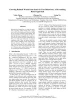
and additional segment of sigmoid colon. The foreign
body, which had been found to be protruding through
the perforation, consisted of a sliver of bone measuring
2.6 × 0.2 cm (Fig ure 1A) and the boney nature of this
foreign body was confirmed microscopically (Figure 1B).
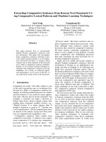
The segment of sigmoid colon had a 5.5 × 4.4 cm cir-
cumferential ulcerate d mass with marked luminal
Table 1 Reported cases of colon perforation resulting from foreign body and previously undiagnosed carcinoma
Case
No.
Age/
Sex
Colon
Site
Carcinoma Morphology Foreign
Body
Outcome Reference
No/Year
1 78/F Sigmoid Large carcinoma Chicken
bone
Full recovery 4/1985
2 64 Sigmoid 6.5 cm long circular ulcerated moderately differentiated
adenocarcinoma without stenosis
pT3 N0 M0
Poultry
bone
Full recovery 5/1996
3 57/M Sigmoid Exophytic infiltrating moderately differentiated adenocarcinoma
pT4 N1 M0
Metallic
staple
Unknown 6/1997
4 69/M Sigmoid Polypoid mass, adenocarcinoma Chicken
bone
Unknown 7/2001
5 86/M Sigmoid 5.5 × 4.4 cm circumferential ulcerated mucinous/signet ring
adenocarcinoma
pT3 N2 MX
Chicken
bone
Died 2 days postop
from sepsis
2010
Figure 1 Foreign body, found intraoperatively to be protruding through the colonic perforation. (A) Gross, consistent with sliver of bone,
(B) Microscopic, confirming the boney nature of the foreign body.
McGregor et al. World Journal of Surgical Oncology 2011, 9:24
/>Page 2 of 4
obstruction and a 0.2 × 0.2 cm perforation 1.0 cm prox-
imal to the mass. (Figure 2A, B)
Microscopically, the colonic mass distal to the perfora-
tion, was a poorly differentiated adenocarcinoma, signet
ring cell type (histologic grade 4), with invasion through
the muscularis propria into subserosal adipose tissue
(Figure2C,D),andthereweremetastasesin20of35
pericolic lymph nodes (pathologic stage T3 N2 MX).
The colonic perforation was found to be subacute, with
extensive acute and organizing peritonitis. 100% heavy
growth of Escherichia coli was cultured from peritoneal
fluid. Postoperatively, the patient remained septic and
hypotensive, and he expired two days later.
Conclusions
Colo nic perforation is usually due to extrins ic or intrin-
sic obstruction, but occasionally other factors such as
foreign bodies may be involved. We report here a case
of sigmoid colon perforation which resulted from an
ingested chicken bone penetrating the colonic wall due
to obstruction by a previously undiagnosed sigmoid
colonic adenocarcinoma. This appears to be the fifth
reported case of colonic perforation resulting from for-
eign body perforation due to previously undiagnosed
adenocarcinoma.
Table 1 outlines the basic specifics of these five cases.
Not surprisingly, all of these obstructing colon cancers
Figure 2 Segment of colon. (A) Gross, with probe through site of perforation and obstructing ulcerated mass to the left of (distal to) the
perforation, (B) Gross, with longitudinally sectioned colon showing relationship between the perforation (with probe) on the right and the
obstructing ulcerated mass on the left, (C) Microscopic, with the proximal perforation on the right and the distal transmurally invasive
adenocarcinoma on the left (H&E, 1×), (D) Microscopic, same section as (C), showing the mucinous nature of the carcinoma (mucicarmine, 1×) 14.
McGregor et al. World Journal of Surgical Oncology 2011, 9:24
/>Page 3 of 4
werelargedeeplyinvasiveadenocarcinomasandtheir
locations were the anatomical ly distal and relatively nar-
row sigmoid colon. The foreign bodies included 3
chicken/poultry bones (as in the present case 5) and a
metallic staple. The clinical outcomes were full recovery
(cases 1 and 2), postoperative death due to sepsis (case
5) and unknown (cases 3 and 4).
The above case report and four previous cases show
the s imilarities among t hese five cases - highly unusual
examples of a potentially lethal malignant n eoplasm
being clinically revealed by a usually (but not always)
innocuous event, the ingestion of a small foreign body.
Consent
Written informed consent was obtained from the
patient’ s next o f kin for publication of this case report
and any accompanying images. A copy of the written
consent is available for review by the Editor-in-Chief o f
this journal.
Acknowledgements
The authors thank Mr. Dennis Friesen for photographic assistance, Ms. Peggy
Knaus for secretarial assistance, and Ms. Inga Barringer for translation
assistance.
Author details
1
Department of Pathology and Laboratory Medicine, University of Kansas
Medical Center, Kansas City, Kansas, USA.
2
Pathology and Laboratory
Medicine Service, Veterans Affairs Medical Center, Kansas City, Missouri, USA.
3
Surgical Care Service, Veterans Affairs Medical Center, Kansas City, Missouri,
USA.
4
Department of Surgery, University of Missouri - Kansas City, Kansas
City, Missouri, USA.
Authors’ contributions
DHM and XL conceived the idea of the manuscript, conducted a literature
search and drafted the manuscript. OU edited the manuscript and assisted
in the submission process. KDP and SLS performed the sigmoid segmental
resection.
Authors’ information
Douglas H. McGregor is Professor of Pathology at the University of Kansas
Medical Center and Director of Surgical Pathology at the Kansas City
Veterans Affairs Medical Center, and he has been a manuscript reviewer for
the World Journal of Surgical Oncology. Xiaoying Liu was Pathology
Resident and Cytopathology Fellow at the University of Kansas Medical
Center when this manuscript was conceived and developed, and she is
currently Assistant Professor at Dartmouth-Hitchcock Medical Center,
Lebanon, New Hampshire. Ozlem Ulusarac is Assistant Professor of
Pathology at the University of Kansas Medical Center and Director of
Microbiology/Immunology and Chemistry at the Kansas City Veterans Affairs
Medical Center. Kimberly D. Ponnuru is Assistant Clinical Professor of Surgery
at the University of Missouri - Kansas City and Staff Surgeon at the Kansas
City Veterans Affairs Medical Center. Stephanie L. Schnepp was Surgery
Resident at the University of Missouri - Kansas City at the time of the
patient’s surgery and currently practices general surgery with Bellevue
Surgical Associates, Saint Louis, Missouri.
Competing interests
The authors declare that they have no competing interests.
Received: 26 August 2010 Accepted: 18 February 2011
Published: 18 February 2011
References
1. Akhtar S, McElvanna N, Gardiner KR, Irwin ST: Bowel perforation caused by
swallowed chicken bones - a case series. Ulster Med J 2007, 76:37-38.
2. Rasheed AA, Deshpande V, Slanetz PJ: Colonic perforation by ingested
chicken bone. Am J Roentgenol 2001, 176:152.
3. Mohanty AK, Flannery MT, Johnson BL, Brady PG: Clinical problem-solving.
A sharp turn right. N Eng J Med 2006, 355:500-5.
4. Osler T, Stackhouse CL, Dietz PA, Guiney WB: Perforation of the colon by
ingested chicken bone leading to diagnosis of carcinoma of the
sigmoid. Dis Colon Rectum 1985, 28:177-9.
5. Wunsch M, Nagy GC, Merkle N: Detection of an asymptomatic sigmoid
carcinoma after extramural foreign body perforation. Chirurg 1996,
67:766.
6. Stiefel D, Muff B, Neff U: Intestinal foreign body with sigmoid perforation
in an area of carcinomatous stenosis: incidental finding or etiology.
Swiss Surg 1997, 3:100-3.
7. Vardaki E, Maniatis V, Chrisikopoulos H, Papadopoulos A, Roussakis A,
Kavadias S, Stringaris K: Sigmoid carcinoma incidentally discovered after
perforation caused by an ingested chicken bone. Am J Roentgenol 2001,
176:153-4.
doi:10.1186/1477-7819-9-24
Cite this article as: McGregor et al.: Colonic perforation resulting from
ingested chicken bone revealing previously undiagnosed colonic
adenocarcinoma: report of a case and review of literature. World Journal
of Surgical Oncology 2011 9:24.
Submit your next manuscript to BioMed Central
and take full advantage of:
• Convenient online submission
• Thorough peer review
• No space constraints or color figure charges
• Immediate publication on acceptance
• Inclusion in PubMed, CAS, Scopus and Google Scholar
• Research which is freely available for redistribution
Submit your manuscript at
www.biomedcentral.com/submit
McGregor et al. World Journal of Surgical Oncology 2011, 9:24
/>Page 4 of 4