báo cáo khoa học: "Surgical perspectives from a prospective, nonrandomized, multicenter study of breast conserving surgery and adjuvant electronic brachytherapy for the treatment of breast cancer" pps
Bạn đang xem bản rút gọn của tài liệu. Xem và tải ngay bản đầy đủ của tài liệu tại đây (881.69 KB, 10 trang )
RESEARCH Open Access
Surgical perspectives from a prospective,
nonrandomized, multicenter study of breast
conserving surgery and adjuvant electronic
brachytherapy for the treatment of breast cancer
William C Dooley
1*
, Ozer Algan
2
, Kambiz Dowlatshahi
3
, Darius Francescatti
4
, Elizabeth Tito
5,6
, J David Beatty
7
,
Art G Lerner
8
, Betsy Ballard
9
, Susan K Boolbol
10
Abstract
Background: Accelerated partial breast irradiation (APBI) may be used to deliver radiation to the tumor bed post-
lumpectomy in eligible patients with breast cancer. Patient and tumor characteristics as well as the lumpectomy
technique can influence patient eligibility for APBI. This report describes a lumpectomy procedure and examines
patient, tumor, and surgical characteristics from a prospective, multicenter study of electronic brachytherapy.
Methods: The study enrolled 65 patients of age 45-84 years with ductal carcinoma or ductal carcinoma in situ, and
44 patients, who met the inclusion and exclusion criteria, were treated with APBI using the Axxent
®
electronic
brachytherapy system following lumpectomy. The prescription dose was 34 Gy in 10 fractions over 5 days.
Results: The lumpectomy technique as described herein varied by site and patient characteristics. The balloon
applicator was implanted by the surgeon (91%) or a radiation oncologist (9%) during or up to 61 days post-
lumpectomy (mean 22 days). A lateral approach was most commonly used (59%) for insertion of the applicator
followed by an incision site approach in 27% of cases, a medial approach in 5%, and an inferior approach in 7%. A
trocar was used during applicator insertion in 27% of cases. Local anesthetic, sedation, both or neither were
administered in 45%, 2%, 41% and 11% of cases, respectively, during applicator placement. The prescription dose
was delivered in 42 of 44 treated patients.
Conclusions: Early stage breast cancer can be treated with breast conserving surgery and APBI using electronic
brachytherapy. Treatment was well tolerated, and these early outcomes were similar to the early outcomes with
iridium-based balloon brachytherapy.
Background
The treatment of breast cancer has advanced consider-
ably in the last two decades due to earlier detection,
improved techniques for staging, development of alter-
native surgical approaches and radiation technologies,
and coordination of multidisciplinary teams to imple-
ment multi-faceted treatment programs [1,2]. With the
shift from mastectomy to breast-conserving surgery has
come the reliance on post-operative adjuvant radiation
therapy as an integral part of the local treatment regi-
men to the breast [3-5]. However, studies have shown
that some patients opt for a mastectomy rather than
lose time from family or work traveling to a distant
radiation facility and/or undergoing a lengthy radiation
treatment such as with conventional whole breast irra-
diation (WBI) [6-9]. The development of several techni-
ques of accelerated partial breast irradiation (APBI)
provides an alternative to WBI that reduces treatment
time from weeks to days [6,10-12].
APBI studies using multiple interstitial catheters to
deliver fractionated radiotherapy have demonstrated
good long-term control rates and cosmesis with an
* Correspondence:
1
University of Oklahoma Health Sciences Center, 825 NE 10th Street Suite
4500, Oklahoma City, OK 73104, USA
Full list of author information is available at the end of the article
Dooley et al. World Journal of Surgical Oncology 2011, 9:30
/>WORLD JOURNAL OF
SURGICAL ONCOLOGY
© 2011 Dooley et al; licensee BioMed Central Ltd. This is an Open Ac cess article distributed under the terms of the Crea tive Commons
Attribution License ( g/licenses/by/2.0), which perm its unrestricted use, distribution, and reproduction in
any medium, pro vided the original work is properly cited.
acceptable safety profile at up to 12 years o f follow up
[12-14]. The use of a single balloon catheter for APBI has
demonstrated good control rates, cosmesis and safety at
up to 5 years followup [15-17] The majority of APBI
techniques require the use of an
192
Iridium source, which
in turn requires a heavily-shielded radiation vault and a
high dose rate (HDR) afterloader unit. These facilities are
not present in many geographical areas of the United
States due to the large capital expenditure [18]. An elec-
tronic X-ray source was developed as an alternative to
the
192
Iridium source for APBI. The electronic bra-
chytherapy (EBT) system (Axxent
®
, Xoft, Inc., Sunnyvale,
CA) uses a miniature HDR electronic 50 kV X-ray source
for intracavitary APBI in a minimally shielded environ-
ment [18-20]. The electronic source mimics an
192
Iri-
dium source and provides an equivalent or higher dose
rate with a steeper fall off of dose over distance [20].
In prospective studies of balloon-based APBI that
enrolled patients prior to surgical implantation of the
balloon applicator, approximately 30% of patients were
ineligible for irradiation following implantation [20,21].
Nonconformance of the balloon to the lumpectomy cav-
ity or inadequate margins between the balloon surface
and the skin were the primary reasons for exclusion
from brachytherapy treatment in both the
192
Iridium
and EBT studies. This re port examines surgical techni-
ques used during implantation of the EBT balloon appli-
cator and contains patient data from the first
multicenter EBT study. The initial publication of this
study focused on treatment outcomes and characteristics
of treated patients and tumors omitting critical surgical
aspects of the study [20]. Herein we evaluate character-
istics of both the ineligible and the eligible patients, the
complete listing of adverse events and da ta from patient
questionnaires. This report also provides an illustrated
lumpectomy procedure that details optimal design of
the lumpectomy cavity and overlying skin bridge in pre-
paration for EBT balloon applicator placement.
Methods
Overall results from the initial phase IV, prospective,
multicenter, non-randomized EBT study were reported
by Mehta, et al. [20], and the study methods were
detailed in that publication. Data not presented in that
paper regarding surgical details, characteristics of ineligi-
ble patients, adverse events, and patient questionnaires
will be presented here, and the methods perta ining to
those data are summarized below.
The study enrolled 65 patients at 10 study centers from
March 2007 to Ma rch 2008. The I nstitutional Review
Board at each of the 10 study sites approved the study
protocol. The study was conducted in accordance with
the Declaration of Helsinki and all applicable regulations.
The patient selection criteria were based on the
American Society of Breast Surgeons Consensus State-
ment for Accelerated Partial Breast Irradiation and the
American Brachytherapy Society Breast Brachytherapy
Task Group report [22,23]. Patients were initially
screened for enrollment based on age (greater than 50
years), disease status (completely resected T1 invasive
duc tal cancer or ductal carcinoma in situ, less than 2 cm
in diameter), availability for balloon applicator implanta-
tion within 5 weeks of their lumpectomy, and pathologi-
cally negative surgical margins on permanent section of
at least 1 mm. Exclusion criteria included pregnancy,
breast-feeding, a diagnosis of scleroderma, systemic
sclerosis, active lupus, or a histological diagnosis of infil-
trating lobular cancer. Each patient underwent informed
consent prior to enrollment. After balloon applicator
insertion, geometric conformance of the inflated balloon
to the surgical cavity was verified using computerized
tomography (CT) imaging as was a balloon surface to
epidermal skin surface distance of at least 7 mm. Patients
not meeting these criteria were excluded from treatment.
The EBT system was used to deliver intracavitary APBI
to eligible patients. The EBT system uses an electronic,
high dose rate, low energy (50 keV maximum energy) X-
ray tube integrated into a flexible, multi-lumen catheter
to deliver radiation. A sterile, disposable, single use bal-
loon applicator functions as a guide for the X-ray source,
and a mobile controller allows the X-ray source to be
stepped within the balloon in order to tailor the radiation
dose distribution to the tissue surrounding the balloon. A
drainage system has been integrated into the Balloon
Applicator to allow for suction of air or fluid from the
lumpectomy cavity. The prescription dose delivered was
3.4 Gy t wice daily for 5 days (10 fractions) to a distance
of 1 cm beyond the balloon surface. Additional details
about the system and treatment planning have been
described in prior reports [18-20].
Patient and tumor characteristics were compared
between groups of patients meeting all inclusion criteria
and those ineligib le for trea tment. Factors a ffecting the
success of implanting the balloon applicato r and admin-
istering the prescrib ed radiation the rapy were analyzed.
Patients answered a questionnaire after implantation
regarding their level of pain on a scale (normalized)
from 0 (mild/none) to 6 (severe). Patients also answered
a questionnaire post-treatment regarding their satisfac-
tion with this radiation therapy on a scale from 0 (not
satisfied) to 6 (very satisfied). Patient compliance with
the A PBI regimen and all study procedures was
recorded. Adverse events were categorized by relation-
ship to treatment and using the CTCAE 3.0 grading sys-
tem [24]. Any recurrences and new cancers detected
during the course of normal follow-up were reco rded.
Patients were evaluated at 1, 6 and 12 months and
annually thereafter for up to 5 years.
Dooley et al. World Journal of Surgical Oncology 2011, 9:30
/>Page 2 of 10
Results
In this ph ase IV study, 65 p atients gave informed con-
sent and were fully evaluated for eligibility to participa te
in the study. The inclusion and exclusion criteria were
metby44/65(68%)patients,and21(32%)patients
exited the study without treatment. Reasons for inelig-
ibility included inadequate skin to ball oon surface dis-
tance in 13 patients, balloon-to-cavity nonconformance
in 3, age under 50 years in 1 patient, spontaneous bal-
loon deflation in 2 patients (leading to withdrawal from
the study), a positive axillary lymph no de in 1 patient,
and a positive margin on permanent pathologic analysis
in 1 patient. Patient characteristics of the eligible and
ineligible groups are shown in Table 1, and tumor char-
acteristics of both groups are shown in Table 2. The
majority of patients were post-menopausal Caucasian
women with no prior history of cancer and no family
history of breast cancer. The study was initially designed
to follow patients for 6 months. Six months of follow-up
data have been collected for 43/44 (98%) patients, and 1
patient was lost to follow-up after the 3-month visit.
The protocol w as amended t o follow patients annually
for up to 5 years, and 36/44 patients consented to the
follow up phase of the study. One-year data are available
on 36 patients, with a median duration of follow up of
394 days.
The implantation procedure was successful, and the
eligibility criteria were met in 44 patients with 3 excep-
tions: two patients with tumors of > 2.0 cm and one
who was under the age of 50 were allowed treatment. A
majority of balloon applicators were placed by a surgeon
(91%) in a procedure room of the surgeon’s office (48%),
an outpatient clinic (30%), an operating room (11%) or
another location (11%). A radiation oncologist placed
the balloons i n 9% of patients. A lateral approach was
most commonly used (59%) for insertion of the applica-
tor followed by an incision site approach in 27% of
cases, a medial approach in 5%, and an inferior
approach in 7%. A trocar was used during applicator
insertion in 27% of cases. The procedure lasted a mean
of 32 minutes (range 4-150 minutes) and was done on
average 22 days (range 0-61 days) after the lumpectomy.
Of the 44 patients who underwent balloon applicator
placement, local anesthetic and sedation were adminis-
tered in 18 pat ients at the time of applicator placement,
local anesthetic without sedation in 20 patients, and
sedation only in 1 p atient. Five patients were not given
any local anesthetic or sedation during balloon applica-
tor placement.
The size and shape of each balloon applicator was
predicated to best fit the cavity geometry of each indivi-
dual patient in order to provide uniform contact
between the wall of the applicator and the resultant sur-
gical cavity. The 4-5 cm spherical balloon applicator was
implanted in 84% of patients, the 3-4 cm spherical bal-
loon in 2%, the 5-6 cm s pherical balloon in 9% and the
5 × 7 cm ellipsoidal balloon in 5%. The mean volume of
fluid instilled was 56.5 cc (range 35-110 cc depending
on balloon size). After CT scan, the initial volumes were
adjusted to optimize balloon conformance to the lum-
pectomy cavity. The final adjusted balloon volume was a
mean of 57.7 cc (range 21-125 cc). Balloon conformance
was inadequate in 3 patients leading to exclusion from
the study. In two of these three patients, the physician
attempted to place the balloon applicator, and in one
patient the cavity was assessed by ultrasound and deter-
mined to not be adequate for placement of a balloon
applicator. The time interval from lumpectomy to bal-
loon applicator placement or ultrasound assessment in
these 3 patients was 15, 19 and 16 days.
Assessment of the balloon and measurement of the dis-
tance from balloon sur face to skin surface was evaluated
at the time of implantation as well as prior to the first
treatment. The distance was found to be inadequate in
13 patients leading to exclusion from treatment. Mean
distance from balloon surface to skin surface was 25.4
mm (median 15.0 mm, range 8-96 mm) in the 44 patients
who received treatment. For patients who reported CTC
grade 1 and grade 2 skin toxicities, such as ery thema,
Table 1 Patient Demographics at Baseline
Treated
Patients
Ineligible
Patients
P-Value
Number of Patients 44 21 –
Age: mean (range) 64 years
(45-84)
64 Years
(48-83)
p=NS
Ethnicity: n (%)
Caucasian 38 (86.4%) 19 (90.5%) p = NS
African-American 5 (11.4%) 2 (9.5%)
Asian 1 (2.3%) 0 (0.0%)
Menopausal Status: n (%)
Pre-Menopausal 1 (2.3%) 1 (4.8%) p = NS
Peri-Menopausal 2 (4.6%) 1 (4.8%)
Post-Menopausal 41 (93.2%) 19 (90.5%)
Prior history of cancer: n (%)
Yes 8 (18.2%) 3 (14.3%) p = NS
No 36 (81.8%) 17 (81.0%)
Not reported 0 (0.0%) 1 (4.8%)
Familial History of Breast Cancer:
n (%)
No Family History 28 (64%) 14 (66.7%) p = NS
First Degree Relative With
Breast Cancer
13 (30%) 6 (28.6%)
Second Degree Relative
With Breast Cancer
4 (9%) 0 (0%)
NS = not significant.
Dooley et al. World Journal of Surgical Oncology 2011, 9:30
/>Page 3 of 10
hypopigmentation, ecchymosis, and hyperpigmentation,
the mean skin spacing assessed on CT prior to the first
fraction was 14.8 mm (median 15.0 mm, range 6-28 mm).
The prescription dose was 34 Gy in 10 fractions over
5 days. The mean dwell times were 6.6, 7.8, 8.4, and
10.2 minutes in patients with a balloon size of 3-4 cm,
4-5 cm, 5-6 cm, and 5 × 7 cm, respectively [20].
Patients were asked to rate procedural pain on a scale
of 1 (mild/none) to 6 (severe). In the 5 patients who did
not receive local anesthetic or sedation during balloon
applicator insertion, a mean s core of 1.8 was tabulated.
The mean score was 1.5 for the 20 patients who
received local anesthesia, 0 for the patient administered
sedation, and 2.1 for the 18 patients who received both
local anesthesia and sedation. Patients were also asked
to complete a survey about their participation in the
study. Patient satisfaction was measured on a scale from
0 (not satisfied) to 6 (very satisfied). At one month post-
treatment, the mean score for overall satisfaction with
treatment was 5.8 (range 4-6). The mean score for over-
all satisfaction with study participation was 5.7 (range 2-
6). The most commo n reason given by patient s for
Table 2 Tumor Characteristics
Treated (n = 44) Ineligible (n = 21) P-Value
Tumor Size: mean (range) 1.2 cm (0.2-2.8 cm) 1.2 cm (0.01-5.5 cm) p = NS
Initial volume of excised tissue: mean (range) 63.6 cc (15-180 cc) 69.2 cc (35-144 cc) p = NS
Excised volume after re-excision: mean (range) 32.6 cc (7-60 cc) N/A
Additional surgery to assure negative margins:
Yes 9 (20.5%) 1 (4.8%)
p=NSNo 34 (77.3%) 20 (95.2%)
Not reported or not applicable 1 (2.3%) 0 (0.0%)
AJCC Class: n (%)
p=NS
Tis 12 (27.3%) 4 (19.1%)
T1a 1 (2.3%) 3 (14.3%)
T1b 8 (18.2%) 4 (19.1%)
T1c 21 (47.7%) 8 (38.1%)
T1mic 0 (0.0%) 1 (4.8%)
T2 2 (4.6%) 0 (0.0%)
Not reported 0 (0.0%) 1 (4.8%)
Histopathologic Grade
p=NS
G1 Well Differentiated 12 (27.3%) 5 (23.8%)
G2 Moderately Differentiated 18 (40.9%) 9 (42.9)%
G3 Poorly Differentiated 10 (22.7%) 1 (4.8%)
Grade Not Available 4 (9.1%) 5 (23.8%)
Not reported 0 (0.0%) 1 (4.8%)
Breast Cup Size
p=NS
B 12 (27.3%) 7 (33.3%)
C 16 (36.4%) 4 (19.1%)
D 11 (25.0%) 2 (9.5%)
Not reported 5 (11.4%) 8 (38.1%)
Lesion Location: Side
p=NS
Left Side 27 (61.4%) 14 (66.7%)
Right Side 17 (38.6%) 7 (33.3%)
Lesion Location: Vertical
p=NS
Upper 29 (65.9%) 14 (66.7%)
Lower 10 (22.7%) 3 (14.3%)
Midline 5 (11.4%) 4 (19.1%)
Lesion Location: Horizontal
p=NS
Outer 26 (59.1%) 8 (38.1%)
Inner 10 (22.7%) 6 (28.6%)
Midline 8 (18.2%) 5 (23.8%)
Not reported 0 (0.0%) 2 (9.5%)
cc = cubic centimeters, cm = centimeters, NS = non-significant.
Dooley et al. World Journal of Surgical Oncology 2011, 9:30
/>Page 4 of 10
participating in this study was physician recommenda-
tion (91% of patients) followed closely by a shortened
radiation treatme nt time (86%) and delivery of radiation
to a smaller area of the body (77%). Twelve of the
patients (27%) indicated that having a local treatment
facility was a factor in their decision to participate.
Study centers were all located in or near major cities
with cancer centers (Oklahoma City, OK, Evergreen
Park, IL, Chicago, IL, Seattle, WA, Providence, RI, San
Mateo, CA, Marietta, GA, New Yo rk City, NY, White
Plains, NY, Silver Spring, MD).
Adverse events were gen erally mild and manageable
during treatment and over a median duration of follow
up of 394 days. Table 3 reviews Grade 2-3 adverse
events as reported by Mehta, et al. [20], and provides all
Grade1adverseevents.Therewerenoseriousadverse
events. Four patients had CTC grade 3 toxicities (blister-
ing in 1, breast tenderness in 1, and moist desquamation
in 2) with subsequent resolution in the post-treatment
period as described in detail elsewhere [20,25]. A ll other
adverse events were Grade 1 or 2.
Discussion
The American Society of Breast Surgeons and the
American Brachytherapy Society have published guide-
lines for the screening and selection of patients for
APBI [22,23], and these g uideline s formed th e basis f or
patient selection in the EBT multicent er study [20]. Of
the 65 patients that met the initial screening require-
ments, 44 patients met all eligibility criteria and were
treated. The majority of the 21 patients not eligible for
treatment were disqualified at the time of implantation
for inadequate balloon conformance to the tumor cavity
or inadequate distance from skin to balloon surface. In
this study, patients were enrolled and screened post-
lumpectomy. For patients undergoing lumpectomy with
the intention of pursuing APBI, a surgeon should be
able to determine at the time of lumpectomy whether a
patient is likely to meet the eligibility requirements for
successful post-operative balloon implantation [11]. The
surgical technique used at t he time of lumpectomy c an
help promote successful balloon spacing and help the
patient meet the eligibility criteria. Careful attention to
the depth of the lesion from the skin using ultrasound
measurements is needed for optimal design of the lum-
pectomy and the post-lumpectomy cavity, which will
determine balloon position. Many patients have tumors
too close to the skin or more extensive than appreciated
on pre-op and intra-op imaging. These patients end up
withanarrowskinbridgeorpositivemarginsand
would not be candidates for APBI. With rather simple
modifications of certain oncoplastic techniques, the
Table 3 Adverse Events
Adverse Event Grade 1 Grade 2 Grade 3
Blistering 2 (4.5%) 0 1 (2.3%)
Bruising 1 (2.3%) 0 0
Desquamation, Dry 1 (2.3%) 1 (2.3%) 0
Desquamation, Moist 1 (2.3%) 0 2 (4.5%)
Drainage, Serosanguinous 1 (2.3%) 0 0
Dry Skin (Breast) 2 (4.5%) 0 0
Ecchymosis 1 (2.3%) 0 0
Erythema, redness/rash 19 (43.2%) 8 (18.2%) 0
Fatigue 2 (4.5%) 4 (9.1%) 0
Fibrosis 1 (2.3%) 1 (2.3%) 0
Firmness (Breast tissue) 2 (4.5%) 0 0
Firmness (Skin) 2 (4.5%) 0 0
Hyperpigmentation / Hypopigmentation / Skin Discoloration 6 (13.6%) 3 (6.8%) 0
Induration 3 (6.8%) 0 0
Infection 0 2 (4.5%) 0
Itching / Pruritis 4 (9.1%) 0 0
Mass, 2.5 cm, non-calcified 1 (2.3%) 0 0
Pain (Rib) 1 (2.3%) 0 0
Pain / Tenderness / Discomfort 7 (15.9%) 5 (11.4%) 1 (2.3%)
Seroma 0 2 (4.5%) 0
Swelling 2 (4.5%) 0 0
Wound complication, non-infection 1 (2.3%) 0 0
Number (%) of treated patients reporting each adverse event by CTC grade (N = 44).
CTC = Common Terminology Criteria, cm = centimeter.
Dooley et al. World Journal of Surgical Oncology 2011, 9:30
/>Page 5 of 10
overlying skin can be excised or margin width increased
in such a way that the deeper 270 degrees of the lum-
pectomy base is still ideally radiated and treated opti-
mally with APBI. This requires joint pre-op planning by
the surgical and radiation teams. This methodology
increases dramatically the number of acceptable APBI
candidates and decreases poor balloon pla cement and
conformity issues.
An illustrated lumpectomy procedure that details opti-
mal design of the l umpec tomy cavity and overlying skin
bridge in preparation for EBT balloon applicator place-
ment was developed at this site during this trial and is
included as an example; these procedures may need to
be modified given individual differences between
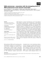
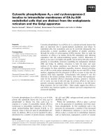
patients. Figure 1 illustrates a lesion just superior to the
areola. Since a standard lumpectomy would remove
approximately 1 cm of normal breast surrounding the
lesion, the overlying skin bridge would be too thin for
balloon-based APBI. The EBT balloon appears to be
slightly thicker than other APBI balloons. When the bal-
loon is inflated, the tissue bridge superficial to the bal-
loon tends to compress to a greater degree with the
Axxent balloon than with other APBI balloons. Co nse-
quently the overlying skin bridge should be a minimum
of 1.2-1.5 cm. In this example it is necessary to excise
all breast and skin within an area of less than 2.5 cm of
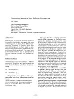
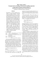
the skin surface. As seen in Fi gure 2, this can re adily be
accomplished by using modifications of the standard
oncoplastics incisions. Since the lesion in this case was
close to the areolar edge, we chose a bat wing masto-
pexy. With this approach for APBI, we are also inter-
ested in the tissue depth between the back side of the
balloon and the underlying ribs and lung. By not carry-
ing the excision to the full thickness commonly illu-
strated in oncoplastics descriptions [26], we preserve
some breast tissue to add to the posterior spacing and
offer more lung protection.
Retractors were not used in order to avoid beveling
toward the skin. Ins tead of retractors, two prolene
stitches were placed lateral and medial to the lesion, and,
as these were pulled upward, electrocautery was used to
cut toward the lesion. The flat superficial surface is
where the skin island is located. Beveling outward is
minimal, and the bottom side of the removed tissue “V"s
downward, resulting in a shape similar to that of a typical
solitaire cut diamond. It is important to have supporting
breast tissue structure, especially in older women.
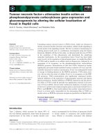
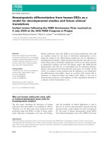
Optimal closures (Figure 3) were performed in three
layers and began deeper than conventional closure, at
least 12-15 mm from the skin, making sure to take a
generous thickness of tissue. The dense superficial fascia
of the breast and superficial dense breast tissue when
present will hold the sutures for this layer most effec-
tively. An option for a resilient closure, which will not
collapse during balloon inflation, is the use of a run-
ning barbed suture, such as a Quill suture, for each of
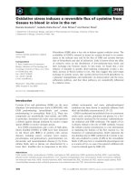
these layers. An inflated balloon exerts pressure against
the skin, and an adequate skin bridge will maintain the
distance from the balloon to the skin surface. The
greater the volume of the balloon, with the attendant
compression of adjacent breast tissue, the more fully
the target breast tissue will be irradiated to achieve
margin sterilization. These techniques usually place the
center of the inflated balloon slightly deeper than the
original tumor center but maintain all residual breast
tissues within 1 cm of the lumpectomy tightly com-
pressed to the balloon surface for optimal therapy (Fig-
ure 4). In some patients, implantation of the balloon
may be possible at the time of lumpectomy. Alterna-
tively some surgeons have used a removable place-
holder device, a cavity evaluation device (CED), to
preserve the cavity while fashioning an easily accessible
tract between the cavity and the skin surface [11]. This
can facilita te balloon implantation in the post-operative
period. Post-operative antibiotic coverage is used in
this circumstance to lessen the risk of infection. As
with other devices, the surgical technique should be be
discussed with the radiation oncologist to enable
proper treatment planning.
Figure 1 Side view of a small breast cancer just superior to the
areolar edge.
Dooley et al. World Journal of Surgical Oncology 2011, 9:30
/>Page 6 of 10
In this study, balloon applicators were implanted up to
61 days post-lumpectomy. In the 3 patients with inade-
quate balloon conformance to cavity geometry, the time
from lumpectomy to implantation for each was 15, 16
and 19 days, which was less than the mean for the study
group. Tumor characteristics were similar between the
eligible group and ineligible group. The age range of
patients was 45-84 years, with a range of lesion sizes
from 0.2-2.8 cm. Breast cup sizes were evenly distribu-
ted between B, C, and D. No apparent trends were
noted between eligible and ineligible patients although
the sample size limits statistical analysis.
The prescription dose of 34 Gy was delivered in 42/44
patients, and 2/44 patients received total doses of 30.6 and
33.96 Gy [20]. Treatment was well tolerated, and adverse
events were similar to adverse events with other forms of
APBI. Complications associated with the imp lantation of
the balloon applicator are similar to complications
reported during the insertion of a post surgical drain [27].
This type of complication has also been r eported with
192
Ir-based balloon brachytherapy [11,15,16]. During this
EBT study one patient had incisional redness/drainage at
3 months post-treatment, 2 patients had infection at 3 and
6 months, respectively, and 2 pati ents had seromas at or
within 4 weeks of treatment. The patients who were
Figure 2 Modified bat wing approach . This approach allows
excision of the tumor and a thin skin bridge but preserves posterior
breast tissue for lung spacing.
Figure 3 Closed incision with several layers of running barbed
suture.
Figure 4 Electronic brachytherapy balloon inflated with source
active in place with adequate skin spacing.
Dooley et al. World Journal of Surgical Oncology 2011, 9:30
/>Page 7 of 10
Table 4 Comparison electronic brachytherapy (EBT) and
192
Iridium brachytherapy (IBT)
Patient/Tumor Characteristics EBT IBT
Number of Patients Enrolled 65 75
Number of Patients Treated 44 (67.7%) 43 (57.3%)
Age: mean (range) 64 years (45-84) 69 years (50-90)
Menopausal Status: n (%)
Pre-Menopausal 1 (2.3%) 0
Peri-Menopausal 2 (4.6%) 2 (5%)
Post-Menopausal 41 (93.2%) 41 (95%)
Tumor Size: mean (range) 1.2 cm (0.2-2.8 cm) 1.0 cm
AJCC Class: n (%)
Tis 12 (27.3%) 0
T1a 1 (2.3%) 9 (21%)
T1b 8 (18.2%) 16 (37%)
T1c 21 (47.7%) 18 (42%)
T2 2 (4.6%) 0
Histopathologic Grade
G1 Well Differentiated 12 (27.3%) 17 (40%)
G2 Moderately Differentiated 18 (40.9%) 16 (37%)
G3 Poorly Differentiated 10 (22.7%) 6 (14%)
Grade Not Available 4 (9.1%) 4 (9%)
Minimum Distance from Balloon to Skin Surface 6 mm 5 mm
Breast Cup Size
A 0 1 (2%)
B 12 (27.3%) 9 (21%)
C 16 (36.4%) 15 (35%)
D+ 11 (25.0%) 11 (26%)
Not reported 5 (11.4%) 7 (16%)
Cosmesis EBT IBT
Good - Excellent cosmesis at 1 month 35/44 (80%) 38/43 (88%)
Good - Excellent cosmesis at 1 year 24/32 (75%) –
Good - Excellent cosmesis at 5 years – 35/43 (81%)
Adverse Events in > 2 patients EBT
a
IBT
b
Blistering 3 (6.8%) 2 (3.7%)
Bruising / hematoma 1 (2.3%) 3 (5.6%)
Catheter Site Drainage 0 28 (51.9%)
Desquamation, Dry 2 (4.5%) 7 (13.0%)
Desquamation, Moist 3 (6.8%) 3 (5.6%)
Dry Skin (Breast) 2 (4.5%) 6 (11.1%)
Ecchymosis 1 (2.3%) 17 (31.5%)
Edema (breast) 0 8 (14.8%)
Erythema, redness/rash 27 (61.4%) 31 (57.4%)
Fibrosis 2 (4.5%) 3 (5.6%)
Hyperpigmentation / Hypopigmentation 9 (20.5%) 5 (9.3%)
Induration 3 (6.8%) 1 (1.9%)
Infection 2 (4.5%) 2 (3.7%)
Itching / Pruritis 4 (9.1%) 5 (9.3%)
Pain, tenderness, discomfort 13 (29.5%) 23 (42.6%)
Rash 0 4 (7.4%)
Seroma 2 (4.5%) 6 (11.1%)
Skin Irritation 0 3 (5.6%)
Patient and tumor characteristics at baseline, cosmesis at 1 month and 1 or 5 years, and adverse events (breast and skin symptoms and signs) following EBT at 6
months as reported herein and by Mehta, et al.,
20
or IBT at 1 month as reported by Keisch, et al.
21
a
Adverse events in 1 or 2 patients in the EBT study (not listed above) included firmness of breast tissue, skin firmness, non-calcified mass (2.5 cm), rib pain,
serosanguinous drainage, swelling, and wound complication (non-infection).
b
Adverse events in 1 or 2 patients in the IBT study (not listed above) included abscess, eschar, mastitis, serosanguinous drainage, telangiectasia, and
vasodilatation.
24
Dooley et al. World Journal of Surgical Oncology 2011, 9:30
/>Page 8 of 10
diagnosed with infection were treated with oral antibiotics,
and the infections resolv ed. Study sites used standard
wound care procedures at the skin exit site, which
included the use of topical antibiotic ointment and/or
hydrogen peroxide. Patients were instructed to wear a sur-
gical bra and avoid showering during the 5-day treatment
period. Patient compliance with the treatment regimen
was excellent. Patients expressed satisfaction with the con-
duct of the study as well as the delivery of the radiotherapy
based on a questionnaire given at follow-up visits. Mehta,
et al., reported cosmesis to have been evaluated as good to
excellent by 80% of patients at 6 months [20].
Our initial experience with both a new balloon applica-
tor and a novel radiation source parallels t he initia l
experience with the
192
Ir-based balloon brachytherapy
[17,21], and the two studies are similar in enrollment,
subsequent treatment eligibility, patient characteristics,
tumor characteristics, and cosmetic results following
EBT and
192
Ir-based brachytherapy as summarized in
Table 4 [17,20,21]. The range and frequency of adverse
events were similar for erythema, pain, and infection.
However, catheter site drainage was reported in 52% of
patients following
192
Ir- based brachytherapy but was not
reported with EBT. This may be due to differences in the
design of the balloon applicators. The most significant
difference with the EBT system is the use of an electronic
high dose rate, low energy X-ray source to generate
radiation, which eliminates the i ssues involved in the
handling and storage of radioactive isotopes [18]. Many
radiation treatment centers as well as community hospi-
tals across the United States lack the funds and infra-
structure to maintain isotopes o r build heavily shielded
treatment rooms that are required for the delivery of
HDR brachytherapy. T he EBT system does not require a
heavily shielded treatment room or an HDR brachyther-
apy afterloader. The EBT system should provide a means
that will allow women not currently able to travel to
radiation facilities with HDR brachytherapy afterloaders
to be treated within their local communities and receive
state of the art breast radiotherapy as an adjunct to mod-
ern breast conserving surgery.
Conclusions
Early stage breast cancer can be treated with breast con-
servin g therapy and accelerated partial breast irradiation
using electronic brachytherapy. Treatment was well tol-
erated, and these early outcomes were similar to the
early outcomes with iridium-based balloon
brachytherapy.
Abbreviations
APBI: accelerated partial breast irradiation; CED: cavity evaluation device; CT:
computerized tomography; CTC: Common Terminology Criteria; cm:
centimeter; EBT: electronic brachytherapy; Gy: gray; HDR: High dose rate; Inc:
incorporated; Ir: Iridium.
Acknowledgements
The authors wish to thank all research staff who supported this research.
The authors would like to acknowledge Leslie Todd for assistance with this
manuscript on behalf of Xoft, Inc.
Author details
1
University of Oklahoma Health Sciences Center, 825 NE 10th Street Suite
4500, Oklahoma City, OK 73104, USA.
2
Department of Radiation Oncology,
University of Oklahoma Health Sciences Center, 825 North East 10th Street
Suite 1430, Oklahoma City, OK 73104, USA.
3
Rush University Medical Center,
60 E Delaware Place Suite 1400, Chicago, IL 60611, USA.
4
Rush University
Medical Center, 1725 West Harrison Street Suite 810, Chicago, IL 60612, USA.
5
Rhode Island Hospital, Providence, RI 02903, USA.
6
Enterprise Surgical, 91
Washington St Unit 302, Taunton, MA 02780, USA.
7
Swedish Cancer Institute,
Comprehensive Breast Center, Swedish Medical Center, 1600 East Jefferson
St. Suite 305, Seattle, WA 98122, USA.
8
Dickstein Cancer Center, White Plains
Hospital, White Plains, 4 Longview Ave, NY 10601, USA.
9
Holy Cross Medical
Center, 2101 Medical Parks Drive Suite 304, Silver Spring, MD 20902, USA.
10
Beth Israel Medical Center, 10 Union Square East, New York City, NY 10003,
USA.
Authors’ contributions
All authors contributed to treatment of patients, collection of data, review of
results and manuscript, and approval of the final draft.
Competing interests
Darius Francescatti is a paid consulting surgical medical director for Xoft Inc.
All other authors declare that they have no competing interests.
Received: 17 August 2010 Accepted: 7 March 2011
Published: 7 March 2011
References
1. Hulvat MC, Hansen NM, Jeruss JS: Multidisciplinary care for patients with
breast cancer. Surg Clin North Am 2009, 89:133-176.
2. Hunt KK, Buchholz TA, Hortobagyi GN: Now and Later? The sticky
question of lymph node management in patients receiving preoperative
chemotherapy. Ann Surg Oncol 2005, 12:683-685.
3. Whelan TJ, Julian J, Wright J, Jadad AR, Levine ML: Does locoregional
radiation therapy improve survival in breast cancer? A meta-analysis. J
Clin Oncol 2000, 18:1220-1229.
4. Clarke M, Collins R, Darby S, Davies C, Elphinstone P, Evans E, Godwin J,
Gray R, Hicks C, James S, MacKinnon E, McGale P, McHugh T, Peto R,
Taylor C, Wang Y, Early Breast Cancer Trialists’ Collaborative Group
(EBCTCG): Effects of radiotherapy and of differences in the extent of
surgery for early breast cancer on local recurrence and 15-year survival:
an overview of the randomized trials. Lancet 2005, 366:2087-2106.
5. Fisher B, Anderson F, Redmond C, Wolmark N, Wickerham DL, Cronin WM:
Reanalysis and results after 12 years of follow-up in a randomized
clinical trial comparing total mastectomy and lumpectomy with or
without irradiation in the treatment of breast cancer. N Eng J Med 1995,
333:1456-1461.
6. Pawlik TM, Buchholz TA, Kuerer HM: The Biologic Rational for and
Emerging Role of Accelerated Partial Breast Irradiation for Breast Cancer.
J Am Coll Surg 2004, 199:479-492.
7. Schroen AT, Brenin DR, Kelly MD, Knaus WA, Slingluff CL Jr: Impact of
patient distance to radiation therapy on mastectomy use in early-stage
breast cancer patients. J Clin Oncol 2005, 23:7074-7080.
8. Athas WF, Adams-Cameron M, Hunt WC, Amir-Fazli A, Key CR: Travel
distance to radiation therapy and receipt of radiotherapy following
breast conserving surgery. J Natl Cancer Inst 2000, 92:269-271.
9. Hébert-Croteau N, Brisson J, Latreille J, Blanchette C, Deschênes L:
Compliance with consensus recommendations for the treatment of early
stage breast carcinoma in elderly women. Cancer 1999, 85:1104-1113.
10. Vicini FA, Baglan KL, Kestin LL, Mitchell C, Chen PY, Frazier RC,
Edmundson G, Goldstein NS, Benitez P, Huang RR, Martinez A: Accelerated
treatment of breast cancer. J Clin Oncol 2001, 19:1993-2001.
Dooley et al. World Journal of Surgical Oncology 2011, 9:30
/>Page 9 of 10
11. Beitsch PD, Hodge CW, Dowlat K, Francescatti D, Gittleman MA, Israel P,
Nelson JC, Potruch T, Snider HC Jr, Whitworth P, Zannis VJ, Patel RR: The
surgeon’s role in breast brachytherapy. Breast J 2009, 15:93-100.
12. Sauer R, Sautter-Bihl ML, Budach W, Feyer P, Harms W, Souchan R,
Wollwiener D, Kreienberg R, Wenz F: Accelerated partial breast irradiation:
Concensus statement of 3 German oncology societies. Cancer 2007,
110:1187-1194.
13. Ott OJ, Hildebrandt G, Pötter R, Hammer J, Hindemith M, Resch A, Spiegl K,
Lotter M, Uter W, Kortmann RD, Schrauder M, Beckmann MW, Fietkau R,
Strnad V: Accelerated partial breast irradiation with interstitial implants:
risk factors associated with increased local recurrence. Int J Radiat Oncol
Biol Phys 2010.
14. Polgár C, Major T, Fodor J, Sulyok Z, Somogyi A, Lövey K, Németh G,
Kásler M: Accelerated partial-breast irradiation using high-dose-rate
interstitial brachytherapy: 12-year update of a prospective clinical study.
Radiother Oncol 2010, 94:274-279, Epub 2010 Feb 22.
15. Khan AJ, Vicini F, Beitsch P, Haffty B, Quiet C, Keleher A, Garcia D, Snider H,
Gittleman M, Zannis V, Kuerer H, Whitacre E, Whitworth P Jr, Fine R: Local
control, toxicity, and cosmesis in women younger than 50 enrolled onto
the American Society of Breast Surgeons Mammosite Radiation Therapy
System Registry Trial. Ann Surg Oncol 2009, 16:1612-1618.
16. Jeruss JS, Vicini FA, Beitsch PD, Haffty BG, Quiet CA, Zannis VJ, Keleher AJ,
Garcia DM, Snider HC, Gittleman MA, Whitacre E, Whitworth PW, Fine RE,
Arrambide S, Kuerer HM: Initial outcomes for patients treated on the
American Society of Breast Surgeons MammoSite clinical trial for ductal
carcinoma-in situ of the breast. Ann Surg Oncol 2006, 13:967-976.
17. Benitez PR, Keisch ME, Vicini F, Stolier A, Scroggins T, Walker A, White J,
Hedberg P, Hebert M, Arthur D, Zannis V, Quiet C, Streeter O, Silverstein M:
Five-year results: the initial clinical trial of Mammosite balloon
brachytherapy for partial breast irradiation in early-stage breast cancer.
Am J Surg 2007, 194:456-462.
18. Dickler A, Kirk MC, Seif N, Griem K, Dowlatshahi K, Francescatti D,
Abrams RA: A dosimetric comparison of MammoSite high-dose-rate
brachytherapy and Xoft Axxent® electronic brachytherapy. Brachytherapy
2007, 6:164-168.
19. Dickler A: Xoft Axxent® electronic brachytherapy: a new device for
delivering brachytherapy to the breast. Nat Clin Pract Oncol 2009,
6:138-142.
20. Mehta VK, Algan O, Griem KL, Dickler A, Haile K, Wazer DE, Stevens RE,
Chadha M, Kurtzman S, Modin SD, Dowlatshahi K, Elliott KW, Rusch TW:
Experience with an electronic brachytherapy technique for intracavitary
accelerated partial breast irradiation. Am J Clin Onc 2010, 33:327-335.
21. Keisch M, Vicini F, Kuske RR, Hebert M, White J, Quiet C, Arthur D,
Scroggins T, Streeter O: Initial clinical experience with the Mammosite
breast brachytherapy applicator in women with early-stage breast
cancer treated with breast-conserving therapy. Int J Radiat Oncol Biol Phys
2003, 55:289-293.
22. Pawlik TM, Perry A, Strom EA, Babiera GV, Buchholz TA, Singletary E,
Perkins GH, Ross MI, Schecter NR, Meric-Bernstam F, Ames FC, Hunt KK,
Kuerer HM: Potential applicability of balloon catheter-based accelerated
partial breast irradiation after conservative surgery for breast carcinoma.
Cancer 2004, 100:490-498.
23. Arthur DW, Vicini FA, Kuske RR, Wazer DE, Nag S, American Brachytherapy
Society: Accelerated partial breast irradiation: an updated report from
the American Brachytherapy Society. Brachytherapy 2003, 2:124-130.
24. Trotti A, Colevas AD, Setser A, Rusch V, Jaques D, Budach V, Langer C,
Murphy B, Cumberlin R, Coleman CN, Rubin P: CTCAE v3.0: development
of a comprehensive grading system for the adverse effects of cancer
treatment. Semin Radiat Oncol 2003, 13:176-181.
25. Chen SS, Strauss JB, Shah AP, Rao RD, Bernard DA, Griem KL: Radiation
recall reaction with Docetaxel administration after accelerated partial
breast irradiation with electronic brachytherapy. Brachytherapy 2009,
8:331-334.
26. Clough KB, Kaufman GJ, Nos C, Buccimazza I, Sarfati IM: Improving breast
cancer surgery: a classification and quadrant per quadrant atlas for
oncoplastic surgery. Ann Surg Oncol 2010, 17:1375-1391.
27. Chadha NK, Cumming S, O’Connor R, Burke M: Is discharge home with
drains after breast surgery producing satisfactory outcomes? Ann R Coll
Surg Engl 2004, 86:353-357.
doi:10.1186/1477-7819-9-30
Cite this article as: Dooley et al.: Surgical perspectives from a
prospective, nonrandomized, multicenter study of breast conserving
surgery and adjuvant electronic brachytherapy for the treatment of
breast cancer. World Journal of Surgical Oncology 2011 9:30.
Submit your next manuscript to BioMed Central
and take full advantage of:
• Convenient online submission
• Thorough peer review
• No space constraints or color figure charges
• Immediate publication on acceptance
• Inclusion in PubMed, CAS, Scopus and Google Scholar
• Research which is freely available for redistribution
Submit your manuscript at
www.biomedcentral.com/submit
Dooley et al. World Journal of Surgical Oncology 2011, 9:30
/>Page 10 of 10