báo cáo khoa học: "A Review of Metallothionein Isoforms and their Role in Pathophysiology" ppsx
Bạn đang xem bản rút gọn của tài liệu. Xem và tải ngay bản đầy đủ của tài liệu tại đây (288.61 KB, 7 trang )
REVIEW Open Access
A Review of Metallothionein Isoforms and their
Role in Pathophysiology
N Thirumoorthy
1*
, A Shyam Sunder
2
, KT Manisenthil Kumar
3
, M Senthil kumar
3
, GNK Ganesh
4
and
Malay Chatterjee
5
Abstract
The Metallothionein (MT) is a protein which has several interesting biological effects and has been demonstrated
increase focus on the role of MT in various biological systems in the past three decades. The studies on the role of
MT were limited with few areas like apoptosis and antioxidants in selected organs even fifty years after its
discovery. Now acknowledge the explo ration of various isoforms of MT such as MT-I, MT-II, MT-III and MT-IV and
other isoforms in various biological systems.
Strong evidence exists that MT modulates complex diseases and the immune system in the body but the primary
function of MT still remains unknown. This review’s main objective is to explore the capability to specifically
manipulate MT levels in cells and in animals to provide answers regarding how MT could impact those complex
disease scenarios.
The experimental result mentioned in this review related among MT, zinc, cadmium, diabetic, heart disease, bone
retardation, neuro toxicity, kidney dysfunction, cancer, and brain suggest novel method for exploration and
contribute significantly to the growing scientist to research further in this field.
Keywords: Metallothionein Isoforms MT-I, MT-II, MT-III, MT-IV
Introduction
The metallothionein (MT) was first isolated in 1957
from the cortex of horse kidney as a cadmium binding
protein [1]. This protein was first report ed by Kagi and
Vallee in 1960 and by Kojima in 1976 as cysteine-rich
(33 mol %), low molecular weight (7 kDa), heat-stable
and metal binding protein.
There are at least ten known closely related metal-
lothionein proteins expressed in the human body. In
humans, large quantities are synthesized primarily in the
liver and kidneys, however they have been found at a
number of other sites as well. Its production is depen-
dent on availability of the dietary minerals zinc and sele-
nium, and the amino acids histidine and cysteine
present in the body.
This protein has properties like detoxification of heavy
metals like mercury and cadmium, homeostasis o f
essential metals including copper and zinc, antioxidation
against reactive oxygen species, protect against DNA
damage, oxidative stress, cell survi val, angiogenesis,
apoptosis, as well as increase proliferation, etc in the
body [2]. In general the MT is known to modulate three
fundamental processes: 1) the release of gaseous media-
tors such as hydroxyl radical or nitric oxide; 2) apopto-
sis, and 3) the binding and exchange of heavy metals
such as zinc, cadmium or copper.
MT and Its Isoforms
MT isoforms are classified based on various factors like
molecular weight, metal which bind, encoded genes,
chromosomes, binding atoms, amino acids environment
etc. Broadly it is classified as major and minor groups.
The major groups are MT-1 and MT-2; these are the
unique structure which is identical for the two major
isoforms binds 7 g atoms of divalent metals like zinc
and cadmium. The MT-3 and MT-4 are minor isoforms
which are normally found in specialized cells. The MT-
3 protein was first isolated as a growth inhibiting factor
(GIF)frombrainneurons,andtheMT-4proteinwas
found in stratified epithelium [3].
* Correspondence:
1
Dept. of Pharmaceutics, Cherraan’s College of Pharmacy, 521 Siruvani Road,
Perur, Coimbatore-39, TN, India
Full list of author information is available at the end of the article
Thirumoorthy et al. World Journal of Surgical Oncology 2011, 9:54
/>WORLD JOURNAL OF
SURGICAL ONCOLOGY
© 2011 Thiru moorthy et al; licensee BioMed Central Ltd. This is an Open Access article distributed under the terms of the Creative
Commons Attribution License ( nses/by/2.0), which permits unrestricted use, distribution, and
reproduction in any medium, provided the original work is properly cited.
In human, the MT proteins are encoded by a family of
genes which are located on chromosome 16q13, and
may involve at least 10 identified functional genes [3].
This main isomers are again subdivided as MT I a, MT
I b, MT I c etc or any other resemble pattern.
Another study classified MT I and II based on the
sequences and that do not consider either the state of
the thiols or the cargo, namely which metal is associated
with these protein but there is a scarcity of functional
studies that would relate them to class I MTs.
The four isoforms of MT are identified in mammals,
three of which, MT-I, II and III are found in the central
nervous system. The expression of MT-I/II is mainly loca-
lized i n g lias and is induced by exposure to metals including
Hg, C d, Cu and Zn, cytokines and ROS. On t he other hand,
the MT-III isoform are mainly present in neurons and are
not easily i nduced by exposure to the above agen ts [4].
Four classes of MTs have been characterized in mam-
mals. The MT-I and II genes are expressed in many tis-
sues, and at a particularly high level in liver and kidney.
Expression of MT-III is restricted to the brain and to
male reproductive organs, while that of MT-IV is speci-
fic to stratified squamous epitheli a, since mice that can-
not synthesize either MT-I or MT-II and reproduce
normal ly. Mice lacking MT-III do not reveal any neuro-
logical or behavioral deficiencies [5].
A another classification based on a large family of eukar-
yote and some are in prokaryote refers to three classes: (i)
proteins with sequences related to mammalian MT, (ii)
proteins with sequences not related to mammalian MT,
and (iii) peptides that are not genetically encoded [6]. All
above classification are listed in below table 1.
General task of MT isoforms in the Body
Four primary MT proteins and their important role in
the body
• MT-I and II are present in all cells throughout the
body. They regulate copper and zinc, are involved in
cell transcription, detoxify heavy metals, play a role
in immune function, and are involved in a variety of
G.I. tract functions.
• MT-III is found primarily in the brain and func-
tions as a growth inhibitory factor in the brain. MT-
III is located primarily in the central nervous system
with small amounts present in the pancreas and
intestines. It plays a major role in the development,
organization and programmed death of brain cells.
• MT-IV is found in the skin and upper G.I. tract.
They help regulate stomach acid pH, taste and tex-
ture discrimination of the tongue and help protect
against sunburn and other skin traumas.
Structure of MT
Spectroscopic Characterization
Few studies have revealed the structure and characteri-
zation of MT. The three-dimensional protein structure
of this was reported by both X-ray crystallography and
NMR spectroscopy in the 1990s. Structural studies have
shown that this unusual protein with 61 amino acids
(mammalian MT) can bind with both essential metals
(zinc and copper) and toxic metals (cadmium and mer-
cury) in two distinct cluster structures within the mole-
cule. One cluster is closer to the N-terminal and three
metal atoms are b ound to nine cysteines with three
bridging sulfur atoms, while in the second cluster closer
to the C-terminal and four metal atoms are bound to 11
cysteines with five bridging sulfur atoms [7].
Fluorescent probes for the structure of MT
Fluorescence methods for demonstrate the structure of
human metallothionein in vivo which depends on the
presence of metal ions and the redox environment of
amino acids. Attachment of fluorescent labels generated
metallothionein FRET (fluorescence resonance energy
transfer) sensors for investigating its structure and func-
tion in living cells differential chemical modifications of
Table 1 Classification of MT-Isomers
S.No Based on Classification
1. Metals Present Major -MT-1 &MT-2
Minor - MT-3 &MT-4
2. Proteins are encoded by a family of genes which are located on chromosome 16q13 MT I a, MT I b, MT I c etc
3. Biological system Central nervous system MT-I, MT-II and MT-III
Neurons MT-III
4. Family of eukaryote and some are in prokaryote Proteins with sequences related to mammalian MT MT- i
Proteins with sequences not related to mammalian MT MT- ii
Peptides that are not genetically encoded MT- iii
5. Expressed in tissues Liver and kidney MT-I and MT-II
Brain and to male reproductive organs MT-III
Specific to stratified squamous epithelia MT-IV
Thirumoorthy et al. World Journal of Surgical Oncology 2011, 9:54
/>Page 2 of 7
its cysteine thiols with fluorescent probes allowed three
states in structure: 1)Metallothionein (zinc-bound
thiolate), 2) Thionein (free thiols), and 3) Thionin
(disulfides). Interrogation of this zinc-binding property
with fluorescent chelating agents revealed that the
affinities of the seven zinc ions over four orders of
magnitude [6].
MT Isomers and Cancer
In Breast Cancer
These isoforms are expressed in a tissue specific pattern
and may play distinct roles in the different cell types.
There are several reports on the expression of certain
specific isoforms in various human tumors. The mRNA
ofMT-1seriesnamedasA,E,F,G,H,XandMT-3
isoforms but not MT-1B and MT-4 isoforms have been
detected in breast cancer tissues. The MT-2A mRNA
trans cript which has been reported to be highest among
all the functional isoforms detected in breast tissues and
is positively correlated with cell proliferation and histo-
logical grade. Expression of MT-1F isoform has also
been found to influence histological differentiation in
invasive breast cancer since estrogen is known to play
important role in b reast cancer tumorgenesis, the MT-
1E isoform has been postulated to participate in alterna-
tive processes that replace the function of estrogen. It
has also been reported that MT-3 isoform overexpres-
sion is associated with a poor prognosis for patients
with breast cancer [3].
In Renal Tumor
The renal cell cancer tissue shows three different type of
expression as up-regulation of MT-2A, down-reg ulati on
of MT-1A and MT-1G transcripts. Expressi on of the
MT-3 isoform h as been reported in the tubules of nor-
mal kidney and also in renal cell carcinoma along with
other isoforms of MT. The expression of the MT-3 iso-
form in cancerous bladder tissues which was absent in
normal bladder tissues, and suggested its use as a poten-
tial biomarker for bladder cancer. They have also shown
high levels of MT-1X mRNA expressio n in bladder can-
cer. The MT-3 isoform which was originally reported as
specific to brain has been demonstrated in normal
human kidney, renal carcinoma, bladder cancer and pro-
static adenocarcinoma [3].
In Prostate Cancer
In normal prostate tissue, the MT-I A, E, X and MT-2A
isoforms were present but there was a down-regulation
of the MT-IX isoform in advanced prostate cancer. It
wasreportedthatMT-1andMT-2isoformsmaybe
related to the proliferative activity of breast, colon and
prostate human cancers [3].
In Papillary Thyroid Cancer
MT isoforms have not been much studied in papillary
thyroid cancer. The function of MT1 and MT2 isoforms
in papillary thyroid cancer cells (KAT5) demonstrated
that KAT5 cells expressed eight functional MT1 and
MT2 isoforms induced by cadmium. Elevated calcium
and activated ERK1/2 pre dated MT expression. The
alternation in cell cycle disappeared when the expression
of MT isoforms was blocked by calcium inhibitor or
ERK1/2 inhibitor. Collectively, KAT5 cells express eight
functional MT1 and MT2 isoforms in a pathway con-
trolled by calcium and ER K1/2. The elevation of the
MT isoforms contributes to the decreased G0/G1 but
increased G2-M phase revealed a novel pathway for the
expression of the functional MT in papillary thyroid
cancer. Bone thyroid cancers are classified as papillary,
follicular, medullary, and undifferentiated or anaplastic
[8]. Table 2 is to express the role of MT isoforms on
cancer.
Bone Growth Retardation and MT Isomers
Bone growth retardation, zinc and its binding protein
MT are important in regulating growth and develop-
ment of bone. A study on relationship between dietary
Zn and MT interact in regulatin g bone growth were
reported that the MT mice, having lower Zn concentra-
tions in plasma and long bone, showed growth retarda-
tion as demonstrated by lower body length gain, shorter
and smaller tibia/femur, lower chondrocyte prolifera-
tion, reduced metaphysis heights, but increased osteo-
clast densities on trabecular bone, particularly in mice
fed Zn low diet (Zn-L). The mRNA expression of MT-
I&II was induced in mice fed with the Zn-L diet possi-
bly compensating for Zn limitation that interact
between dietary Zn and endogenous MT is important
for maximal bone growth, and particularly important in
the regulation of Zn pool for bone growth during mod-
erate Zn limitation [9].
Role in Oxidative Stress
Recent experiments have shown that thiolate ligands in
MT confer redox activity on zinc clusters. This strongl y
suggests that MT would control the cellular zinc distri-
bution as a function of the cellular energy state [2]. A
review of report have proved that the anti oxidant prop-
erty of MT enhances in presence of zinc. The zinc
redox-dependent functions of MT are important for the
regulation of physiological processes that depend on
zinc and the pathological processes in which oxidative
stress mobilizes zinc. The decrease of zinc availability
from MT suggests that the mutant MT is either less
reactive towards nitric oxide or it is in an oxidized state
and does not bind sufficient amounts of zinc [6].
Thirumoorthy et al. World Journal of Surgical Oncology 2011, 9:54
/>Page 3 of 7
A DNA microarray used to examine genes induce d by
gallium nitrate in specified cells named as CCRF-CEM
cells. This study found that gallium increased MT-2A and
heme oxygenase-1 (HO-1) gene expression and altered the
levels of other stress-related genes. Gallium nitrate
increased the phosphorylation of p38 mitogen-activated
protein kinase and activated Nrf-2, a regulator of HO-1
gene transcription. Gallium induced Nrf-2 activation and
HO-1 expression were diminished by a p38 MAP kinase
inhibitor. This concludes that gallium nitrate induces cel-
lular oxidative stress that triggers the expression of HO-1
and MT2A through different pathways [10].
Protection in cadmium toxicity
Cadmium (Cd) is an environmental pollutant ranked
eighth in the top 20 hazardous substances and the
human activity has markedly increased the distribution
of Cd in the global environment. Cd is toxic to number
of tissues in body. Prolonged exposure to Cd produces
nephrotoxicity, osteotoxicity, and immunotoxicity. this is
also classified as a human carcinogen causing genitour-
inary disorders like tumors of the lung, prostate, injec-
tion site, and other tissues. Most of Cd in the body is
bound to a small, cysteine-rich, metal binding protein
MT [11].
This protein expression in Cd-induced tumors varies
depending on the type and the stage of tumor develop-
ment. High levels of MT are detected in Cd-induced
sarcomas at the injection site and sarcomas metastases
are devoid of MT suggest the critical role for protecting
human health from Cd toxicity either by neither detoxi-
fication nor heavy metal binding [12].
MT in Kidney
The prevalence of cadmium-related kidney dysfunction
among population groups residing in cadmium contami-
nated areas in China report reveal a dose-response
relationships between urinary-Cd and renal tubular dys-
function such as urinary beta-2-microglobulin or N-
acetyl-beta-D-glucosaminidase-NAG or urinary albumin,
a biomarker of glomerular kidney dysfunction. Since
long term cadmium exposure in occupational and gen-
eralenvironmentsmaygiverisetokidneydysfunction.
These dose-response relationships include:
1) MT- mRNA levels in peripheral blood
lymphocytes,
2) Biomarker of the ability of each person, and
3) To synthesize metallothionein (a protein known
to provide intracellular protection against cadmium
toxicity [13].
In The Central Nervous System
ThetransgenicmodelsofMTexpressionwereestab-
lished using various experimental approaches. This
important protein plays a major role in the defense
against neurodegenerative disorders and other injuries,
influencetissuearchitectureandcognition,finallypro-
tect against mercury neurotoxicity [14].
Nicotine treatment, which can improve working mem-
ory, eliminated the impairment associated with the dele-
tion of the MT-1 and MT-2 genes in a dose-related
fashion after acquisition t raining in the aging adult
mice. These have been suggested its roles in metal phy-
siology or cellular protection are involved in spatial
learning and memory function. These studies conclude
that MT has important functions in the central nervo us
system and brain because MT-1 and MT-2 protect the
central nervous system from damage induced by inter-
leukin, 6-aminonicotinamide, kainic acid, and physical
injury [15].
This investigates also studied that, transition metals
have been associated with impaired neurological devel-
opment, and neurobehavioral activity. The role of MT
Table 2 Showing the Isoforms of MT on Tumorgenesis
S.
NO
CANCER ISOFORMS ROLE
1 Breast Cancer MT-1 A,E,F,X, H,G, MT-3,
MT-2A.
1) MT-1E: Alternative processes that replace the function of estrogen i;e breast cancer
tumorgenesis.
2) MT-3: Poor prognosis for patients with breast cancer.
3) MT-2A: Detected in breast tissue is positively correlated with cell proliferation and histological
grade.
4) MT-1F: It found to influence histological differentiation in invasive breast cancer.
2 Renal Cancer MT-1A, MT-1G, MT-IX,
MT-3.
1) MT-3: Normal kidney & also in renal cell carcinoma, cancerous bladder tissues,(which was
absent in normal bladder tissues)prostatic adenocarcinoma
2) MT-IX:Bladder cancer.
3 Prostate Cancer MT-1A, MT-1E, MT-1X,
MT-2A, MT-1, MT-2.
1) MT-IX:Advanced prostate cancer.
2) MT-1, MT-2: proliferative activity of breast, colon & prostate human cancer.
4 Papillary
Thyroid Cancer
MT-1, MT-2. Thyroid Cancer cells (KAT5 cells) + Calcium elevated ERK1/2 ® 8 functional MT-1&MT2 induced
By cd ® MT expression+ Decrease in G0, G1 phase, Increase in G2-M phase ®
Novel pathway for Functional MT expression.
Thirumoorthy et al. World Journal of Surgical Oncology 2011, 9:54
/>Page 4 of 7
in learning and memory of mice with deletions of two
metalloth ione in genes (MT-1 and MT-2) were trained
on a win-shift task in an 8-arm radial maze. The paren-
tal strain of these mice learned the maze at a normal
rate over an 18-session acquisition period. In contrast,
the MT-1/MT-2-null mice, which had a similar choice
accuracy level at the beginning of training, showed a
poorer rate of learning during the training period. In
addition, the MT-1/MT-2-null mice showed significantly
less choice of accuracy than the parental strain [15].
MT in Heart Disease
MT-IIA in heart-derived cell line in human confers
oxidative protection
MT is a metal binding protein and cardio protective. In
order to understand the molecular mechanisms underly-
ing the role of MT in the heart a study established a
stable MT-IIA over-expressing cardiac cell line, and
evaluated its anti-oxidative property. The transfected
cell line (H9c2MT7) exhibited similar growth kinetics
and morphology.
The western blotting analysis of this study showed
that H9c2MT7 had a remarkable increase in MT protein
level compared with the parent cell line H9c2. Upon
addition of 25 M ZnSO
4
showed an undetectable effect
on the induction of endogenous MT, but it likely stabi-
lized the MT protein that is expressed only in
H9c2MT7 cells. In addition, transfection of MT con-
ferred cellular resistance to cadmium toxicity have
established a stable human MT-IIA over-express ing car-
diac cell line; and this cell line showed a markedly
increased oxidative protection and would be useful for
dissection of the mechanisms of MT in the cardiac pro-
tection [16].
MT and Diabetes
Apoptosis and Pathological Remodeling in the Diabetic
Heart
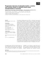
A preclinical research in 2008 concluded the result that
the acute angiotensin II administration to WT mice or
neonatal cardiomyocytes increased cardiac apoptosis,
nitrosative damage, and membrane translocation of the
nicotinamide adenine dinucleotide phosphate oxidase
(NOX) isoform p47phox. Prolonged administration of
suppressor doses of Ang II (0.5 mg/kg every other day
for 2 weeks) also induced apoptosis and nitrosative
damage in both diabetic and non-diabetic WT hearts,
but not in diabetic and non-diabetic MT-TG hearts.
Long-term follow-up (1 to 6 months) of both WT and
MT-TG mice aft er discontinuing Ang II administration
revealed progressive myocardial fibrosis, hypertrophy,
and dysfunction in WT mice but not in MT-TG mice.
This study finalize MT suppresses Ang II-induced
NOX-dependent nitrosative damage and cell death in
bothnon-diabeticandadiabeticheartearlyinthetime
course of injury and prevent the late development of
Ang II-induced cardiomyopathy and the same has been
expressed in the figure 1[17].
MT, Zinc and Diabetes
Diabetes and polymorphisms in human genes control
the cellular availability of zinc ions. One protein is the
zinc transporter ZnT-8 that supplies pancreatic b-cells
with zinc. The other is MT 1A, a member of a protein
family that links zinc and redox metabolism. Changes in
the availability of zinc ions modulate insulin signaling
and redox processes. Both zinc and MT protect cells
against the redox stress that occurs in diabetes and con-
tributes to its progression towards diabetic complica-
tions, including heart disease.
Zinc an insulinomimetic
Figure 2 shows that, the MT in presence of zinc can
able to reduce diabetic by insulinmimetic activity
through phosphorylation thereby diabetic induced hea rt
disease are also controlled.
In diabetes, these functions of MT come to bear on
insulin signaling and coronary heart disease. Insulin and
zinc ions have potent stimulatory effects on lipogenesis
and glucose uptake. Zinc-deficient animals are less sen-
sitive to insulin. Zinc can replace insulin in mammalian
cells cultured in serum-free media [18]. The actions of
zinc are intracellular because zinc increases the phos-
phorylation state of the insulin receptor, and hence, pro-
tein phosphorylation downstr eam in the insulin
signaling pathways [19]. It has been suggested that zinc
inhibition of protein tyrosine phosphatase 1B, the major
phosphatase controlling the phosphorylation state of the
insulin receptor, is responsible for these insulinomimetic
effects of zinc [20]. Coronary heart disease is the leading
cause of mortality in type 2 diabetes. The diabetics with
are C-allele carriers are more likely to develop cardio-
vascular complications might indicate a role for MT and
Administration
Acute angiotensin II
Short term
(2 weeks)
Dose0.5mg/kg
WT mice heart
Diabetic and non-
diabetic WT mice
hearts
Effected to
Apoptosis and
Nitrosative damage
MT-TG mice heart
Diabetic and non-
diabetic MT-TG mice
hearts
Not effected
Long term
(1to 6 months)
Dose 0.5mg/kg
WT mice
Diabetic and non
diabetic WT mice
hearts
Effected to
myocardial fibrosis,
Hypertrophy
MT-TG mice
Diabetic and non
diabetic MT-TG mice
hearts
Not effected
Figure 1 A Chart Explain a study on Apoptosis and
Pathological Remodeling In the Diabetic Heart.
Thirumoorthy et al. World Journal of Surgical Oncology 2011, 9:54
/>Page 5 of 7
zinc in the pathogenesis of diabetic heart disease. In
mice, zinc supp lementation prevents the development of
diabetic cardi omyopathy through inducti on of MT,
which has an antioxidant function in the heart [21].
Low serum zinc in type 2 diabetics is significantly cor-
related with mortality from coronary heart disease [22].
Despite the presence of extensive zincuria in ty pe 2 dia-
betics, there is still no consensus about whether or not
the zinc in their blood plasma reflects a generally
reduced zinc status [23]. Also, a recent review of the lit-
era ture concludes that there is no eviden ce of zinc sup-
plementation being effective in preventing diabetes [24].
Numerous studies have been proved the antidiabeto-
genic properties of zinc supplementation in both dia-
betic laboratory animals and in humans. The above
findings indicate that differences in the cellular availabil-
ity of zinc in both insulin producing b-cells and in insu-
lin target cells are associated with risk for diabetes in
specific human populations [25,26].
Discussion
The past decades have demonstrated increased focus on
its isoforms like MT-I, MT-II, MT-III and MT-IV. MT-
I and II were mainly focused in oncogenesis, tumor pro-
gression, therapy response, and patient prognosis. Stu-
dies have reported increased expression of MT-I and II
mRNA and protein in various human tumors; such as
breast, kidney, lung, nasopharynx, ovary, prostate, sali-
vary gland, testes, urinary bladder, cervical, endometrial,
skin, and pancreatic cancers, as well as in melanoma
and all, where in some cases MT-I and II expression
correlates with tumor grade/stage, chemotherapy/radia-
tion resistance, and poor prognosis.
However, MT is down-regulated in certain tumors
such as hepatocellular carcinoma and liver adenocarci-
noma. Hence, the expression of MT is not universal to
all human tumors, but may depend on the differentiation
status and proliferative index of tumors, along with other
tissue factors and gene mutations. In certain tumors such
as germ cell carcinoma, the expression of MT is closely
related to the tumor grade and proliferative activity [3].
All this studies confirm the direct or indirect link
between MT isoforms and tumor prognosis.
The expression of MT-I and MT-II (MT-I/II) isoforms
were measured together with Western blotting, copper
level, and l ipid peroxides amounts increased in an age-
dependent manner in the spinal cord, the region respon-
sible for motor paralysis concluded, MTs could have a
disease modifying property [4].
The finding on kidney correlates MT as a biomarker
of glomerular kidney dysfunction by dose-response rela-
tionships between urinary-Cd and the pre valence of
increased levels of biomarkers in urine of renal tubular
dysfunction such as urinary beta-2-microglob ulin or
N-acetyl-beta-D-glucosaminidase - NAG or urinary
albumin [13].
The relationships among MT, zinc, and oxidative
stress suggest many new areas for exploration, with
the expectation that results forthcoming from experi-
ments designed on the basis of these new findings will
contribute significantly to our understanding of the
role of zinc in diabetes and to the prevention and
treatment of diabetes and its complications in suscepti-
ble populations [6]
Conclusion
Commonly it is understood that, MT is an endogenous
substance which regulates m etal level in animal and
human body. The MT has got fingers like arrangement
and when metal level is elevated in the body these fin-
gers are triggered to bind with them. If metal level is
low in the body, it becomes vice- versa. All together the
expression pattern for MT will be a changed in the
body when any diseases caused due to metal factor.
This review article conclude that, many independent
groups of investigators found direct casual relationships
between MT and pathophysiology but more pronounced
reasons among those was endogenous and exogenous
stimuli including glucocorticoids, interferon, interleukin-
1, progesterone, vitamin D
3
endotoxins, serum factors,
heavy metals, storage of metal ions and regulation of
cellular zinc etc. may trigger the expression of MT in
human and animal’s body.
However more research is required to test the impor-
tant hypothesis of either MT could consider as biomar-
ker, alter toxicity and susceptibility of humans.
diabetic mice
with
Matallothionein+
Zn
No diabetic by
insulinomimetica
effects of zinc and
phosphorylation stase
prevents the development of
diabetic cardiomyopathy through
induction of MT
No heart disease by
Antioxidant function in
heart
Figure 2 Mechanisms of MT and Zinc as an insulinomimetic in
mice.
Thirumoorthy et al. World Journal of Surgical Oncology 2011, 9:54
/>Page 6 of 7
Author details
1
Dept. of Pharmaceutics, Cherraan’s College of Pharmacy, 521 Siruvani Road,
Perur, Coimbatore-39, TN, India.
2
Dept. of Pharmacology, College of
Pharmacy and Nursing, University of Nizwa, Sultanate of Oman.
3
Dept. of
Pharmacology, K.M.C.H. College of Pharmacy, Kovai Estate, Kalapatti Road,
Coimbatore -35, TN, India.
4
Dept. of Pharmaceutics, JSS College of Pharmacy,
Ooty, TN, India.
5
Dept. of Pharmaceutical Technology, Jadavpur University,
Kolkatta. WB, India.
Received: 22 November 2010 Accepted: 20 May 2011
Published: 20 May 2011
References
1. Margoshes M, Vallee BL: A cadmium protein from equine kidney cortex. J
Am Chem Soc 1957, 79:4813-4814.
2. Higashimoto Minoru, Isoyama Naohiro, Ishibashi Satoshi, Inoue Masahisa,
Takiguchi Masufumi, Suzuki Shinya, Ohnishi Yoshinari, Sato Masao: Tissue-
dependent preventive effect of metallothionein against DNA damage in
dyslipidemic mice under repeated stresses of fasting or restraint. Life
Sciences 2009, 84:569-575.
3. Cherian M George, Jayasurya A, Bay Boon-Huat: Metallothioneins in
human tumors and potential roles in carcinogenesis. Mutation Research
2003, 533:201-209.
4. Tokuda Eiichi, Ono Shin-Ichi, Ishige Kumiko, Naganumad Akira, Ito Yoshihisa,
Suzuki Takashi: Metallothionein proteins expression, copper and zinc
concentrations, and lipid peroxidation level in a rodent model for
amyotrophic lateral sclerosis. Toxicology 2007, 229:33-41.
5. Michèle Durliat, Muller Jean-Pierre, André Michèle, Wegnez Maurice:
Expression of the Xenopus laevis metallothionein gene during
ontogeny. Int J Dev Biol 1999, 43:575-578.
6. Maret Wolfgang: Fluorescent probes for the structure and function of
metallothionein. Journal of Chromatography B Analyt Technol Biomed Life
Sci 2009, 877(28):3378-83, 15.
7. Schicht Oliver, Freisinger Eva: Spectroscopic characterization of Cicer
arietinum metallothionein 1. Inorganica Chimica Acta 2009, 362:714-724.
8. Liua Zhi-Min, van Hasselt C Andrew, Song Fang-Zhou, Alexander CVlantis,
Cheriand MGeorge, Koropatnicke James, Chena GGeorge: Expression of
functional metallothionein isoforms in papillary thyroid cancer. Molecular
and Cellular Endocrinology 2009, 302:92-98.
9. Fong Laura, Tan Kim, Tran Cuong, Cool Johanna, Scherer AMichaela,
Elovaris Rachel, Coyle Peter, Foster KBruce, Rofe MAllan, Xian JCory:
Interaction of dietary zinc and intracellular binding protein
metallothionein in postnatal bone growth. Bone 2009, 44:1151-1162.
10. Yang Meiying, Chitambar RChristopher: Role of oxidative stress in the
induction of metallothionein-2A and heme oxygenase-1 gene
expression by the antineoplastic agent gallium nitrate in human
lymphoma cells. Free Radical Biology & Medicine 2008, 45:763-772.
11. Curtis DKlaassen, Liu Jie, Diwan ABhalchandra: Metallothionein protection
of cadmium toxicity. Toxicology and Applied Pharmacology 2009,
238:215-220.
12. Lee Jane-Dar, Wu Su-Mei, Lu Lieng-Yi, Yang Ya-Tang, Jeng Shaw-Yeu:
Cadmium Concentration and Metallothionein Expression in Prostate
Cancer and Benign Prostatic Hyperplasia of Humans. J Formos Med Assoc
2009, 108(7):555-559.
13. Nordberg FGunnar, Jin Taiyi, Wu Xunwei, Lu Jian, Chen Liang, Lei Lijian,
Hong Feng, Nordberg Monica: Prevalence of kidney dysfunction in
humans - Relationship to cadmium dose, metallothionein. immunological
and metabolic factors, Biochimie 2009, 91:1282-1285.
14. West KAdrian, Hidalgo Juan, Eddins Donnie, Levin DEdward,
Aschner Michael:
Metallothionein in the central nervous system: Roles in
protection, regeneration and cognition. Neuro Toxicology 2008,
29:489-503.
15. Levin DEdward, Perraut Charles, Pollard Ninitia, Freedman HJonathan:
Metallothionein expression and neurocognitive function in mice.
Physiology & Behavior 2006, 87:513-518.
16. Xue Wanli, Liu Qiuqu, Cai Lu, Wang Zhilun, Feng Wenke: Stable
overexpression of human metallothionein-IIA in a heart-derived cell line
confers oxidative protection. Toxicology Letters 2009, 188:70-76.
17. Zhou Guihua, Li Xiaokun, Hein WDavid, Xiang Xilin, Marshall PJames,
Prabhu DSumanth, Cai Lu: Metallothionein suppresses angiotensin II-
Induced nicotinamide adenine dinucleotide phosphate oxidase
activation, nitrosative stress, apoptosis, and pathological remodeling in
the diabetic heart. 2008, 52:655-66.
18. Wong VVT, Nissom PM, Sim SL, Yeo JHM, Chuah SH, Yap MGS: Zinc as an
insulin replacement in hybridoma cultures. Biotech Bioeng 2005,
93:553-563.
19. Tang X, Shay NF: Zinc has an insulin-like effect on glucose transport
mediated by phosphoinositol-3-kinase and Akt in 3T3-L1 fibroblasts and
adipocytes. J Nutr 2001, 131:1414-1420.
20. Haase H, Maret W: Protein tyrosine phosphatases as targets of the
combined insulinomimetic effects of zinc and oxidants. BioMetals 2005,
18:333-338.
21. Wang J, Song Y, Elsherif L, Song Z, Zhou G, Prabhu SD, Saari JT, Cai L:
Cardiac metallothionein induction plays the major role in the prevention
of diabetic cardiomyopathy by zinc supplementation. Circulation 2006,
113:544-554.
22. Soinio M, Marniemi J, Laakso M, Pyorala K, Lehto S, Ro¨nnemaa T: Serum
zinc level and coronary heart disease events in patients with type 2
diabetes. Diabetes Care 2007, 30:523-528.
23. Taylor CG: Zinc, the pancreas, and diabetes: Insights from rodent studies
and future directions. BioMetals 2005, 18:305-312.
24. Beletate V, El Dib RP, Atallah N: Zinc supplementation for the prevention
of type 2 diabetes mellitus (Review). Cochrane Database of Systematic
Reviews 2007, 1, Art. No. CD005525.
25. Roussel AM, Kerkeni A, Zouari N, Mahjoub S, Matheau JM, Anderson RA:
Antioxidant effects of zinc supplementation in Tunisians with type 2
diabetes mellitus. J Am Coll Nutr 2003, 22:316-321.
26. Schott-Ohly P, Lgssiar A, Partke HJ, Hassan M, Friesen N, Gleichmann H:
Prevention of spontaneous and experimentally induced diabetes in
mice with zinc sulfate-enriched drinking water is associated with
activation and reduction of NF-jB and AP-1 in islets, respectively. Exp Biol
Med 2004, 229:1177-1185.
doi:10.1186/1477-7819-9-54
Cite this article as: Thirumoorthy et al.: A Review of Metallothionein
Isoforms and their Role in Pathophysiology. World Journal of Surgical
Oncology 2011 9:54.
Submit your next manuscript to BioMed Central
and take full advantage of:
• Convenient online submission
• Thorough peer review
• No space constraints or color figure charges
• Immediate publication on acceptance
• Inclusion in PubMed, CAS, Scopus and Google Scholar
• Research which is freely available for redistribution
Submit your manuscript at
www.biomedcentral.com/submit
Thirumoorthy et al. World Journal of Surgical Oncology 2011, 9:54
/>Page 7 of 7