báo cáo khoa học: "Synchronous double primary malignant tumor of the gallbladder and liver: a case report" potx
Bạn đang xem bản rút gọn của tài liệu. Xem và tải ngay bản đầy đủ của tài liệu tại đây (434.05 KB, 4 trang )
CAS E REP O R T Open Access
Synchronous double primary malignant tumor of
the gallbladder and liver: a case report
Ji Won Kim
1
, Jae Woong Han
1
, So Young Jung
1
, Jae Pil Jung
1*
and Jeong Won Kim
2
Abstract
We report a case of synchronous double primary tumor of gallbladder and liver. A 63-year-old male was admitted
to the hospital complaining of abdominal discomfort. Enhanced computed tomography of the abdomen showed
acute cholecystitis with tiny gallbladder stones and a 2.2 cm size enhanced nodule in the left lobe of the liver.
Under the impression of acute cholecystitis with gall bladder stones and hepatocellular carcinoma of the left Liver,
the patient underwent a laparotomy. At laparotomy, a mass was palpated on the surface of the neck portion of
the gall bladder. Intraoperative frozen diagnosis revealed adenocarcinoma of the gall bladder. The patient was
diagnosed as having gall bladder cancer and hepatocellular carcinoma, so extended cholecystectomy with
dissection of regional lymph nodes and left hemihepatectomy were performed. Histological examination revealed
moderated differentiated adenocarcinoma of gallbladder and hepatocellular carcinoma of liver. To our knowledge,
the simultaneous occurrence of primary malignant tumor of the gallbladder and liver has never been published
before. The patient is doing well with no evidence of recurrence 17 months after surgery.
Keywords: Hepatocellular carcinoma, Gallbladder cancer, synchronous double primary malignant tumor
Background
Synchronous double primary malignant neoplasms are a
secondary malignancy o ccurring at the same time or
within 6 months after the first malignancy. Improvement
of survival rates for patients with malignancy due to early
diagnosi s and new treatments has enabled more pat ients
to survive long enough to develop the subsequent primary
malignancy, and development of more sophisticated diag-
nostic tools has made possible the detection of synchro-
nous occult malignancies. Synchronous double primary
malignant neoplasms in a single patient have been well-
documented in the literature. But, synchronous double
primary malignant tumor of gallbladder and liver has
never been reported. Herein, the authors report a case of a
63-year-old male patient with double primary cancer of
gallbladder and liver.
Case presentation
In February 2010, a 63-year-old male patient visited our
hospital with the chief complaint of abdominal discomfort
in right upper quadrant for 1 year. In 2008, the patient
had been diagnosed with acute cholecystitis at our hospi-
tal. There was no remarkable family history. On admis-
sion, vital signs (blood p ressure , heart rate, respiration
rate, and body temperature) were within normal limits.
The patient was in good general health and had no signifi-
cant weight loss. On physical examination, the conjunctiva
was anemic. The abdomen was soft but tender in the right
upper quadrant. Slight resistance, but no rigidity, was
recognized in the tender area. Complete blood count and
serum biochemistry data on admission showed the follow-
ing: white blood cell, 16,120/uL; hemoglobin, 8.5 g/dl;
hematocrit, 25.3%; platelet, 178000/mm
3
;bloodglucose,
209 mg/dl; total bilirubin, 0.5 mg/dl; alkaline phosphatase
(ALP), 41 IU/l; aspartate aminotransferase (AST), 193 IU/
l; alanine aminotransferase (ALT), 146 IU/l, and amylase
212 IU/l; C-reactive protein 76.7 mg/l. Viral markers were
hepatitis B surface antigen [HBsAg(+)], anti-HBs(-) and
anti-hepatitis C virus(-). Tumor marker assays showed
alpha-fetoprotein was 17.9 n/ml (normal 0-8.1), carci-
noembryonic antigen (CEA) was 4.2 ng/ml (no rmal 0-5),
carbohydrate antigen 19-9 (CA 19-9) was 112.5 U/ml
(normal 0-37). A computed tomography (CT) scan of the
abdomen showed distension of the gallbladder with
* Correspondence:
1
Department of Surgery, Kangnam Sacred Heart Hospital, Hallym Medical
Center, 948-1, Daerim-1Dong, Yeongdeunpo-gu, Seoul 150-950, Korea
Full list of author information is available at the end of the article
Kim et al. World Journal of Surgical Oncology 2011, 9:84
/>WORLD JOURNAL OF
SURGICAL ONCOLOGY
© 2011 Kim et al; licensee BioMed Central Ltd. This is an Open Access article distributed under the terms of the Creative Commons
Attribution License ( which permits unrestr icted use, distribution, and reproductio n in
any medium, provided the original work is properly cited.
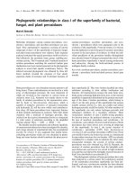
gallbladder stones and gallbladder wall thickening,
suggesting acute cholecystitis (Figure 1a), cirrhosis of liver
and heterogenously enhanced tumorous lesion in the left
lobe of liver (Figure 1b). Thus, the preoperative diagnosis
was hepatocellular carcinoma and acute cholecystitis
accompanied by gallstones. At laparotomy, the gallbladder
was slightly distended and showed wall thickening. There
was a palpable mass felt on the surface of the gallbladder
neck portion. The patient underwent surgical resection of
the gallbladder and the left lobe of the liver. Intraoperative
histologic examination revealed adenocarcinoma of gall-
bladder with invasion to the perimuscular connective tis-
sue. The patient was diagnosed with synchronous double
primary cancer of the gallbladder and liver, so additional
resection of extrahepatic bile duct and segment 5 of liver,
with a dissection of regional lymph nodes, was performed.
Biliary reconstruction was performed by Roux-en-Y hepa-
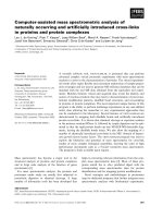
ticojejunostomy. The resected gallbladder was 12 cm in
length and 13 cm in greatest circumference, and contained
multiple black pigment stones (Figure 2a). The mass in the
neck portion of gallbladder was 5 × 3 × 1.5 cm in size and
microscopic findings revealed mod erate aden ocarcinoma
with prominent desmoplastic response infiltrating the gall-
bladder wall. There were invasive micropapillary compo-
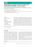
nents ( Figure 2b). The tumor in the left lobe of liv er was
about 2.2 × 2.0 × 1.5 cm in size, the cut section of which
revealed a brownish, ovoid, and highly-circumscribed
mass (Figure 3a). Microscopically, the tumor in the lateral
segment of liver was a trabecular hepatocellular carcinoma
with grade II-III cellular atypism in the Edmondson classi-
fication. Neither portal or hepatic vein infiltration, nor bili-
ary duct infiltration was found (Figure 3b). There were no
microscopically evident malignant cells in surgical
resection margins (extended cholecystectomy, Lt. hemihe-
patectomy). The postoperative course was uneventful
except for postoperative ascites, which was successfully
treated conservatively. The patient was discharged on
postoperative day 31. We planned adjuvant chem oradia-
tion therapy because the gallbladder cancer presented as a
T2 lesion with perineural invasion, but it was not per-
formed due to the patient’s refusal. The patien t has been
under regular follow-up with clinical examination and
liver function test every two months, with surveillance for
tumor markers (AFP, CA19-9, CEA) and abdominal CT
scan being done at 4 months intervals (last follow-up June
2011). The patient is doing well with no evidence of local
or distant recurrence more than 17 months after surgery.
Discussion
Multiple primary malignant tumors in a single patient are
relatively rare. In reviews of the literature regarding multi-
ple primary malignant tumors, the overall occurrence rate
of multiple primary malignancies is between 0.73% and
11.7% [1]. Multiple primary cancers have become more
common because of an increase in the number of elderly
patients and advancement in diagnostic techniques. Three
diagnostic criteria have been proposed for multiple pri-
mary malignancy: 1) each tumor must present definite fea-
tures of malignancy, 2) each must be dist inct, and 3) the
chance of one being a metastasis of the other must be
excluded [2]. Multiple primary cancers may be synchro-
nous or metachronous depending on the interval between
their diagnosis. Synchronous cancers are second tumors
occurring simultaneously or within 6 months after the
first malignancy, while metachronous multiple malignan-
cies are secondary cancers that developed after more than
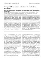
Figure 1 CT imaging results. a) A distended gallbladder with wall thickening and tiny gallstones in the gallbladder (black arrow) is evident. b)
A tumorous lesion heterogeneously enhanced by contrast media in the left lobe of the liver (white arrow) is evident.
Kim et al. World Journal of Surgical Oncology 2011, 9:84
/>Page 2 of 4
6 months after from the first malignancy [3]. Multiple pri-
mary malignancies are classified into four types: 1) multi-
centric, if the two distinct carcinoma arise in the same
organ o r ti ssue; 2) system ic, if they arise on anatomically
or functionally allied organs of the same system (colon
and rectum cancers), 3) paired organs, as in the breasts,
and 4) random, if they occur as a co-incidental or acciden-
tal association in unrelated sites [4]. In our patient, the
malignant features were histopathologically proven in each
tumor. Each tumor was pathologically categorized as a dif-
ferent type; namely, the one detected in the gall bladder
was a moderately differentiated adenocarcinoma, the one
in the liver was a hepatocellular carcinoma. These findings
might also support the fact that these two cancers
occurred in a random and synchronous manner. There
was one case reported in the literature who had synchro-
nous triple primary cancers of gallbladder, common bile
duct and liver in a women with primary biliary cirrhosis
[5]. In our case, two different type malignant tumors, ade-
nocarcinoma of gallbladder and hepatocellular carcinoma,
were observed in a male patient without primary biliary
cirrhosis. Hepatocelullar carcinoma is known to be
Figure 2 Gr oss and histological appearance of the gall bladder. a). Gross appearance of gall bladder. Mass was palpated on the surface of
neck portion of gall bladder (black arrow). b). Moderately differentiated adenocarcinoma with prominent desmoplastic response (right side)
infiltrating the gallbladder wall. Invasive micropapillary components (left side) are evident. (Hematoxylin and eosin stain, × 100).
Figure 3 Gross and hi stologi cal appearance of the liver. a). The cut section, showing a brownish, o void, and highly circumsc ribed mass
measuring 2.2 × 2.0 cm (black arrow). The remaining parenchyma is diffusely cirrhotic, and in which the cirrhotic nodules measure from 0.2 cm
to 1 cm in diameter. b). Hepatocellular carcinoma with trabecular pattern is noted. Tumor cell cords are separated by sinusoid-like blood spaces.
(Hematoxylin and eosin stain, × 200).
Kim et al. World Journal of Surgical Oncology 2011, 9:84
/>Page 3 of 4
pathogenically associated with liver cirrhosis, chronic
hepatitis virus infection, and abuse of alcohol [6]. Gallblad-
ders containing stones or infectious agents develop cancer
as a result of recurrent trauma and chronic inflammation.
Although the mechanism involved in the development of
multiple primary cancer has not been clarified, some fac-
tors such as heredity, constitution, environmental and
immuno logical factors, carcinogenic, viruses, radiological
and chemical treatments have been implicated [7]. In the
present case, gallbladder stones and chronic hepatitis B
virus infection may have played an imp ortant rol e in the
pathogenesi s of gallbladder adenocarcinoma and hepato-
cellular carcinoma, respectively. The prognosis of patients
with multiple primary malignant tumors can be deter-
mined independently by the stage of each malignancies. In
the present case, adenocarcinoma of gall bladder was
T2N0M0 (stage II) and hepatocellular carcinoma was
T1N0M0 (stage I). The surgical treatment of choice for
synchronous multiple primary malignancies is curative
resection of each malignant tumors [8].
Conclusions
We report the first case of synchronous double primary
malignancies of gallbladder and liver. The possibility of
synchronous multiple primary malignancies should be
noted in the treatment of elderly patients with malig-
nant tumor. Multiplicity of primary malignancies itself
does not necessarily indicate a poor prognosis as long as
adequate diagnosis and treatment are performed.
Consent
Written informed consent was obtained from the patient
for publication of this case report and any accompany-
ing images. A copy of the written consent is available
for review by the Editor-in-Chief of this journal.
Author details
1
Department of Surgery, Kangnam Sacred Heart Hospital, Hallym Medical
Center, 948-1, Daerim-1Dong, Yeongdeunpo-gu, Seoul 150-950, Korea.
2
Department of Pathology, Kangnam Sacred Heart Hospital, Hallym Medical
Center, 948-1, Daerim-1Dong, Yeongdeunpo-gu, Seoul 150-950, Korea.
Authors’ contributions
Ji Won Kim, Jae Pil Jung, Jae Woong Han, So Young Jung and Jeong Won
Kim made up the surgical and pathological team involved in the case. Jae
Pil Jung and Ji Won Kim wrote and edited the manuscript. All authors read
and approved the final manuscript.
Competing interests
The authors declare that the y have no competing interests.
Received: 18 April 2011 Accepted: 3 August 2011
Published: 3 August 2011
References
1. Demandante CG, Troyer DA, Miles TP: Multiple primary malignant
neoplasms: case report and a comprehensive review of the literature.
Am J Clin Oncol 2003, 26(1):79-83.
2. Warren S, Gates O: Multiple primary malignant tumors: A survey of the
literature and a statistical study. Am J Cancer 1932, 16:1358-1414.
3. Suzuki T, Takahashi H, Yao K: Multiple primary malignancies in the head
and neck: a clinical review of 121 patients. acta otolaryngol suppl 2002,
547:88-92.
4. Moertel CG: Multiple primary malignant neoplasms: historical
perspectives. Cancer 1977, 40(4 Suppl):1786-1792.
5. Imada J, Hoshino H, Nishimura D, Morita K, Yoshida N, Katada N, et al: Case
report: multiple cancers: hepatocellular carcinoma and adenocarcinomas
of the common bile duct and the gall-bladder in a woman with primary
biliary cirrhosis. J Gastroenterol Hepatol 1996, 11(6):546-550.
6. Ohwada S, Yoshihiro O, Iwazaki S, Tanahashi Y, Sawada T, Takeyoshi I, et al:
Double cancer in different hepatic lobes: hepatocellular and
cholangiocellular carcinoma. Hepatogastroenterology 1995, 42(4):411-414.
7. Tamura M, Shinagawa M, Funaki Y: Synchronous triple early cancers
occurring in the stomach, colon and gallbladder. Asian J Surg 2003,
26(1):46-48, discussion 9.
8. Yoshino K, Asanuma F, Hanatani Y, Kumai K, Ishibiki K: Statistical studies on
multiple primary cancers including gastric cancers. Gan No Rinsho 1984,
30(12 Suppl):1514-1523.
doi:10.1186/1477-7819-9-84
Cite this article as: Kim et al.: Synchronous double primary malignant
tumor of the gallbladder and liver: a case report. World Journal of
Surgical Oncology 2011 9:84.
Submit your next manuscript to BioMed Central
and take full advantage of:
• Convenient online submission
• Thorough peer review
• No space constraints or color figure charges
• Immediate publication on acceptance
• Inclusion in PubMed, CAS, Scopus and Google Scholar
• Research which is freely available for redistribution
Submit your manuscript at
www.biomedcentral.com/submit
Kim et al. World Journal of Surgical Oncology 2011, 9:84
/>Page 4 of 4