Báo cáo khoa học: "Gastric glomus tumor: A case report" pptx
Bạn đang xem bản rút gọn của tài liệu. Xem và tải ngay bản đầy đủ của tài liệu tại đây (768.8 KB, 5 trang )
CAS E REP O R T Open Access
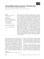
Gastric glomus tumor: A case report
Ioannis Vassiliou
1*
, Aliki Tympa
2
, Theodosios Theodosopoulos
1
, Nikolaos Dafnios
1
, Georgios Fragulidis
1
,
Andreas Koureas
3
, Evi Kairi
4
Abstract
Gastric glomus tumors are rare mesenchymal tumors of the gastrointestinal tract. We describe a 72-year-old patient
who presented with episodes of melena and was subsequently investigate d for a tumor of the antrum of the sto-
mach. Surgical resection revealed a 2 × 2 × 1.7 cm well circumscribed submucosal tumor, extending into the mus-
cularis propria. The histopathologic examination of the specimen demonstrated a glomus tumor of the stomach.
We discuss the preoperative investigation, the diagnostic problems and the surgical treatment of the patient with
this rare submucosal lesion.
Background
Glomus tumors are benign neoplasms of well-differen-
tiated mesenchymal cells. Glomus tumors of the sto-
mach are rare lesions, arising in the intramuscular layer.
They typically present as a solitary submucosal nodule
in the region of the antrum and pylorus. Preoperative
diagnosis of gastric glomus tumors is difficult and
require s a multi-faculty medical approach. We present a
rare case of a glomus tumor of the stomach along with
the investigative procedures and the surgical treatment.
Case Presentation
Two months ago, a 72-year-old woman presented to her
primary care physician with an e pisode of melena that
was suggestive of hemorrhage of the upper gastrointes-
tinal tract. Upon presentation the patient w as hemody-
namically stable with normal laboratory tests and no
evidence of active bleeding in the last 48 hours. Hospita-
lization was not required and the evaluation was com-
pleted in the outpatient department.
The patient was subjected to further investigation.
Upper gastrointestinal endoscopy revealed mild, diffuse
oesophagitis and a small sliding hiatal hernia. At the
antrum of the stomach, a 5 cm, well circumscribed sub-
mucosal mass with normal overlying mucosa was
observed (Figure 1). Multiple regular b iopsies were
taken and some histo logical features of leiomyoma were
identified. An endoscopic ultrasound confirmed the sub-
mucosal lesion which originated from the muscularis
propria, measured 1.9 × 2. 4 cm and was extending in
the second, third and fourth layer of the stomach.
The patient was subsequently referred for surgical
consultation. Physical examination revealed a 72-year-
old female who was awake and alert, appeared healthy
and looked younger than her stated age. Her abdomen
was soft, non-distended, without palpable masses. The
stool was negative for occult blood. The hemoglobin
level was 13.1 g/dL with normal biochemical profile.
Tumor markers were within reference ranges. Abdom-
inal radiography showed normal amount and distribu-
tion of gas within the bowel. An abdominal computer
tomography scan demonstrat ed a 3 cm localized, prepy-
loric enhancing mass at the lesser c urvatu re of the sto-
mach (Figures 2, 3). Lymphadenopathy was not
observed. The differential diagnosis invol ved mesenchy-
mal and other benign gastrointestinal stromal tumors.
The patient was taken to the operative room elec-
tively. She was subjected to antrectomy and Roux-en-Y
anastomosis. The stomach contained a 2 × 2 × 1.7 cm
well circumscribed tumor. (Figures 4, 5). The histo-
pathologic findings of the lesion were c haracteristi c of
glomus tumor of the antrum. In detail, cut surface of
the specimen, demonstrated a grayish-white nodular
tumor, arising from the submucosa and extending
through the muscularis of the stomach, without invol-
ving the s erosal surface. Histologically, the tumor was
composed of sheets of glomus cells, without nuclear
pleomorphism and no mitotic figures. The cells had
eosinophilic and focally clear cytoplasm. Throughout
the tumor telengiectatic vessels were observed and some
contained aggregates of glomus cells in their walls
* Correspondence:
1
Second Department of Surgery, Athens Medical School, Aretaieion Hospital,
76 Vassilisis Sofias Avenue, 11528, Athens, Greece
Vassiliou et al. World Journal of Surgical Oncology 2010, 8:19
/>WORLD JOURNAL OF
SURGICAL ONCOLOGY
© 2010 Vassiliou et al; licensee BioMed Centr al Ltd. This is an Open Access article distributed under the terms of the Creative
Commons Attribution License (ht tp://creativecomm ons.org/licenses/by/2.0), which permits unrestricted use, distri bution, and
reproduction in any medium, provided the original wor k is properly cited.
(Figure 6). Immunohistohemically, the tumor cells were
positive for smooth muscle actin (Figure 7) and vimen-
tin and negative for desmin, CD34, CD117, S-100 pro-
tein and cytokeratins (AE1/3, CAM 5,2). The
proliferating marker Ki-67 was < 5%. The residual gas-
tric mucosa showed atrophic gastritis with focal intest-
inal metaplasia in the pylorus reg ion. Five lymph nodes
retrieved from the major omentum were free of meta-
static tumor. The patient recovered uneventfully and
was discharged 5 days after surgery.
Discussion
Gastric glomus tumor is a benign mesenchymal neo-
plasm arising from the neuromyoarterial glomus. The
glomus apparatus consists of three vascular components:
an afferent artery separated from an efferent venole by
convoluted channels. Multiple layers of epithelioid cells
along with nerve fibers surround these channels [1].
Glomus has also been described as an arteriovenous
shunt that may contract or expand [2]. Glomus tumors
are commonly observed in the dermis or the subcutis.
They have also been described in the bone and joints,
skeletal muscle, soft tissue, mediastinum, trachea, kid-
ney, uterus and vagina [3].
The first case of gastric glomus tumor was reported in
1951 by Key et al. [4] and since then, few cases have
been reported. Vascular tumors of the gastrointestinal
tract are rare (accounting for less than 2% of benign
tumors), but according to Miettinen et al. [3] the
Figure 1 Glomus tumor of the stomach as featured on upper
gastrointestinal endoscopy: a well circumscribed submucosal
mass with normal overlying mucosa.
Figure 2 Glomus tumor of the stomach in a 72 year-old
woman: unenhanced computer tomography scan shows the
well-circumscribed mass (arrow) in the gastric antrum.
Figure 3 Glomus tumor of the stomach in a 72 year-old
woman: On a contrast-enhanced computer tomography scan,
the mass is greatly enhanced (arrow).
Figure 4 The prepyloric mass of the stomach at the lesser
curvature.
Vassiliou et al. World Journal of Surgical Oncology 2010, 8:19
/>Page 2 of 5
frequency of gastric glomus tumors is estimated to be
1% of that of gastrointestinal stromal tumors. Glomus
tumors of the stomach have a marked predominance i n
females [5-8] although older studies [9] showed nearly
equal sex distribution. Moreover, they usually occur in
the fifth or sixth decade of life. However, in a clinico-
pathologic study among Korean population, the age of
onset ranged from 30 to 68 years old [7].
Gastric glomus tumors present with a variety of symp-
toms. Epigastric discomfort (intermittent or continuous),
hematemesis, melena and occasionally nausea and
vomiting can occur. Overt gastrointestinal bleeding has
also been reported [3,7], in cases of ulcerated overlying
mucosa. From our literat ure search, gastric glomus
tumors rarely are incidental findings.
Glomus tumors are usually solitary. There is only one
case report of multiple gastric glomus tumors [10]. Six
glomus tumors were observed in the stomach wall and
the perigastric adipose tissue of a 75-year-old black man
presenting with hematemesis. Furthermore, gastric glo-
mus tumors are small and have a greater incidence on
the greater curvature of the stomach [7,9,11]. In our
case, as well as in the r eport by Yan et al. [12], the
tumor occurred in the lesser curvature.
Glomus tumors have to be differentiated from other
lesions, such as gastrointestinal stromal tumors (GISTs)
and mesenchymal tumors. Preoperative diagnosis of glo-
mus neoplasms is difficult. Glomus tumors grossly
appear as red-blue nodules that originate from the mus-
cularis propria [13,14]. In barium studies, most reported
cases are localized at the greater curvature side of the
antrum and they appear as smooth submucosal masses
with or without ulceration. On CT, they manifest as
well-circumscribed submucosal masses with homoge-
neous density on unenhanced study and may contain
tiny flecks of calcifications. After contrast medium
administration, these tumors show strong enhancement
on arterial phase images and persistent enhanceme nt on
portal venous phase images, which reflects their hyper-
vascular nature. However, imaging techniques fail to dif-
ferentiate glomus tumors from other stromal or
mesenchymal lesions. The above mentioned ima ging
features can also be se en with other gastric tumors
(endocrine tumors or GISTs). Endoscopic ultrasound
findings suggest that gastric glomus tumors are hetero-
genous, hypoechoic circumscribed masses, with few tub-
ular structures [12,15,16]. They usually originate from
the fourth endoscopic ultrasound layer . On Power Dop-
pler sonography, hypervascularity is typical of glomus
Figure 7 Glomus tumor of the stomach. Positive staining for
smooth muscle actin (× 100).
Figure 5 The specimen of the stomach.
Figure 6 Trabeculae of tumor cells distributed around dilated
and ectactic blood vessels (Hematoxylin & Eosin staining ×100).
Vassiliou et al. World Journal of Surgical Oncology 2010, 8:19
/>Page 3 of 5
tumors [3,17]. On the contrary, no turbulent pulsatile
flow within leiomyomas was observed [18].
Endoscopic biopsies may fail to provide sufficient
amounts of material or representative samples of the
submucosal lesion and deeper submucosal lesions can-
not be reached adequately [17]. Fine needle aspiration
(FNA), performed during endoscopy or endoscopic
ultrasound may not contribute to the preoperative diag-
nosis. In our case, FNA was misleading. Biopsies from
the lesion were positive for leiomyoma. Kapur et al. had
similar FNA biopsy results [19]. In addition, Lorber et
al. [6] reported that FNA biopsy in their case, suggested
a well differentiated neuroendocrine tumor, possibly car-
cinoid. Nevertheless, surgical resection of the tumor and
histopathologic examination, demonstrated gastric glo-
mus tumor.
Although glomus tumors of the stomach are usually
benign, malignant behavior cannot be excluded. Folpe et
al. [13] proposed the following classification criteria for
malignant glomus tumors: a) deep location and size
more than 2 cm or b) pre sence of atypical mitotic figure
or c) combination of moderate to high nuclear grade
and mitotic activity (5 mitoses/50 high-power fields). It
should also be mentioned that the classification criteria
have been established for supe rficial or deep soft tissue
glomus tumors. However, due to lack of evidence in the
current literature, we suggest that the above mentioned
criteria should be used by convention for gastric glomus
tumors. Only one case of metastatic gastric glomus
tumor has been described [3]. The tumor measured 6.5
cm and on histological analysis mild atypia (1-3
mitoses/HPF) was observed.
Histomorphology of benign gastric glomus tumors is
distinctive. Benign glomus tumors consist of small uni-
form rounded glomus cells that are located in the walls
of dilated vessels. The tumor cells have small uniform
nuclei, show positive immunoreactivity for smooth mus-
cle actin and are outlined by PAS-positive basement
membranes [13]. Glomus tumors are also calponin posi-
tive and lack the C-KIT mutation seen with GIST
tumors [20]. Immunohistochemistry is essential in the
differential diagnosis of glomus tumors. Immunohisto-
chemical staining for actin is negative in gastrointest inal
endocrine tumors, but positive in about half of the
GISTs. Gastric epithelioid GISTs are usually positive for
C-KIT (CD117) [3]. Leiomyomas a nd leiomyosarcomas
are differentiated from GISTs by positive immunoreac-
tivity for desmin and smooth muscle actin and negative
immunoreactivity for C-KIT (CD117) and CD34 [8,16].
Finally, operative intervention should be carefully
planned in cases of submucosal ga stric masses. All the
patients with gastric glomus tumors reported in the lit-
erature were operated [1-7], [9-16,19]. Lymph node
metastases were not common. As gastric glomus tumors
are mesenchymal tumors with potential malignant beha-
vior, wedge resection with negative margins should be
the treatment of choice [21]. Enucleation is not recom-
mended due to the high recurrence rates [21]. Gastric
glomus tumors should always be included in the differ-
ential diagnosis of submucosal gastric lesions, keeping in
mind that preoperative investigation of these patients
often yields misleading results.
Conclusions
Preoperative diagnosis of gastric glomus tumor is diffi-
cult. Despite their distinct histological appearance, their
clinicopathologic, radiology and upper endoscopy fea-
tures overlap with more common gastric tumors. The
diagnostic gold standard for such lesions is the histolo-
gical examination and the immunohistohemical markers.
A multi-faculty medical approach of the patient opti-
mizes the chances for an accurate preoperative diagnosis
and leads to a targeted surgical intervention.
Consent
Written informed consent was obtained from the patient
for the publication of this case report. A copy of the
written consent is available for review by the Editor-in-
Chief of this journal
Author details
1
Second Department of Surgery, Athens Medical School, Aretaieion Hospital,
76 Vassilisis Sofias Avenue, 11528, Athens, Greece.
2
First Department of
Anesthesiology, Athens Medical School, Aretaieion Hospital, 76 Vassilisis
Sofias Avenue, 11528, Athens, Greece.
3
First Department of Radiology,
Athens Medical School, Aretaieion Hospital, 76 Vassilisis Sofias Avenue ,
11528, Athens, Greece.
4
Department of Pathology, Athens Medical School,
Aretaieion Hospital, 76 Vassilisis Sofias Avenue, 11528, Athens, Greece.
Authors’ contributions
IV, TT and ND carried out the surgical procedures and contributed to the
design of the study. AT gathered the data, drafted the manuscript and
critically revised it. AK performed the computed tomography scanning and
provided figures for the manuscript along with their interpretation. EK
performed the histological analysis of all surgical specimens and provided
histological sections as figures of the manuscript. IV revised and finally
approved the manuscript for publication.
Competing interests
The authors declare that they have no competing interests.
Received: 26 December 2009 Accepted: 22 March 2010
Published: 22 March 2010
References
1. Kumbel JM: Glomus tumor: A benign gastric neoplasm. Mil Med 1988,
153:417-418.
2. Pack GT: Unusual tumors of the stomach. Ann NY Acad Sci 1964,
114:985-1011.
3. Miettinen M, Paal E, Lasota J, Sobin LH: Gastrointestinal glomus tumors: a
clinicopathologic, immunohistochemical, and molecular genetic study of
32 cases. Am J Surg Pathol 2002, 26:301-11.
4. Key S, Callaahn WP, Murray MR: Glomus tumor of the stomach. Cancer
1951, 4:726-736.
5. Enzinger FM, Weiss SW: Perivascular tumors. Soft tissue tumors St Louis,
MD: MosbyEnzinger FM, Goldblum JR , 4 2001, 985-1003.
Vassiliou et al. World Journal of Surgical Oncology 2010, 8:19
/>Page 4 of 5
6. Lorber J, Kalish J, Farraye FA, Cerda S, Babineau TJ: Glomus tumor of the
gastric antrum: case report. Curr Surg 2005, 62:436-8.
7. Lee HW, Lee JJ, Yang DH, Lee BH: A clinicopathologic study of glomus
tumor of the stomach. J Clin Gastroenterol 2006, 40:717-20.
8. Applman HD: Mesenchymal tumors of the gastrointestinal tract.
Pathology of the gastrointestinal tract Baltimore, MD: Williams and
WilkinsMing SC, Goldman H , 2 1998, 361-398.
9. Kanwar YS, Manaligod JR: Glomus tumor of the stomach. An
ultrastructural study. Arch Pathol 1975, 99:392-7.
10. Haque S, Modlin IM, West AB: Multiple glomus tumors of the stomach
with intravascular spread. Am J Surg Pathol 1992, 16:291-9.
11. Debol SM, Stanley MW, Mallery S, Sawinski E, Bardales RH: Glomus tumor
of the stomach: cytologic diagnosis by endoscopic ultrasound-guided
fine-needle aspiration. Diagn Cytopathol 2003, 28:316-21.
12. Yan SL, Yeh YH, Chen CH, Yang CC, Kuo CL, Wu HS: Gastric glomus tumor:
a hypervascular submucosal tumor on power Doppler endosonography.
J Clin Ultrasound 2007, 35:164-8.
13. Folpe AL, Fanburg-Smith JC, Miettinen M, Weiss SW: Atypical and
malignant glomus tumors: analysis of 52 cases, with a proposal for the
reclassification of glomus tumors. Am J Surg Pathol 2001, 25:1-12.
14. Tsai TL, Changchien CS, Hu TH, Hsiaw CM: Demonstration of gastric
submucosal lesions by high-resolution transabdominal sonography.
J Clin Ultrasound 2000, 28:125-32.
15. Imamura A, Tochihara M, Natsui K, Murashima Y, Suga T, Yaosaka T,
Fujinaga A, Koito K, Miyakawa H, Higashino K, et al: Glomus tumor of the
stomach: endoscopic ultrasonographic findings. Am J Gastroenterol 1994,
89:271-2.
16. Agawa H, Matsushita M, Nishio A, Takakuwa H: Gastric glomus tumor.
Gastrointest Endosc 2002, 56:903.
17. Wielch T, Walch A, Werner M: Histopathological Classification of
nonneoplastic and neoplastic gastrointestinal submucosal lesions.
Endoscopy 2005, 37:630-634.
18. Iwase H, Kusugamp K, Suga S, Kyokane K, Yamaguchp T: Color Doppler-
enhanced endoscopic ultrasonographic diagnosis of upper submucosal
lesions. Dig Endosc 2007, 9:116-121.
19. Kapur U, Hobbs CM, McDermott E, Mooney EE: Gastric glomus tumor. Ann
Diagn Pathol 2004, 8:32-5.
20. Porter PL, Bigler SA, McNutt M, Gown AM: The immunophenotype of
hemangiopericytomas and glomus tumors, with special reference to
muscle protein expression: an immunohistochemical study and review
of the literature. Mod Pathol 1991,
4:46-52.
21. Pidhorecky I, Cheney RT, Kraybill WG, Gibbs JF: Gastrointestinal stromal
tumors: current diagnosis, biologic behavior and management. Ann Surg
Oncol 2000, 7:705-12.
doi:10.1186/1477-7819-8-19
Cite this article as: Vassiliou et al.: Gastric glomus tumor: A case report.
World Journal of Surgical Oncology 2010 8:19.
Submit your next manuscript to BioMed Central
and take full advantage of:
• Convenient online submission
• Thorough peer review
• No space constraints or color figure charges
• Immediate publication on acceptance
• Inclusion in PubMed, CAS, Scopus and Google Scholar
• Research which is freely available for redistribution
Submit your manuscript at
www.biomedcentral.com/submit
Vassiliou et al. World Journal of Surgical Oncology 2010, 8:19
/>Page 5 of 5