Báo cáo khoa học: "Stage II/III rectal cancer with intermediate response to preoperative radiochemotherapy: Do we have indications for individual risk stratification?" pdf
Bạn đang xem bản rút gọn của tài liệu. Xem và tải ngay bản đầy đủ của tài liệu tại đây (1.13 MB, 11 trang )
WORLD JOURNAL OF
SURGICAL ONCOLOGY
Sprenger et al. World Journal of Surgical Oncology 2010, 8:27
/>Open Access
RESEARCH
BioMed Central
© 2010 Sprenger et al; licensee BioMed Central Ltd. This is an Open Access article distributed under the terms of the Creative Commons
Attribution License ( which permits unrestricted use, distribution, and reproduction in
any medium, provided the original work is properly cited.
Research
Stage II/III rectal cancer with intermediate response
to preoperative radiochemotherapy: Do we have
indications for individual risk stratification?
Thilo Sprenger*
1
, Hilka Rothe
2
, Klaus Jung
3
, Hans Christiansen
4
, Lena C Conradi
1
, B Michael Ghadimi
1
, Heinz Becker
1
and Torsten Liersch
1
Abstract
Background: Response to preoperative radiochemotherapy (RCT) in patients with locally advanced rectal cancer is
very heterogeneous. Pathologic complete response (pCR) is accompanied by a favorable outcome. However, most
patients show incomplete response. The aim of this investigation was to find indications for risk stratification in the
group of intermediate responders to RCT.
Methods: From a prospective database of 496 patients with rectal adenocarcinoma, 107 patients with stage II/III
cancers and intermediate response to preoperative 5-FU based RCT (ypT2/3 and TRG 2/3), treated within the German
Rectal Cancer Trials were studied. Surgical treatment comprised curative (R0) total mesorectal excision (TME) in all
cases. In 95 patients available for statistical analyses, residual transmural infiltration of the mesorectal compartment,
nodal involvement and histolologic tumor grading were investigated for their prognostic impact on disease-free (DFS)
and overall survival (OS).
Results: Residual tumor transgression into the mesorectal compartment (ypT3) did not influence DFS and OS rates (p
= 0.619, p = 0.602, respectively). Nodal involvement after preoperative RCT (ypN1/2) turned out to be a valid prognostic
factor with decreased DFS and OS (p = 0.0463, p = 0.0236, respectively). Persistent tumor infiltration of the mesorectum
(ypT3) and histologic tumor grading of residual tumor cell clusters were strongly correlated with lymph node
metastases after neoadjuvant treatment (p < 0.001).
Conclusions: Advanced transmural tumor invasion after RCT does not affect prognosis when curative (R0) resection is
achievable. Residual nodal status is the most important predictor of individual outcome in intermediate responders to
preoperative RCT. Furthermore, ypT stage and tumor grading turn out to be additional auxiliary factors. Future clinical
trials for risk-adapted adjuvant therapy should be based on a synopsis of clinicopathologic parameters.
Background
Multimodal treatment strategies and optimized surgical
procedures with total mesorectal excision (TME) led to a
significant improvement in rectal cancer therapy within
the last 15 years [1-5]. Nevertheless, a postulation of
more individualized approaches in rectal cancer treat-
ment exists for some time. To some extent this postula-
tion is realized in stage dependant therapy as
preoperative RCT is recommended only in locally
advanced (stage II/III) rectal cancer [6,7].
After preoperative RCT, therapy-induced downsizing
effects have widely been described as important prognos-
tic factors [8,9]. Local response to neoadjuvant long-term
RCT is very heterogeneous and varies between no mor-
phologic alteration and complete shrinkage with patho-
logic complete response (pCR). Anyway, in most patients
a moderate local response with variable residual tumor
infiltration depth (ypT2/3) results [10]. This group of
patients with intermediate response is of particular inter-
est as it represents the largest subcategory, which prog-
nostically is difficult to classify. Within this group, tumor
* Correspondence:
1
Department of General and Visceral Surgery, University Medical Center
Göttingen, Georg-August-University, Göttingen, Germany
Full list of author information is available at the end of the article
Sprenger et al. World Journal of Surgical Oncology 2010, 8:27
/>Page 2 of 11
transgression of the actual rectal wall and infiltration of
the mesorectal compartment (≥ ypT3) constitutes a dis-
tinction with unknown impact on prognosis. Subclassifi-
cation of pT3 rectal cancers has already turned out to be a
reliable risk factor for cancer recurrence in patients
undergoing primary surgery [11-13] but its prognostic
relevance after preoperative RCT is still unclear.
According to TNM classification [14], tumor invasion
depth of the mesorectal compartment is divided into sub-
groups depending on the precise infiltration depth:
(y)pT3a to (y)pT3d. Therefore (y)pT3 category spans the
invasion of only a few tumor cells beyond the muscularis
propria to a complete infiltration of the mesorectum,
nearby reaching the visceral peritoneum or contiguous
organs [14].
A risk-adapted stratification of patients after preopera-
tive RCT and TME-based surgery is crucial for adjuvant
treatment strategies in individual patients. Currently, a
beneficial impact of adjuvant chemotherapy (CT) is dis-
cussed controversely [15,16]. To date, standardized appli-
cation of adjuvant CT is guaranteed only within
randomized clinical trials and clinicopathologic indica-
tions for risk stratification in patients after multimodal
therapy are extensively missing.
In this study we investigated 107 patients with interme-
diate local response to preoperative 5-FU based RCT
(ypT2/3) and curative (R0) surgery. The aim of this inves-
tigation was to clarify the impact of residual tumor infil-
tration of the mesorectal compartment (≥ ypT3b), nodal
status (ypN) and histologic tumor grading on DFS and
OS and to evaluate their relevance within an individual
risk stratification model in intermediate responders to
RCT.
Methods
Eligibility
This study included patients with locally advanced rectal
cancer (stage II/III) and moderate RCT-induced histo-
pathologic tumor regression (TRG 2 and 3 according to
the Dworak classification[17]) and concomitant residual
ypT2/3 status. All tumors were located not more than 16
cm from the anal verge, measured by rigid rectoscopy.
Patients with clinical evidence of distant metastatic dis-
ease were excluded from the actual investigation and
received individual multimodal treatment.
Clinical Assessments
Pretherapeutical staging procedures consisted of rigid
rectoscopy, flexible colonoscopy, endorectal ultrasound
(ErUS), magnetic resonance imaging (MRI) of the pelvis
and computed tomography (CT) scans of chest, liver and
pelvis. Staging results were conferred and interdisciplin-
ary discussed before initiation of multimodal treatment.
Clinical tumor stages (cT, cN, cUICC) were determined
by ErUS, pelvic MRI, and CT scans.
Multimodal Treatment
Preoperative treatment included fractional radiation with
cumulative 50.4 Gy (28 × 1.8 Gy) in 3- or 4-field tech-
nique. Concomitant chemotherapy consisted of either 5-
Fluorouracil (5-FU) monotherapy in 84 patients or a
combined 5-FU + Oxaliplatin regime in 23 patients. Six
weeks after completion of neoadjuvant treatment all
patients underwent standardized TME-based surgery.
Subsequently, postoperative systemic therapy was applied
according to the preoperative treatment regimen (5-FU
monotherapy or combined 5-FU + Oxaliplatin) and the
actual study protocol.
Pathologic Assessment
Quality assessment of the surgical specimens was per-
formed according to the MERCURY criteria[18] and was
followed by standardized pathological diagnostics of the
specimens by an experienced gastrointestinal pathologist.
The complete tumor area and all detectable mesorectal
lymph nodes were paraffin-embedded and investigated
using hematoxylin and eosin staining.
Pathological Staging/Grading
Pathological staging included ypTNM stage according to
the current TME classification[14], tumor differentiation
grading, evaluation of proximal, distal and circumferen-
tial resection margins and intra- and extramural vascular
and perineural invasion. Nodal staging included histolog-
ical evaluation of all detected lymph nodes and statement
of lymph node ratio in all cases with regard to the consen-
sual minimum number of 12 nodes per specimen [14,19].
RCT-induced tumor regression was denoted on the basis
of a semi-quantitative 5 point grading system according
to established methods [10,17]. Subdivision of ypT3 sta-
Table 1: Subdivision of yp T3 status
ypT3a Residual Tumor Infiltration
into the Mesorectum < 1 mm
ypT3b Residual Tumor Infiltration
into the Mesorectum > 1 - 5
mm
ypT3c Residual Tumor Infiltration
into the Mesorectum > 5 - 15
mm
ypT3d Residual Tumor
Manifestation into the
Mesorectum > 15 mm
Sprenger et al. World Journal of Surgical Oncology 2010, 8:27
/>Page 3 of 11
tus was performed in accordance to subdivision of pT3
status [20] and is shown in Table 1.
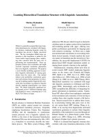
Histopathologic differentiation of residual tumor cells
was evaluated after preoperative RCT and subdivided
into two categories (Figure 1):
High Grade Differentiation: well and moderate differ-
entiated residual tumor cell clusters with preserved glan-
dular growth pattern
Low Grade Differentiation: less and poorly/undifferen-
tiated residual tumor cell clusters with non-glandular
growth pattern
Follow-up
Follow-up assessments included measurement of blood
parameters including serum carcinoembryonic antigen
and abdominal ultrasound every 3 months. Rectoscopy
and CT scans were performed every 6 months within the
first 3 years and every 12 months thereafter. Local recur-
rence was defined as cancer relapse within the pelvic
region or the site of the anastomosis. Distant metastatic
disease appeared as any tumor manifestation outside the
pelvis.
Statistical Methods
DFS and OS probabilities were estimated by the Kaplan-
Meier method and compared between the different levels
of clinicopathologic parameters (ypT, ypN, cN and tumor
differentiation grading) by a Cox proportional hazards
regression model. The ypT and ypN parameters were
additionally evaluated in a multivariate analysis.
The distributions of ypN status within the two sub-
groups of ypT (ypT2/3a and ypT3b-d) were compared by
Fisher's exact test. The number of detected lymph nodes
between nodal positive and negative patients was com-
pared with the Mann-Whitney-U test. The significance
level was set to α = 5% for all tests. All analyses were per-
formed with the free software R (version 2.6,http://
www.r-project.org).
Results
Patient Population
Between January 1998 and June 2008 496 patients with
histologically confirmed adenocarcinoma of the rectum
were treated at our department. Of these, 153 patients
with locally advanced (stage II/III) rectal cancer received
preoperative RCT within the German rectal cancer trials
(CAO/ARO/AIO-94 [6], XelOx [21] and the ongoing
CAO/ARO/AIO-04 trial) and underwent quality assessed
curative (R0) TME-based surgery. The approval from the
medical ethics committee of the University of Göttingen
and informed consent from all subjects were obtained
prior to enrolment into the respective study. Following
TME, 107 patients (70%) were defined as intermediate
responders to neoadjuvant RCT. Of these, 95 were
included in the present analysis (Figure 2).
At the time of surgery, 3 patients without previous evi-
dence of distant metastatic disease presented with syn-
chronous liver metastases (stage IV), as detected by
manual liver palpation and intraoperative ultrasound.
These patients likewise had evidence of residual mesorec-
tal lymph node metastases within the surgical specimen
and were excluded from survival analysis.
During a median follow-up period of 42 months (range:
4 - 126 month), 9 of the 107 patients died of non-cancer-
related disease and were excluded from cancer specific
survival analyses. Seventeen (15.9%) patients had cancer
relapse, with 15 cases of separate distant metastatic dis-
ease and 2 cases of local recurrence combined with syn-
Figure 1 Histopathologic differentiation: well/moderate differentiated residual tumor cell clusters after RCT with preserved glandular
growth pattern (High Grade Tumors; Figure 1a). Poorly differentiated residual tumor cells with non-glandular formations (Low Grade Tumors; Fig-
ure 1b).
Sprenger et al. World Journal of Surgical Oncology 2010, 8:27
/>Page 4 of 11
chronous distant metastases. No isolated local
recurrence occurred. Three patients failed statistical
analyses due to occult synchronous hepatic metastases
detected during surgery (ypUICC IV). In summary, 95
patients were included into survival analysis.
Patient characteristics, pretherapeuthical staging
results and treatment procedures of all 107 intermediate
responders to preoperative RCT are presented in Table 2.
The postsurgical and pathological staging results are
summarized in Table 3.
Pathological Staging Results
When comparing pretherapeuthical clinical staging with
pathological staging results, RCT-induced T-Level down-
sizing was achieved in 42% of patients (n = 45). Eight
tumors, initially staged as cT4 were downsized to ypT2 (n
= 2), ypT3b (n = 2), ypT3c (n = 3) and ypT3d (n = 1).
Thirty-seven tumors, previously staged as cT3 were
downsized to ypT2 status. Nodal downstaging from
cUICC III to ypUICC II stage was achieved in 41% of
patients (n = 44).
The median number of detected and histopathologi-
cally evaluated lymph nodes per specimen was 21 (range:
6 - 79). In 68% of specimens, lymph node yield accounted
for ≥ 18 nodes. Fewer than 12 nodes, which is the consen-
sual number according to TNM criteria, were found in a
total of 5 specimens (4.7%).
In patients with extended lymph node recovery, lymph
node metastases were detected more frequently. How-
ever, this finding was not statistically significant (p =
0.06). In detail, the median number of lymph nodes found
in the ypN0 group was 19 (range: 6 - 79) compared to 24
(range: 7 - 77) in the ypN1/2 group.
Of 95 patients included in cancer specific survival anal-
yses, 63 (66.3%) were classified as node negative (ypN0),
and 32 (33.7%) patients presented with residual lymph
node metastases (ypN1/2) after RCT. Fifty patients
(54.3%) had intramural tumor infiltration with a maximal
infiltration of ≤ 1 mm beyond the muscularis propria
(ypT2/3a). Forty-two patients (45.7%) had advanced ypT
status with distinct (>1 mm) tumor invasion into the
mesorectal compartment (ypT3b-d).
Pathologic Staging Parameters: Correlation with Survival
Compared to the ypT2/3a stage, advanced residual infil-
tration into the mesorectal compartment (ypT3b-d) after
preoperative RCT was not associated with a significantly
decreased DFS (77% vs. 85%, p = 0.619) and OS (84% vs.
94%, p = 0.602) (Figure 3a and 3c). However, residual
nodal involvement after preoperative RCT (ypN1/2)
appeared as an important parameter for abbreviated DFS
(88% vs. 64%, p = 0.0463) as well as OS (95% vs. 80%, p =
0.0236) (Figure 3b and 3d). In multivariate analyses, a
persistent positive nodal status could be confirmed as an
independent factor for poor DFS (p = 0.035). For OS, the
significance failed however in the multivariate approach
(p = 0.053) (Table 4).
The probability of cancer relapse and distant metasta-
ses was stage-dependent. There was no significant differ-
ence within the group of nodal-negative patients with
stage I and II disease (ypT2/3a N0 and ypT3b-d N0: 91%
and 88%) or within the group of stage III patients (ypT2/
3a N+ and ypT3a-d N+: 55% and 67%).
Residual mesorectal tumor infiltration (ypT3b-d) -
though without immediate impact on survival - was sig-
nificantly associated with occurrence of metastatic lymph
node involvement after preoperative RCT (p < 0.001),
which itself is an independent prognostic factor for sur-
vival.
Histologic tumor differentiation grading after RCT had
a significant influence on DFS (p = 0.04), whereas
patients with well and moderate tumor differentiation
(high grade residual tumors) showed a tendency for pro-
longed OS (94% vs. 71%, p = 0.09). Furthermore, histo-
logic tumor differentiation grading after RCT correlated
with residual lymph node metastases (p < 0.001) as well
as mesorectal tumor infiltration (ypT3b-d) (p = 0.0001).
ypN0: Relevance of Pretherapeuthical Nodal Status?
When evaluating the 63 patients with ypN0 status for
their pretherapeuthical nodal status (cN), staged by ErUS
and MRI, 29% (n = 18) of patients had previous cN0 sta-
tus and 71% (n = 45) presented with cN+ status. DFS and
OS did not significantly differ in patients who initially
presented with clinical evidence of mesorectal lymph
node involvement but resulted in ypN0 after RCT (p =
0.46 and p = 0.54, respectively). Patients with clinically
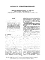
Figure 2 Distribution of ypT stage in 153 patients treated with
preoperative RCT within clinical phase II/III trials. 107 patients
(70%) manifested as intermediate responders with irradiation-induced
tumor regression (TRG 2/3)[17] and ypT2 and ypT3 category.
Sprenger et al. World Journal of Surgical Oncology 2010, 8:27
/>Page 5 of 11
Table 2: Clinical findings and treatment procedures
Feature Number of Patients (n = 107) %*
Gender
Male 83 78
Female 24 22
Age (years)
Median 62.3
Range 36 - 81
Tumor Distance from Anal Verge (cm)
0-6 53 50
>6-12 47 44
>12-16 7 7
cT Stage
100
211
39892
487
cN Stage
Positive 82 77
Negative 25 23
cUICC Stage
I00
II 25 23
III 82 77
IV 0 0
Neoadjuvant Treatment
50.4 Gy + standard 5-FU 84 79
50.4 Gy + intensified 5-FU/Oxaliplatin 23 21
Surgical Procedure (including TME)
Low Anterior Resection (incl.
laparoscopic)
63 (2) 59 (2)
Abdominoperineal Resection (incl.
laparoscopic)
43 (1) 40 (1)
Hartmann's Procedure 1 1
Sprenger et al. World Journal of Surgical Oncology 2010, 8:27
/>Page 6 of 11
Table 3: Pathological findings
Feature Number of Patients (n = 107) %
ypT Stage
23936
3a 16 15
3b 30 28
3c 20 19
3d 2 2
ypN Stage
06964
12826
2109
ypUICC Stage
26964
33533
4 * 3 3
Resection Status
R0 107 100
R1 0 0
Tumor Regression Grading
17
Grade 2 59 55
Grade 3 48 45
Circumferential Resection Margin
Negative 107 100
Positive 0 0
Histologic Differentiation Grading after
RCT
High Grade Differentiation 73 68
Low Grade Differentiation 34 32
Nodal Yield (nodes)
0-12 5 5
12-18 30 28
18-30 50 47
>30 22 21
Sprenger et al. World Journal of Surgical Oncology 2010, 8:27
/>Page 7 of 11
staged III rectal cancers therefore showed no higher risk
of cancer relapse and cancer-related death than initially
node-negative patients, as long as sterilization of lymph
node metastases can be achieved with RCT.
Discussion
Recent results from the randomized multicenter trial
CAO/ARO/AIO-94 showed an enhanced local control
and sphincter preservation with concurrently decreased
toxicity after preoperative long-term RCT compared to
postoperative RCT [6]. These results led to the recom-
mendation of preoperative RCT in locally advanced
(stage II/III) rectal cancers [7]. Preoperative RCT results
in a very heterogeneous tumor response, which can be
measured by various response parameters such as T-level
downsizing, tumor downstaging, elimination of lymph
node metastases, and pathomorphologic tumor regres-
sion.
Of 153 patients with stage II/III rectal cancer who
received standardized preoperative RCT within random-
ized clinical trials, pCR as a major response criterion, was
achieved in 16% (n = 10) of patients. pCR rates vary
between 10 and 20% and were associated with a favorable
outcome [8,10]. Nevertheless the majority of rectal can-
cers (70% of the actual collective) show intermediate
response with residual tumor either within (ypT2) or
beyond (ypT3) the rectal wall (Figure 2).
It remains unclear which subgroup of patients with
intermediate response can be considered as cured after
preoperative RCT and subsequent TME surgery. Conver-
sly, it is of enormous clinical interest to know which sub-
group necessitates adjuvant systemic therapy.
Involvement of circumferential resection margins
(CRM) has recently been described as a very strong prog-
nostic factor after preoperative short term radiation [22].
Although this is distinctly reasonable, fortunately only a
considerable small group of patients is affected by posi-
tive CRM after preoperative long-term RCT. In our study,
7% of patients (n = 8) presented with cT4 status and
potential CRM involvement in pretherapeuthical imag-
ing. RCT-induced tumor downsizing was achieved in all
cases, resulting in a maximal residual mesorectal infiltra-
tion of ≥ 1.5 cm (ypT3d) in 2 patients (2%). Pathologically
confirmed complete (R0) resection with negative (>1
mm) CRM after RCT was accomplished in all patients
including those previously classified as high-risk for posi-
tive CRM.
Cancer Recurrence
Total 17 16
Local 0 0
Local + Distant 2 2
Distant 15 14
* Patients were not included in statistical analysis
Table 3: Pathological findings (Continued)
Table 4: Comparison of DFS and OS with respect to ypT, ypN status and Tumor Grading
Parameter Variable Estimated 5 Year
DFS Probability
(%)
p-value:
*univariate
**multivariate
Estimated 5 Year
OS Probability
(%)
p-value:
*univariate
**multivariate
ypT 2/3a 85 *0.6 94 *0.6
3b-d 77 **0.56 84 **0.7
ypN 0 88 *0.04 95 *0.02
1/2 64 **0.03 80 **0.053
Residual Tumor
Differentiation
High 88 *0.04 94 *0.09
Low 62 71
Sprenger et al. World Journal of Surgical Oncology 2010, 8:27
/>Page 8 of 11
Prior to implementation of neoadjuvant strategies for
rectal cancer, a tumor invasion of ≥ 5 mm into the
mesorectal compartment, besides circumferential
involvement, was described as a significant prognostic
factor [23]. The decision to apply postoperative radiation
or radiochemotherapy, was based on tumor invasion as
well as a positive nodal status, and led to reduced recur-
rence rates and prolonged survival [24].
We therefore evaluated the impact of intramural depth
of tumor invasion (ypT2) together with minimal (<1 mm)
transgression of the muscularis propria (ypT3a) com-
pared to a distinct transmural tumor invasion into the
mesorectum (>1 mm; ypT3b-d). Since patients with
ypT3a status show only an extremely marginal infiltration
of the mesorectal compartment (<1 mm) we consider
them to prognostically belong to the ypT2 group rather
than to the tumors with distict mesorectal infiltration.
Our results underline this assumption showing an
increased incidence of nodal metastases in ypT3b-d
patients compared to ypT2/3a patients.
In the patients presenting with previous cT3/4 rectal
cancers (only 1 patient had cT2 N+ status, according
stage III) the RCT-induced regression of tumor invasion
depth to ypT2/3a status had no impact on prolonged DFS
and OS. Thus, residual tumor transgression into the
mesorectum after preoperative RCT showed no signifi-
cant influence on cancer recurrence, providing that com-
plete resection with negative CRM is achieved by
adequate TME surgery.
Tumor downsizing from the extramural mesorectal
compartment into the actual rectal wall therefore seems
to be of importance only when tumor-free CRM and R0-
resection cannot be guaranteed (former T3d/4 status).
In contrast to ypT, nodal status after preoperative CRT
(ypN) significantly influenced cancer recurrence and
overall survival in stage II/III rectal cancer patients with
Figure 3 DFS in patients with rectal cancer and intermediate response to preoperative RCT stratified by ypT stage (3a) and ypN stage (3b).
OS in patients with rectal cancer and intermediate response to preoperative RCT stratified by ypT stage (3c) and ypN stage (3d).
Sprenger et al. World Journal of Surgical Oncology 2010, 8:27
/>Page 9 of 11
intermediate response within our investigation. This
finding coincides with previous results and supports
recent investigations with considerable numbers of
patients [25,26] but it is based on a collective of patients
with highly standardized diagnostic and treatment proce-
dures according to the protocols of the respective clinical
phase II and III trials of the German Rectal Cancer Study
Group.
In agreement with other authors [25,27], we observed
that pretherapeutical nodal involvement (cN+) has no
impact on the prognosis of patients, in which ypN0 status
can be achieved. Patients with evidence of lymph node
involvement in pretreatment staging can therefore not
categorically be considered as high risk for cancer relapse.
Anyway, patients with ypN+ status should be consid-
ered for upcoming trials with intensified adjuvant CT
regimes as this might be more efficient in preventing sys-
temic tumor relapse. Nonetheless, mesorectal tumor
invasion (ypT3b-d) was significantly associated with
residual lymph node metastases after RCT in our study (p
< 0.001). We interpret this finding with a generally lower
response to RCT regarding both downsizing of the pri-
mary tumor and sterilization of lymph node metastases.
This might be due to improved biological behavior and
enhanced resistance to RCT in individual cancers. The
prognostic impact of mesorectal tumor infiltration
remains unclear. We could not show straight effects on
tumor recurrence and survival but are well aware that
this might be due to the relative small number of patients
underlying this investigation.
Neoadjuvant RCT has repeatedly been accused of
reducing lymph node yield in rectal cancer specimens
[28-31]. It has also been reported that the number of
detected nodes in stage II rectal cancer patients influ-
ences survival [32-34]. Within our investigation, we eval-
uated a median number of 21 lymph nodes per specimen.
In contrast to other investigations [35], we found no sig-
nificant difference in lymph node yield between ypN0
patients with and those without subsequent development
of distant metastases and tumor-related death. This
might be explained by the implementation of extensive
lymph node recovery at our institution and a minor vari-
ance of evaluated lymph node numbers between both
groups.
While histologic tumor grading in colorectal cancers
after primary surgery has been ascertained as a prognos-
tic factor [13], its prognostic relevance following preoper-
ative RCT remains unclear and currently does not belong
to standard pathologic staging in rectal cancer speci-
mens. Our results show that histologic grading of residual
tumor cells is a reliable parameter, which correlates with
advanced tumor biology and has straight impact on DFS
despite RCT-induced histomorphologic alterations of the
tumor. Thus, histopathologiclogic grading of residual
tumor cells should be considered within risk stratification
in rectal cancers after RCT.
Not unexpectedly, lymph node status displays as the
major criterion for therapy stratification after application
of preoperative RCT within our study and several recent
investigations and might subdivide patients with need of
intensified adjuvant treatment from those who can be
considered as cured after surgery. In contrast Collette et
al. [15], who reported the results of the EORTC 22921
trial, underlined that only patients with RCT-induced
tumor downsizing to ypT1/2N0 status benefited from
adjuvant CT. They interpret their results with an
increased sensitivity to preoperative RCT as well as post-
operative CT in this subgroup. However, 5-FU mono-
therapy was used in both, neoadjuvant and adjuvant
setting in this trial. This might explain the failure of adju-
vant CT in patients with a minor response to preopera-
tive RCT. Thus, in the adjuvant setting an intensified or
combined CT should be applied with different anti-
tumoral mechanisms (e.g. FOLFOX/FOLFIRI regime ±
targeted therapy) in patients with minor response to neo-
adjuvant treatment.
To date, most patients with positive nodal status after
preoperative RCT will intuitively get adjuvant CT. Pro-
spective randomized clinical trials should therefore clar-
ify the impact of adjuvant treatment in patients
undergoing preoperative RCT and radical surgery. For
ypN0 patients 5-FU based adjuvant CT was shown as a
potential overtreatment and had no significant effect on
survival [36,37].
Nevertheless, in our actual study population, 7 patients
with ypN0 status developed distant metastases during
follow-up. All 7 had poorly differentiated residual tumors
(low grade). Poor differentiation of residual tumor cell
clusters after RCT and advanced invasion depth turned
out to be predictors of lymph node metastases and may
be indicators of occult nodal (micro-) metastases in
patients classified as ypN0. Both parameters should thus
be taken into account in ypN0 patients, particularly in
cases of minor lymph node recovery, and might have
influence on the decision for adjuvant CT.
Although this investigation is based on a homogeneous
collective of patients treated within randomized clinical
trials with replicable and standardized diagnostic and
therapeutic procedures, its principal limitations are the
retrospective character and the relatively small number of
patients. Thus this study does not want to claim to ulti-
mately answer the question which subgroup of patients
need adjuvant CT after preoperative multimodal treat-
ment and subsequent R0-resection. Prospective random-
ized trials will have to clarify the debatable role of
postoperative CT in rectal cancer patients after preopera-
tive RCT and radical TME surgery. The clinicopathologic
parameters investigated in this study might give indica-
Sprenger et al. World Journal of Surgical Oncology 2010, 8:27
/>Page 10 of 11
tions to stratify patient groups with lower and higher
individual risk of tumor relapse and tumor-related death
within future clinical trials.
Competing interests
The authors declare that they have no competing interests.
Authors' contributions
TS prepared the study design, assembled and analysed the data and drafted
the manuscript. HR carried out the pathological diagnostics of the rectal can-
cer specimens and reviewed the manuscript. KJ carried out the statistical anal-
yses. HC contributed the radiation therapy data and reviewed the manuscript.
LC participated in assembling of the data and reviewed the manuscript. BMG
and HB reviewed the manuscript. TL supervised the study and data assembling
and critically reviewed the manuscript. All authors read and approved the final
manuscript.
Acknowledgements
This work was supported by the Deutsche Forschungsgemeinschaft (KFO 179:
Biological basis of individual tumor response in patients with rectal cancer)
The authors would like to thank Birgit Jünemann for excellent technical and
organisational assistance.
Author Details
1
Department of General and Visceral Surgery, University Medical Center
Göttingen, Georg-August-University, Göttingen, Germany,
2
Department of
Pathology, University Medical Center Göttingen, Georg-August-University,
Göttingen, Germany,
3
Department of Medical Statistics, University Medical
CenterGöttingen, Georg-August-University, Göttingen, Germany and
4
Department of Radiotherapy and Radiooncology, University Medical Center
Göttingen, Georg-August-University, Göttingen, Germany
References
1. Heald RJ, Husband EM, Ryall RD: The mesorectum in rectal cancer
surgery - the clue to pelvic recurrence? Br J Surg 1982, 69:613-6.
2. MacFarlane JK, Ryall RD, Heald RJ: Mesorectal excision for rectal cancer.
Lancet 1993, 341:457-60.
3. Kapiteijn E, Marijnen CA, Nagtegaal ID, Putter H, Steup WH, Wiggers T,
Rutten HJ, Pahlman L, Glimelius B, van Krieken JH, Leer JW, Velde CJ van
de, Dutch Colorectal Cancer Group: Preoperative radiotherapy
combined with total mesorectal excision for resectable rectal cancer.
N Engl J Med 2001, 345:638-46.
4. Improved survival with preoperative radiotherapy in resectable rectal
cancer. Swedish Rectal Cancer Trial. N Engl J Med 1997, 336:980-7.
5. Lavery IC, Fazio VW, Lopez-Kostner F: Radiotherapy for rectal cancer. N
Engl J Med 1997, 337:346-7. author reply 347-8
6. Sauer R, Becker H, Hohenberger W, Rödel C, Wittekind C, Fietkau R, Martus
P, Tschmelitsch J, Hager E, Hess CF, Karstens JH, Liersch T, Schmidberger
H, Raab R, German Rectal Cancer Study Group: Preoperative versus
postoperative chemoradiotherapy for rectal cancer. N Engl J Med 2004,
351:1731-40.
7. Schmiegel W, Reinacher-Schick A, Arnold D, Graeven U, Heinemann V,
Porschen R, Riemann J, Rödel C, Sauer R, Wieser M, Schmitt W, Schmoll HJ,
Seufferlein T, Kopp I, Pox C: [Update S3-guideline "colorectal cancer"
2008]. Z Gastroenterol 2008, 46:799-840.
8. Theodoropoulos G, Wise WE, Padmanabhan A, Kerner BA, Taylor CW,
Aguilar PS, Khanduja KS: T-level downstaging and complete pathologic
response after preoperative chemoradiation for advanced rectal
cancer result in decreased recurrence and improved disease-free
survival. Dis Colon Rectum 2002, 45:895-903.
9. Kaminsky-Forrett MC, Conroy T, Luporsi E, Peiffert D, Lapeyre M, Boissel P,
Guillemin F, Bey P: Prognostic implications of downstaging following
preoperative radiation therapy for operable T3-T4 rectal cancer. Int J
Radiat Oncol Biol Phys 1998, 42:935-41.
10. Rodel C, Martus P, Papadoupolos T, Füzesi L, Klimpfinger M, Fietkau R,
Liersch T, Hohenberger W, Raab R, Sauer R, Wittekind C: Prognostic
significance of tumor regression after preoperative
chemoradiotherapy for rectal cancer. J Clin Oncol 2005, 23:8688-96.
11. Miyoshi M, Ueno H, Hashiguchi Y, Mochizuki H, Talbot IC: Extent of
mesorectal tumor invasion as a prognostic factor after curative surgery
for T3 rectal cancer patients. Ann Surg 2006, 243:492-8.
12. Willett CG, Badizadegan K, Ancukiewicz M, Shellito PC: Prognostic factors
in stage T3N0 rectal cancer: do all patients require postoperative pelvic
irradiation and chemotherapy? Dis Colon Rectum 1999, 42:167-73.
13. Steel MC, Woods R, Mackay JM, Chen F: Extent of mesorectal invasion is
a prognostic indicator in T3 rectal carcinoma. ANZ J Surg 2002, 72:483-7.
14. Sobin LH: TNM, sixth edition: new developments in general concepts
and rules. Semin Surg Oncol 2003, 21:19-22.
15. Collette L, Bosset JF, den Dulk M, Nguyen F, Mineur L, Maingon P,
Radosevic-Jelic L, Piérart M, Calais G, European Organisation for Research
and Treatment of Cancer Radiation Oncology Group: Patients with
curative resection of cT3-4 rectal cancer after preoperative
radiotherapy or radiochemotherapy: does anybody benefit from
adjuvant fluorouracil-based chemotherapy? A trial of the European
Organisation for Research and Treatment of Cancer Radiation
Oncology Group. J Clin Oncol 2007, 25:4379-86.
16. Gerard JP, Conroy T, Bonnetain F, Bouché O, Chapet O, Closon-Dejardin
MT, Untereiner M, Leduc B, Francois E, Maurel J, Seitz JF, Buecher B,
Mackiewicz R, Ducreux M, Bedenne L: Preoperative radiotherapy with or
without concurrent fluorouracil and leucovorin in T3-4 rectal cancers:
results of FFCD 9203. J Clin Oncol 2006, 24:4620-5.
17. Dworak O, Keilholz L, Hoffmann A: Pathological features of rectal cancer
after preoperative radiochemotherapy. Int J Colorectal Dis 1997,
12:19-23.
18. Nagtegaal ID, Velde CJ van de, Worp E van der, Kapiteijn E, Quirke P, van
Krieken JH, Cooperative Clinical Investigators of the Dutch Colorectal
Cancer Group: Macroscopic evaluation of rectal cancer resection
specimen: clinical significance of the pathologist in quality control. J
Clin Oncol 2002, 20:1729-34.
19. Greene FL, Brierley J, O'Sullivan B, Sobin LH, Wittekind C, International
Union Against Cancer and American Joint Committee on Cancer: On the
use and abuse of X in the TNM classification. Cancer 2005, 103:647-9.
20. UICC: TNM Supplement 2001. In A commentary on uniform use 2nd
edition. Edited by: Wittekind Ch, Henson DE, Hutter RVP, Sobin LH. John
Wiley & Sons, New York; 2001.
21. Rodel C, Liersch T, Hermann RM, Arnold D, Reese T, Hipp M, Fürst A,
Schwella N, Bieker M, Hellmich G, Ewald H, Haier J, Lordick F, Flentje M,
Sülberg H, Hohenberger W, Sauer R: Multicenter phase II trial of
chemoradiation with oxaliplatin for rectal cancer. J Clin Oncol 2007,
25:110-7.
22. Gosens MJ, Klaassen RA, Tan-Go I, Rutten HJ, Martijn H, Brule AJ van den,
Nieuwenhuijzen GA, van Krieken JH, Nagtegaal ID: Circumferential
margin involvement is the crucial prognostic factor after
multimodality treatment in patients with locally advanced rectal
carcinoma. Clin Cancer Res 2007, 13:6617-23.
23. Merkel S, Mansmann U, Papadopoulos T, Wittekind C, Hohenberger W,
Hermanek P: The prognostic inhomogeneity of colorectal carcinomas
Stage III: a proposal for subdivision of Stage III. Cancer 2001, 92:2754-9.
24. Krook JE, Moertel CG, Gunderson LL, Wieand HS, Collins RT, Beart RW,
Kubista TP, Poon MA, Meyers WC, Mailliard JA: Effective surgical adjuvant
therapy for high-risk rectal carcinoma. N Engl J Med 1991, 324:709-15.
25. Chang GJ, Rodriguez-Bigas MA, Eng C, Skibber JM: Lymph node status
after neoadjuvant radiotherapy for rectal cancer is a biologic predictor
of outcome. Cancer 2009, 115:5432-40.
26. Kim TH, Chang HJ, Kim DY, Jung KH, Hong YS, Kim SY, Park JW, Oh JH, Lim
SB, Choi HS, Jeong SY: Pathologic Nodal Classification Is the Most
Discriminating Prognostic Factor for Disease-Free Survival in Rectal
Cancer Patients Treated with Preoperative Chemoradiotherapy and
Curative Resection. Int J Radiat Oncol Biol Phys 2009 in press.
27. Quah HM, Chou JF, Gonen M, Shia J, Schrag D, Saltz LB, Goodman KA,
Minsky BD, Wong WD, Weiser MR: Pathologic stage is most prognostic of
disease-free survival in locally advanced rectal cancer patients after
preoperative chemoradiation. Cancer 2008, 113:57-64.
28. Baxter NN, Morris AM, Rothenberger DA, Tepper JE: Impact of
preoperative radiation for rectal cancer on subsequent lymph node
evaluation: a population-based analysis. Int J Radiat Oncol Biol Phys
2005, 61:426-31.
Received: 19 January 2010 Accepted: 13 April 2010
Published: 13 April 2010
This article is available from: 2010 Sprenger et al; licensee BioMed Central Ltd. This is an Open Access article distributed under the terms of the Creative Commons Attribution License ( ), which permits unrestricted use, distribution, and reproduction in any medium, provided the original work is properly cited.World Journal of Surgical Oncology 2010, 8:27
Sprenger et al. World Journal of Surgical Oncology 2010, 8:27
/>Page 11 of 11
29. Habr-Gama A, Perez RO, Proscurshim I, Rawet V, Pereira DD, Sousa AH, Kiss
D, Cecconello I: Absence of lymph nodes in the resected specimen after
radical surgery for distal rectal cancer and neoadjuvant
chemoradiation therapy: what does it mean? Dis Colon Rectum 2008,
51:277-83.
30. Wichmann MW, Muller C, Meyer G, Strauss T, Hornung HM, Lau-Werner U,
Angele MK, Schildberg FW: Effect of preoperative radiochemotherapy
on lymph node retrieval after resection of rectal cancer. Arch Surg 2002,
137:206-10.
31. Wijesuriya RE, Deen KI, Hewavisenthi J, Balawardana J, Perera M:
Neoadjuvant therapy for rectal cancer down-stages the tumor but
reduces lymph node harvest significantly. Surg Today 2005, 35:442-5.
32. Tepper JE, O'Connell MJ, Niedzwiecki D, Hollis D, Compton C, Benson AB,
Cummings B, Gunderson L, Macdonald JS, Mayer RJ: Impact of number of
nodes retrieved on outcome in patients with rectal cancer. J Clin Oncol
2001, 19:157-63.
33. Morris EJ, Maughan NJ, Forman D, Quirke P: Identifying stage III
colorectal cancer patients: the influence of the patient, surgeon, and
pathologist. J Clin Oncol 2007, 25:2573-9.
34. Earle CC, Weiser MR, Ter Veer A, Skibber JM, Wilson J, Rajput A, Wong YN,
Benson AB, Shibata S, Romanus D, Niland J, Schrag D: Effect of lymph
node retrieval rates on the utilization of adjuvant chemotherapy in
stage II colon cancer. J Surg Oncol 2009, 100:525-8.
35. Kim YW, Kim NK, Min BS, Lee KY, Sohn SK, Cho CH: The influence of the
number of retrieved lymph nodes on staging and survival in patients
with stage II and III rectal cancer undergoing tumor-specific
mesorectal excision. Ann Surg 2009, 249:965-72.
36. Fietkau R, Barten M, Klautke G, Klar E, Ludwig K, Thomas H, Brinckmann W,
Friedrich A, Prall F, Hartung G, Küchenmeister U, Kundt G: Postoperative
chemotherapy may not be necessary for patients with ypN0-category
after neoadjuvant chemoradiotherapy of rectal cancer. Dis Colon
Rectum 2006, 49:1284-92.
37. Huh JW, Kim HR: Postoperative chemotherapy after neoadjuvant
chemoradiation and surgery for rectal cancer: Is it essential for patients
with ypT0-2N0? J Surg Oncol 2009, 100:387-391.
doi: 10.1186/1477-7819-8-27
Cite this article as: Sprenger et al., Stage II/III rectal cancer with intermediate
response to preoperative radiochemotherapy: Do we have indications for
individual risk stratification? World Journal of Surgical Oncology 2010, 8:27