Báo cáo khoa học: "Advanced moderately differentiated neuroendocrine carcinoma of the rectum with favorable prognosis by postoperative chemoradiation" pps
Bạn đang xem bản rút gọn của tài liệu. Xem và tải ngay bản đầy đủ của tài liệu tại đây (1.07 MB, 4 trang )
WORLD JOURNAL OF
SURGICAL ONCOLOGY
Nojima et al. World Journal of Surgical Oncology 2010, 8:29
/>Open Access
CASE REPORT
BioMed Central
© 2010 Nojima et al; licensee BioMed Central Ltd. This is an Open Access article distributed under the terms of the Creative Commons
Attribution License ( which permits unrestricted use, distribution, and reproduction in
any medium, provided the original work is properly cited.
Case report
Advanced moderately differentiated
neuroendocrine carcinoma of the rectum with
favorable prognosis by postoperative
chemoradiation
Hiroyuki Nojima
1
, Kazuhiro Seike*
1
, Chihiro Kosugi
1
, Takashi Shida
1
, Keiji Koda
1
, Kenji Oda
1
, Shigeyuki Kamata
1
,
Hiroshi Ishikura
2
and Masaru Miyazaki
1
Abstract
Rectal neuroendocrine carcinoma is rare with poor prognosis. We report herein a case of advanced moderately
differentiated neuroendocrine carcinoma of the rectum with relatively favorable prognosis treated by postoperative
adjuvant chemoradiation therapy. A 58-year-old Japanese female was referred and colonofiberscopy revealed an easy-
bleeding irregular tumor in the lower rectum, which was pathologically diagnosed as a neuroendocrine carcinoma.
Surgical treatment consisted of abdominoperineal resection and lymph node dissection. The tumor invaded deeply
into perirectal tissues, and 9 of 11 lymph node metastases were observed. Immunohistochemically, chromogranin A
showed diffuse and strong staining, and the MIB-1 labeling index was 18.3 ± 5.6, supporting the high proliferation of
the tumor. Some nucleus of the tumor showed positive staining for p21/WAF1. A total dose of 46 Gy of radiotherapy
was delivered with 800 mg of daily oral doxifluridine. At 5 years post-surgery, the patient demonstrated no clinical
evidence of intrapelvic recurrence or distant metastases.
Background
Neuroendocrine carcinomas of the colon and rectum are
rare tumors with aggressive behavior and poorer progno-
sis compared with adenocarcinomas, and the reported 3-
year survival rates are 13-15%[1]. These carcinomas are
subclassified into two pathological types, small cell carci-
nomas and moderately differentiated neuroendocrine
carcinomas. Small cell carcinoma of the colon and rec-
tum is virtually indistinguishable from small cell lung
cancer morphologically and immunohistochemically, and
small cell lung carcinoma is sensitive to chemotherapy
and adjuvant chemotherapy after surgery results in pro-
longed survival[2]. Several studies have demonstrated the
efficacy of chemotherapy for colorectal small cell carci-
noma[3]. On the other hand, moderately differentiated
neuroendocrine carcinoma of the colon and rectum has a
similar morphology to large cell lung carcinoma with
neuroendocrine features. Surgery is a mainstay of benefi-
cial treatment although the effect of adjuvant treatment
remains undermined.
We herein report a case of advanced moderately differ-
entiated neuroendocrine carcinoma of the rectum with
relatively favorable prognosis by postoperative adjuvant
chemoradiation therapy.
Case Presentation
Clinical history
A 58-year-old Japanese female was admitted to hospital
with a two-month history of rectal bleeding. Colonofi-
berscopy revealed a tumor in the lower rectum, however,
a biopsied specimen from the tumor showed no malig-
nant findings. She was referred to our institution for fur-
ther examinations.
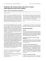
Colonofiberscopy showed an easy-bleeding yellowish
tumor with a relatively regular surface with lateral sub-
mucosal elevation (Fig. 1a) and 5 biopsied specimens
revealed no histological malignancies as in the previous
examination. Computed tomography demonstrated a 40
* Correspondence:
1
Department of General Surgery, Graduate School of Medicine, Chiba
University, Chiba, Japan
Full list of author information is available at the end of the article
Nojima et al. World Journal of Surgical Oncology 2010, 8:29
/>Page 2 of 4
mm diameter tumor on the left side of the lower rectal
wall with regional lymphadenopathy. The laboratory data
were unremarkable expect for elevated circulating levels
of carbohydrate antigen 19-9 (59.1 U/ml; normal value,
<37 U/ml). The examinations were repeated 2 and half
months later. On colonoscopic examination, the tumor
was visualized as more irregular than the previous find-
ings (Fig. 1b) and a biopsied specimen revealed neuroen-
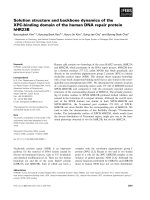
docrine carcinoma. Computed tomography showed a 50
mm-long tumor in the lower rectum with swollen
regional lymph nodes and no distant metastatic lesions
(Fig 2).
Based on these findings, the patient underwent abdom-
inoperineal resection with total mesorectal resection and
bilateral lymph node dissection. The postoperative
course was uneventful. To prevent intrapelvic recurrence,
a total dose of 46 Gy in 2 Gy fractions of radiotherapy was
delivered through a linear accelerator using the 3-field
technique (10 MV), 5 times a week. A daily dose of 800
mg of oral doxifluridine was administered for 5 years
because of patient's rejection to intensive intravenous
chemotherapy. At 5 years post-surgery, the patient dem-
onstrated no clinical evidence of intrapelvic recurrence or
distant metastases.
Methods
Immunohistochemistry
IHC was done on formalin-fixed paraffin-embedded sec-
tions, using labeled streptoavidin-biotin-peroxidase and
microwave antigen retrieval technique. Mouse monoclo-
nal antibodies against chromogranin A (1:50, Dako Cyto-
mation), MIB-1(anti Ki-67,1:50, Dako Cytomation) and
p53 protein(1:50, Dako Cytomation) were used. Goat
polyclonal antibodies against hASH1 (human acetate-
scute homolog 1,1:100, Santa-Cruz, CA, USA) and Neu-
roD(1:400, Santa-Cruz, CA, USA) were used in order to
assess the neuroendocrine differentiation at a transcrip-
tion level. Mouse IgG was used as a negative control, with
dilution of 1:100. Appropriate positive controls known to
contain the antigens in question were processed simulta-
neously.
Pathological findings
Macroscopically, the resected specimen showed a pro-
truding lesion with an irregular surface, 35 × 20 mm in
diameter. The tumor had lateral submucosal elevation
and tumor size including lateral elevation was 60 × 30
mm. Microscopically, the tumor invaded the adjacent
adipose tissue. Nine of 11 lymph node metastases were
observed.
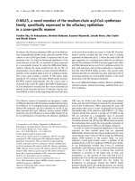
Immunohistochemistry(IHC)
ChromograninA showed a diffuse and strong staning in
the tumor cytoplasm indicating neuroendocrine differen-
tiation. MIB-1(Ki-67 antigen) labeling index showed 18.3
± 5.6 supporting high proliferation of the tumor. Nuclear
staining of p53 was also detected in approximately 10% of
the tumor suggesting the tumor to be an endocrine cell
carcinoma. Strong and diffuse nuclear staining of Neu-
roD and cytoplasmic staining (also in some nucleus) of
hASH1 were also detected (Fig. 3).
Discussion
Neuroendocrine tumors of the colon and rectum repre-
sent a broad clinical-pathologic spectrum with varying
morphologic features and biological behavior, and there
is still much debate concerning their classification. Based
on the WHO classification, neuroendocrine tumor of the
gastrointestinal tract is classified into 3 subtypes: carci-
noid, which is benign or low-grade malignant; malignant
carcinoid, which is low-grade malignant; and poorly dif-
ferentiated neuroendocrine carcinoma, which is high-
grade malignant. Poorly differentiated neuroendocrine
carcinoma is defined as small cell carcinoma, being mor-
phologically similar to small cell carcinoma of the lung[4].
In addition to small cell carcinoma, pathological studies
have shown that moderately differentiated, also known as
large cell or intermediate variant, neuroendocrine carci-
noma should be classified as high-grade malignant
because of its distinct neuroendocrine lineage and bio-
logical aggressiveness[1,4]. Moderately differentiated
neuroendocrine carcinomas are distinguished from small
cell carcinomas by having more vesicular nuclei, more
prominent nucleoi, more abundant cytoplasm, and less
Figure 1 a) Colonoscopic findings, initial evaluation, b) second
evaluation.
Figure 2 CT and MRI findings.
Nojima et al. World Journal of Surgical Oncology 2010, 8:29
/>Page 3 of 4
mitotic activity, morphologically reminiscent of large cell
neuroendocrine carcinoma encountered in the lungs. Ki-
67 antigen labeling index of the present patient showed
18.3 ± 5.6 supporting high proliferation of the tumor. Ki-
67 is expressed by proliferating cells and provides a mea-
surement of the growth fraction in individual tissues and
tumors. Some studies suggest that a relationship exists
between a high proliferative rate, as measured by Ki-67
immunoreactivity, and tumor aggressiveness[5].
Chaudhry et al. demonstrated that patients with gastroin-
testinal neuroendocrine tumors with a low Ki-67 index
have a better prognosis than tumors with a high prolifera-
tive index[6]. According to histopathological findings,
our case would be classified as moderately differentiated
neuroendocrine carcinoma.
Bernick et al. reported that colorectal moderately dif-
ferentiated neuroendocrine carcinomas have a poor
prognosis with a median survival of only 10.4 months,
similar to small cell carcinoma[1]. Patients with neuroen-
docrine cell carcinoma have liver and lymph node
involvement of between 65% and 80% at the time of diag-
nosis[1,7], therefore, they may benefit from treatment
with chemotherapeutic agents. Iyoda et al. showed that
adjuvant chemotherapy based on cisplatin, carboplatin,
or cyclophosphomide prolongs the survival of patients
with large cell carcinoma with neuroendocrine features
only in the early stages[8]. In this report, the patient was
eager to receive oral but not intravenous chemotherapy.
The addition of adjuvant radiotherapy to the primary
treatment of rectal cancer has led to the decreased inci-
dence of local recurrence in several randomized stud-
ies[9], and radiotherapy was therefore offered
postoperatively. Neoadjuvant chemoradiation is consid-
ered as a beneficial option, however, surgery was per-
formed by patient's preference.
5-fluorouracil (5-FU) is a key agent that is widely used
in the treatment of colorectal cancers. TS is an essential
DNA synthetic enzyme that catalyzes the methylation of
dUMP to dTMP[10]. DPD is a rate-limiting enzyme of 5-
FU catabolism, 85% of an administered dose of 5-FU is
degraded to inactive metabolites by DPD[11]. Therefore,
low TS and low DPD activity is reportedly correlated with
high 5-FU chemosensitivity of cancer cells. Doxifluridine
was synthesized by Cook et al[12] and is widely used in
Japan as a prodrug of 5-FU, thus, the efficacy of doxifluri-
dine is influenced by levels of TS and DPD. This tumor
showed scarce staining of TS and negative staining of
DPD, which supports the sensitivity to 5-FU.
p21 is a cyclin dependent kinase inhibitor and its
expression is a marker of tumor radiosensitivity in
patients with rectal cancer[13]. This tumor had positive
staining of p21, indicating the sensitivity to radiation.
This report indicated the difficult histological diagnosis
of neuroendocrine carcinoma by endoscopic biopsied
specimens. The reason for negative biopsies was specu-
lated its submucosal location. Bernick et al. reported that
the sensitivity of preoperative colonoscopic biopsy for
colorectal neuroendocrine carcinoma was approximately
60%[1]. We recommend the re-biopsy of an adequate
thickness of the rectal wall if a malignant tumor is sus-
pected from the clinical findings and radiological exami-
nations.
In conclusion, we experienced a case of advanced neu-
roendcrine carcinoma of the rectum with relatively favor-
able prognosis by postoperative adjuvant chemoradiation
therapy.
Consent
Written informed consent was obtained from the patient
for publication of this case report and any accompanying
images. A copy of the written consent is available for
review by the Editor-in-Chief of this journal.
Figure 3 Pathological findings. HE staining and Immunohistochem-
istry pictures: a; HE staining of the tumor (×200), inset a magnification
of ×400, b; IHC of chromograninA, which shows strong and diffuse
staining in the tumor cytoplasms (×200) c; IHC of hASH1, which shows
diffuse staining in the tumor cytoplasms and also in some nucleus
(×400) d; IHC of NeuroD, which shows strong and diffuse staining in
the tumor nucleus (×400) e; IHC of MIB-1, which shows a high labeling
index (×200) f; IHC of p53, which shows partial staining in the tumor
nucleus (×400).
a b
cd
e
f
cd
f
Nojima et al. World Journal of Surgical Oncology 2010, 8:29
/>Page 4 of 4
Competing interests
The authors declare that they have no competing interests.
Authors' contributions
HN: deta collection, drafting the manuscript. KS: drafting and revising the man-
uscript, surgical management of the patient. CK: surgical management of the
patient and revising the manuscript. TS:pathological review of surgical speci-
mens, preparing histopathological figures. KK: surgical management of the
patient and revising the manuscript. KO: surgical management of the patient
and revising the manuscript. SK: pathological review of surgical specimens,
preparing histopathological figures. HI: pathological review of surgical speci-
mens, preparing histopathological figures. MM: head of the department who
supervised all steps of the work. All authors read and approved final manu-
script.
Author Details
1
Department of General Surgery, Graduate School of Medicine, Chiba
University, Chiba, Japan and
2
Department of Molecular Pathology, Graduate
School of Medicine, Chiba University, Chiba, Japan
References
1. Bernick PE, Klimstra DS, Shia J, Minsky B, Saltz L, Shi W, Thaler H, Guillem J,
Paty P, Cohen AM, Wong WD: Neuroendocrine carcinoma of the colon
and rectum. Dis Colon Rectum 2004, 47:163-169.
2. Perry MC, Eaton WL, Propert KJ, Ware JH, Zimmer B, Chahinian AP, Skarin
A, Carey RW, Kreisman H, Faulkner C: Chemotherapy with or without
radiation therapy in limited small cell carcinoma of the lung. N Eng J
Med 1987, 316:912-918.
3. Okuyama T, Korenaga D, Tamura S, Yao T, Maekawa S, Watanabe A, Ikeda
T, Sugimachi K: The effective of chemotherapy with cisplatin and 5-
fluorouracil for recurrent small cell neuroendocrine carcinoma of the
rectum: report of a case. Surg Today 1999, 29:165-169.
4. Jass JR, Sobin LH: Histological typing of intestinal tumours. In World
Health Organization. International Histological Classification of Tumours 2nd
edition. Berlin: Springer-Verlag; 1989:14-34.
5. Tubiana M, Courdi A: Cell proliferation kinetics in human solid tumors:
Relation to probability of metastatic dissemination and long-term
survival. Radiother Oncol 1989, 15:1-18.
6. Chaudhry A, Oberg K, Wilander E: A study of biological behavior based
on the expression of a proliferating antigen in neuroendocrine tumors
of the digestive system. Tumor Biol 1992, 13:27-35.
7. Godwin JD: Carcinoid tumors: an analysis of 2837 cases. Cancer 1975,
36:560-569.
8. Iyoda A, Hiroshima K, Toyozaki T, Haga Y, Baba M, Fujisawa T, Ohwada H:
Adjuvant chemotherapy for large cell carcinoma with neuroendocrine
features. Cancer 2001, 92:1108-1112.
9. Colorectal Cancer Collaborative Group: Adjuvant radiotherapy for rectal
cancer: a systematic overview of 8507 patients from 22 randomized
trials. Lancet 2001, 358:1291-1304.
10. Van Triest B, Peters GJ: Thymidylate synthase: a target for combination
therapy and determinant of chemotherapeutic response in colorectal
cancer. Oncology 1999, 57:179-194.
11. Fischel JL, Etienne MC, Spector T, Formento P, Renee N, Milano G:
Dihydropyrimidine dehydrogenase: a tumoral target for fluorouracil
modulaion. Clin Cancer Res 1995, 1:991-996.
12. Cook AF, Holman MJ, Kramer MJ, Trown PW: Fluorinated pyrimidine
nucleosides: synthesis and antitumor activity of a series of 5'-deoxy-5-
fluoropyrimidine nucleosides. J Med Chem 1979, 22:1330-1335.
13. Fu CG, Tominaga O, Nagawa H, Nita ME, Masaki T, Ishimaru G, Higuchi Y,
Tsuruo T, Muto T: Role of p53 and p21/WAF1 detection in patient
selection for preoperative radiotherapy in rectal cancer patients. Dis
Colon Rectum 1998, 41:68-74.
doi: 10.1186/1477-7819-8-29
Cite this article as: Nojima et al., Advanced moderately differentiated neu-
roendocrine carcinoma of the rectum with favorable prognosis by postoper-
ative chemoradiation World Journal of Surgical Oncology 2010, 8:29
Received: 30 October 2009 Accepted: 17 April 2010
Published: 17 April 2010
This article is available from: 2010 N ojima et al; l icensee Bi oMed Centra l Ltd. This is an Open Access article distributed under the terms of the Creative Commons Attribution License ( ), which permits unrestricted use, distribution, and reproduction in any medium, provided the original work is properly cited.World Journal of Surgical Oncology 2010, 8:29