Báo cáo khoa học: "Adenosquamous carcinoma of breast in a 19 years old woman: a case report" pot
Bạn đang xem bản rút gọn của tài liệu. Xem và tải ngay bản đầy đủ của tài liệu tại đây (4.63 MB, 3 trang )
WORLD JOURNAL OF
SURGICAL ONCOLOGY
Agrawal et al. World Journal of Surgical Oncology 2010, 8:44
/>Open Access
CASE REPORT
© 2010 Agrawal et al; licensee BioMed Central Ltd. This is an Open Access article distributed under the terms of the Creative Commons
Attribution License ( which permits unrestricted use, distribution, and reproduction in
any medium, provided the original work is properly cited.
Case report
Adenosquamous carcinoma of breast in a 19 years
old woman: a case report
Amit Agrawal*
1
, Shopon Saha
2
, Ian O Ellis
3
and Alache M Bello
4
Abstract
Background: Adenosquamous carcinoma of the breast is a rare form of metaplastic breast carcinoma. We report such
a case in a 19 years old female.
Case presentation: Case notes and histopathology were reviewed. Adenosquamous carcinoma was diagnosed on
wide local excision and patient underwent skin-sparing mastectomy with Latissimus dorsi flap reconstruction.
Conclusions: Adenosquamous carcinoma of the breast is a rare form of metaplastic breast carcinoma. Data on correct
management, follow-up and prognosis are very limited but given the high potential for local recurrence, aggressive
surgery may be the only option.
Background
Adenosquamous carcinoma of the breast is a rare mor-
phological variant of metaplastic breast carcinomas
which was first described in early 1980s [1,2]. Since then
several cases and case series have been reported. We
present a case of adenosquamous carcinoma in a very
young woman aged 19 years. We also present a mini-
review of literature on this known but rare histological
type of breast cancer.
Case presentation
The 19 years old woman presented with approximately
four weeks history of painless 5 × 5 cm breast lump/
lumpiness. There was no history of trauma, nipple dis-
charge or relation with menstrual cycle. There was no
known family history of breast cancer. Palpation revealed
a non-tender, mobile, hard left upper quadrant left breast
lump. Mammogram was not performed given the very
young age of the patient. Ultrasound revealed a promi-
nent diffuse hypoechoic lesion. It appeared to be most
likely benign lesion but as there was an element of uncer-
tainty as to the nature of the lump on ultrasound, an
ultrasound guided core biopsy (14G needle) was per-
formed.
The core biopsy of the concerned area revealed paren-
chymal-rich breast tissue with columnar cell change and
epithelial hyperplasia. In addition, there was fibroblastic
stroma with focal elastosis containing an infiltrative ade-
nosquamous proliferation. Diagnostic excision was rec-
ommended by the breast multi-disciplinary team (MDT)
as the lesion appeared benign but with uncertain malig-
nant potential.
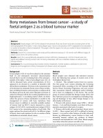
As shown in Figures 1, 2 and 3, subsequent diagnostic
excision biopsy showed an ill-defined area of benign ade-
nosis set in fibroblastic stroma with focal elastosis sug-
gestive of a radial scar or complex sclerosing lesion.
Within this lesion there was an infiltrative proliferation of
angulated glands showing adenosquamous features. A
proportion of these glands lacked definite myoepithelial
layers and the tumour expressed immunoreactivity to p63
gene antibody assay. Both the sclerosing lesion and the
adenosquamous proliferation extended up to the excised
margin. Hence, a wide local excision was deemed neces-
sary.
The wide local excision specimen showed residual
spindle cell lesion with squamoid islands seen in the inf-
ero-lateral shave specimen taken during wide local exci-
sion.
External opinion was sought from international experts
(as acknowledged later) in this uncommon histological
subtype of breast cancer. The consensus view was that it
was low grade adenosquamous carcinoma.
* Correspondence:
1
Professorial Unit of Surgery, City Hospital Campus, University of Nottingham,
Nottingham University Hospitals, Nottingham, NG5 1PB, UK
Full list of author information is available at the end of the article
Agrawal et al. World Journal of Surgical Oncology 2010, 8:44
/>Page 2 of 3
Due to reported low potential for metastasis but high
potential for local recurrence, she underwent Skin-spar-
ing mastectomy with immediate Latissimus Dorsi flap
reconstruction with an implant.
Following mastectomy, no further adjuvant therapy was
deemed necessary due to the completeness of resection
in the specimen histopathology and also due to the
reported low metastatic potential and no known
increased risk of contra-lateral disease. It was decided to
simply follow-up patient clinically annually for five years
due to known high risk of local recurrence and then to
mammogram screening programme.
Conclusions
Metaplastic carcinomas are known to be characterised by
the presence of non-epithelial cellular elements [3]. The
initial core biopsy of our case raised suspicion of ade-
nosquamous carcinoma. However, due to the inherent
limitations of the core biopsy, diagnostic excision was
recommended by the histopathologist. Ho et al [4] have
demonstrated in their small series of four cases that lim-
ited material in fine needle aspiration/core biopsy does
not yield enough material for confirmatory histopatho-
logical diagnosis.
Though diagnostic morphological differentiation may
not be possible in fine needle aspiration/core biopsy but
immuno-expression of myoepithelial marker may be
near-confirmatory. Reis-Filho et al [5] have reported in
their series of 82 archival fine-needle aspirates that p63
marker was demonstrable in majority of such aspirates.
The p63 gene (belonging to the larger p53 gene family) is
expressed in the basal cells of a range of myoepithelial tis-
sues including breast [5-8]. It is now known to be a sensi-
tive and specific myoepithelial marker [9] and has been
reported to be 86.7% sensitive and 99.4% specific for
metaplastic carcinomas [8]. Adenosquamous breast car-
cinomas are known to be immuno-reactive to p63 anti-
body assays. The tumour is this case-report was immuno-
reactive to p63 antibody.
There are data suggesting that it may carry a minimal
risk of metastasis[10]. The exact nature of this tumour is
controversial but it may represent a reactive process.
Complex sclerosing lesion with an adenosquamous pro-
liferation are of uncertain nature but have been reported
to be associated together [11]. This tumour is thought to
carry only a minimal risk for metastatic progression but it
Figure 1 Sclerosing lesion with central fibrosis and radiating pa-
renchyma. A low magnification (×20) view of the lesion. It has the
characteristics of a sclerosing lesion with central fibrosis, entrapped pa-
renchyma and radiating islands of tissue rich in parenchymal struc-
tures.
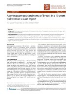
Figure 2 View of one of the radiating arms of the lesion. A view
(×20) of one of the radiating arms of the lesion showing parenchymal
tissue separated by a diagonal band of infiltrative cellular stromal tissue
containing small ductal like structures.
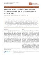
Figure 3 Infiltrative but bland stromal tissue with ductal and epi-
thelial structure. A high magnification view (×40) of the infiltrative
but bland cellular stromal tissue, along with the accompanying ductal
and small epithelial structures. This combination of components is typ-
ical of low grade metaplastic adenocarcinoma.
Agrawal et al. World Journal of Surgical Oncology 2010, 8:44
/>Page 3 of 3
carries a significant risk for local recurrence [12]. There-
fore, complete local excision or mastectomy to achieve
clear margins appear appropriate in light of the fact that
there is paucity of long-term follow-up data in these
patients.
To conclude, adenosquamous carcinomas are a rare
histopathological type of breast cancer. It should be con-
sidered in uncertain sclerosing lesions with an abundance
of epithelial hyperplasia with signs of squamous differen-
tiation in a predominant fibroblastic stroma. Immunore-
activity to p63 antibody appears to be near confirmatory
of adenosquamous differentiation. Although it is not
known to have high metastatic potential but it does have
potential for local recurrence if not suitably excised.
Therefore, aggressive local treatment is presently per-
ceived to be the management of choice until large long-
term follow-up data is available in literature.
Consent
Written informed consent was obtained from the patient
for publication of this case report and any accompanying
images. A copy of the written consent is available for
review by the Editor-in-Chief of this journal.
Competing interests
The authors declare that they have no competing interests.
Authors' contributions
AMB was clinically responsible for patient's care, conceptualised the study and
revised the manuscript. IOE was responsible for pathological interpretation,
images and revised the manuscript. AA and SS drafted the manuscript and
revised it. All authors read and approved the final manuscript.
Acknowledgements
The authors thank the following international experts from whom external
opinion was sought in this uncommon histological subtype of breast cancer:
• Prof PP Rosen, Department of Cancer and Metastasis, Hospital Durani Reynals,
Hospitalet de LLogregat, Barcelona, Spain.
• Prof DL Page, Department of Pathology, Division of Anatomic Pathology, Van-
derbilt University Medical Center, Nashville, Tennessee, USA.
• Prof V Eusebi, Department of Onkology, Ospedale Bellaria, University of Bolo-
gna, Italy.
Author Details
1
Professorial Unit of Surgery, City Hospital Campus, University of Nottingham,
Nottingham University Hospitals, Nottingham, NG5 1PB, UK,
2
Department of
Surgery, City Hospital Campus, Nottingham University Hospitals, Nottingham,
NG5 1PB, UK,
3
Department of Histopathology, City Hospital Campus, University
of Nottingham, Nottingham University Hospitals, Nottingham, NG5 1PB, UK
and
4
Nottingham Breast Institute, City Hospital Campus, Nottingham
University Hospitals, Nottingham, NG5 1PB, UK
References
1. Woodard BH, Brinkhous AD, McCarty KS Sr, McCarty KS Jr:
Adenosquamous differentiation in mammary carcinoma: an
ultrastructural and steroid receptor study. Arch Pathol Lab Med 1980,
104:130-133.
2. Rosen PP, Ernsberger D: Low-grade adenosquamous carcinoma. A
variant of metaplastic mammary carcinoma. Am J Surg Pathol 1987,
11:351-358.
3. Barnes PJ, Boutilier R, Chiasson D, Rayson D: Metaplastic breast
carcinoma: clinical-pathologic characteristics and HER2/neu
expression. Breast Cancer Res Treat 2005, 91:173-178.
4. Ho BC, Tan HW, Lee VK, Tan PH: Preoperative and intraoperative
diagnosis of low-grade adenosquamous carcinoma of the breast:
potential diagnostic pitfalls. Histopathology 2006, 49:603-611.
5. Reis-Filho JS, Milanezi F, Amendoeira I, Albergaria A, Schmitt FC:
Distribution of p63, a novel myoepithelial marker, in fine-needle
aspiration biopsies of the breast: an analysis of 82 samples. Cancer
2003, 99:172-179.
6. Batistatou A, Stefanou D, Arkoumani E, Agnantis NJ: The usefulness of
p63 as a marker of breast myoepithelial cells. In Vivo 2003, 17:573-576.
7. Barbareschi M, Pecciarini L, Cangi MG, Macri E, Rizzo A, Viale G, Doglioni C:
p63, a p53 homologue, is a selective nuclear marker of myoepithelial
cells of the human breast. Am J Surg Pathol 2001, 25:1054-1060.
8. Koker MM, Kleer CG: p63 expression in breast cancer: a highly sensitive
and specific marker of metaplastic carcinoma. Am J Surg Pathol 2004,
28:1506-1512.
9. Stefanou D, Batistatou A, Nonni A, Arkoumani E, Agnantis NJ: p63
expression in benign and malignant breast lesions. Histol Histopathol
2004, 19:465-471.
10. Tse GM, Tan PH, Putti TC, Lui PC, Chaiwun B, Law BK: Metaplastic
carcinoma of the breast: a clinicopathological review. J Clin Pathol
2006, 59:1079-1083.
11. Denley H, Pinder SE, Tan PH, Sim CS, Brown R, Barker T, Gearty J, Elston CW,
Ellis IO: Metaplastic carcinoma of the breast arising within complex
sclerosing lesion: a report of five cases. Histopathology 2000,
36:203-209.
12. Van Hoeven KH, Drudis T, Cranor ML, Erlandson RA, Rosen PP: Low-grade
adenosquamous carcinoma of the breast. A clinocopathologic study of
32 cases with ultrastructural analysis. Am J Surg Pathol 1993, 17:248-258.
doi: 10.1186/1477-7819-8-44
Cite this article as: Agrawal et al., Adenosquamous carcinoma of breast in a
19 years old woman: a case report World Journal of Surgical Oncology 2010,
8:44
Received: 7 October 2009 Accepted: 27 May 2010
Published: 27 May 2010
This article is available from: 2010 Agrawal et al; licensee BioMed Central Ltd. This is an Open Access article distributed under the terms of the Creative Commons Attribution License ( ), which permits unrestricted use, distribution, and reproduction in any medium, provided the original work is properly cited.World Journal of Surgical Oncology 2010, 8:44