Báo cáo khoa học: "PET-CT staging of the neck in cancers of the oropharynx: patterns of regional and retropharyngeal nodal metastasis" potx
Bạn đang xem bản rút gọn của tài liệu. Xem và tải ngay bản đầy đủ của tài liệu tại đây (228.61 KB, 5 trang )
RESEARC H Open Access
PET-CT staging of the neck in cancers of the
oropharynx: patterns of regional and
retropharyngeal nodal metastasis
Marcie Tauzin
1
, Amy Rabalais
1
, Joseph L Hagan
2
, Charles G Wood
3
, Robert L Ferris
4
, Rohan R Walvekar
1*
Abstract
Objective: To study the retropharyngeal lymph node status (RPLN) by pretreatment PET-CT imaging in patients
with squamous cell carcinomas of the oropharynx (OPSCC). Study Design: Retrospective.
Methods: 101 patients with a biopsy proven OPSCC were identified. 53 patients meeting inclusion criteria were
further analyzed.
Results: The frequency of RPLN was 20.8% (11/53). Advanced T stage cancer (OR = 5.6250, 95% CI: 1.06 - 29.80,
p = 0.0410) and advanced clinical N stage cancer (i.e. N2+) had higher odds (OR = 3.9773, 95% CI: 0.9628 -
16.4291) of being RPLN positive as compared to N0-1 patients.
Conclusions: Pre-treatment PET-CT can be used as a staging tool to aid in treatment planning of OPSCC, as rates
of RPLN and nodal metastasis are consistent with those reported in the literature. Advanced T and N stage are
associated with a greater odds ratio of being RPLN positive by PET-CT imaging.
Introduction
Over the last two decades there has been a gradual shift
in the presentation of OPSCC, with an increased inci-
dence in a younger patient population[1,2]. Radiotherapy
or chemoradiotherapy has been advocated as the treat-
ment of choice for oropharyngeal squamous cell carci-
noma (OPSCC) to avoid morbidity of traditional
surgical resection [3,4]. However, both radiotherapy and
chemoradiotherapy also have known severe local adverse
effects and systemic toxicities. Additionally, treatment
with radiotherapy eliminates its future u se for manage-
ment of second head and neck primary cancers (up to
25%) [2,5]. Tra ditional surgical op tions for OPSCC are
not considered the treatment of choice for management
of these tumors due to equivalent survival outcomes
with chemoradiation and also due to associated morbid-
ity with large open resections. However studies have
shown that cancers of the oropharynx, if limited in nat-
ure (e.g. T1-2, N0-1), can be offered minimally invasive
surgical therapy which has lower morbidity and
equivalent margin control, as compared to traditional
surgical opti ons, while also preserving non- surgical
treatment options for the future management of second
primary cancers and/or recurrent tumors [1,6].
Although primary tumor control is achievable in early
tumors with minimally invasive surgery, such as trans-
oral or ro bot-assisted procedures, the management of
the neck is still an important consideration in the treat-
ment of OPSCC. Surgical management of the neck in
patients with OPSCC does not usually involve a dissec-
tion of the RPLNs. However, when RPLNs are treated
surgically, the RPLN dissection is in conjunction with
primary resection and standard neck dissection in
patients with advanced carcinoma of the oropharynx
and hypopharynx. RPLN dissection involves resection of
this nodal basin up to the skull base along with the pri-
mary site in an en bloc fashion, using a mandibular-
splitting procedure in most cases [7-9]. This approach
divides the small nerves of the pharyngeal plexus in the
process of separating the pharyngeal wall from the
structures of the carotid sheath and can be associated
with increased severity of dysphagia [10]. Neck dissec-
tions do not routinely address RPLNs; creating a poten-
tial for recurrence in the retropharynx and the need to
* Correspondence:
1
Department of Otolaryngology Head Neck Surgery, LSU Health Sciences
Center, New Orleans, LA, USA
Full list of author information is available at the end of the article
Tauzin et al. World Journal of Surgical Oncology 2010, 8:70
/>WORLD JOURNAL OF
SURGICAL ONCOLOGY
© 2010 Tauzin et al; licensee BioMed Central Ltd. This is an Open Access article distributed under the terms of the Creative Commons
Attribution License ( which permits unrestricted use, distr ibution, and reproduction in
any medium, provided the original work is properly cited.
address this nodal basin with radiotherapy. This has
been one of the criticisms of primary surgical treatment
for OPSCC.
Thus, in order to individualize treatment strategies for
patients, pre-treatment information regarding the status
of RPLNs would be important. The status of the RPLN
involvement with cancer prior to treatment planning
would be helpful in selecting patients who may benefit
from surgical therapy or staging, i.e. patients with early-
intermediate (T1-2;N0-1) stage OPSCC without RPLN
involvement[2]. The decision to treat with surgery only,
post-operative radiation, or post-operative chemoradia-
tion therapy could then be individualized after evaluating
the surgical specimen pathologically. We have previously
proposed this treatment algorithm in a prior study [2].
This study demon strated that surgical staging in limited
OPSCC can identify patients in whom intensification of
treatment with chemotherapy can be most appropriately
applied, and conversely enables de-intensification of ther-
apy in pathology confirmed stage I-II disease.
The introduction of PET-CT imaging for assessment
of cancers of the head and neck revolutionized cancer
staging and is now routinely performed prior to plan-
ning therapy. However, few studies have evaluated regio-
nal nodal distribution and RPLN assessment by PET-CT
imaging for OPSCC. Our aim was to study the distribu-
tion of regional lymphadenopathy and RPLN status via
pretreatment PET-CT imaging.
Materials and methods
Institutional Review Board approval was obtained before
initiating this retrospective chart review. A hundred and
one patients treated at Mary Bird Perkins Cancer Center
(MBPCC) between September 2002 and March 2008,
with biopsy proven squamous cell carcinoma of the oro-
pharynx were identified by searching the MBPCC
patient data base with appropriate ICD-9 codes. All
patients received primary non-surgical therapy. The
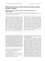
inclusion criteria are listed in Appendix 1. Fifty-three
patients meeting inclusion criteria were further analyzed
for this study after excluding 48 from the analysis for
reasons outlined in Table 1. Fourteen patients with early
and intermediate stage OPSCC who underwent primary
surgical therapy were referred to MBPCC for post-
operative radiation or chemoradiation. These patients
had planning PET-CT scans at MBPCC after initial sur-
gical therapy; therefore, due to lack of pre-treatment sta-
ging PET-CT they were excluded from the study.
Demo graphic data, clinical data that included findings
at physical examination, staging information, and pre-
treatment staging positron emission tomography-com-
puterized tomography data that included features of the
primary tumor and regional metastasis was recorded in
all cases. In a ddition to a review of the initial radiology
report, all pretreatment staging PET-CT scans w ere re-
evaluated with emphasis on evaluating retropharyngeal
lymphadenopathy. Mean standard uptake values (SUV)
and size (in cm) of all FDG avid lesions including pri-
mary tumor site a nd all metastatic lymph nodes were
recorded. PET-CT scans were reviewed for regional
metastatic lymphadenopathy, which was quantified as
ipsilateral or contralateral levels I-V lymphadenopathy.
The cutoff mean SUV was 3.0, with mean SUVs of less
than 3.0 defined as negative and mean SUVs of greater
than 3.0 defined as positive [11]. However, because
SUVs are semi-quantitative, it is not possible to deter-
mine the specific value for reference [11]. All nodal
metastasis was deemed positive if the measured cut-off
value was greater than or equal to 1.0 cm or any suspi-
cious features such as central necrosis were present
[12,13]. Lesions that me t either the size and/or mean
SUV criteria defined above were considered “positive”.
All scans were PET- CT fusion studies and were
obt ained on a single scanner at MBPC C in the majority
of cases (47/53). PET-CT scans were obtained in stan-
dard protocol as previously described in a prior study
from our institution [14].
Follow up data was obtained on all patients in the
study cohort at one month post treatment and at last
follow-up. Data recorded at last follow-up included
information on loco-regional recurrence and distant
metastasis and treatment of the same. Statistical analyses
were performed using the Wilcoxon-Mann-Whitney U
test, exact Pearson Chi-Square test, and odds ratios.
Results
Demographic data
The mean age of the study population was 57.2 year s
(range, 41-88 years) with a male to female ratio of 46:7.
The most common site for cancer within the orophar-
ynx was the tonsil (62.4%; 33/53) followed by b ase of
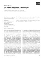
tongue (26.4%; 14/53). The incidence of tumor, nodal,
and overall stages for the cohort is listed in Table 2.
Treatment details
All patients were treated with intensity-modulated radia-
tion therapy (IMRT) at MBPCC with an average dose to
Table 1 Exclusion Criteria
Insufficient data in chart 8
Primary surgical therapy 14
Lost to follow up 5
Did not receive treatment at MBPCC 14
Metastatic disease at presentation 5
Palliative treatment only 1
No pre-treatment PET-CT 2
Total number of excluded patients 48
Tauzin et al. World Journal of Surgical Oncology 2010, 8:70
/>Page 2 of 5
the primary tumor, retropharynx, and gross disease in
the neck of 69 Gy (range, 64-72Gy). Forty-seven patients
(88.7%) received concurrent chemotherapy, 3 patients
(5.7%) additionally received neoa djuvant chemotherapy,
and 1 patient (1.9%) also received post- radiation che-
motherapy. Five patients (9.4%) did not re ceive any che-
motherapy. Patients were then followed on average for
26 months (range, 1.6 to 63 months).
PET-CT results
Patients had pre-treatment PET-CT scan on average 2.5
weeks (range, 0.5–15.8 weeks) prior to treatment. The
primary tumor size was on average 4.4cm(range, 1.4-
12cm) and average SUV of 12.1(range, 2.1-31). The
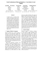
distribution of ipsilateral lymphadenopathy and contral-
ateral lymphadenopathy are illustrated in Table 3.
PET-CT upstaged approximately 43.4% when com-
pared to clinical stage, down staged 5.7%, and did not
change the stage in 50.9%. Examination of the impact
PET-CT scan has on clinical stage indicated that there
was not a significant difference (p = 0.2961) in the T
stage of subjects with upstage compared to those who
were not upstaged by PET-CT. However, there was a
trend toward overall nodal upstaging by PET-CT scan
when compared to clinical stage.
Distribution of regional metastasis by PET-CT
The regional metastatic n odal distribution of late stage
(III and IV) tumors was 10.8% (5/46) N0, 15.2% (7/46)
N1, 10.8% (5/46) N2a, 32.6% (15/46) N2b, 26.1% (12/46)
N2c, 4.8% (2/46) N3. Table 3 illustrates that the greatest
concentration of regional metastasis was in level II d is-
tribution regardless of laterality.
Retropharyngeal Lymphadenopathy
The frequency of retropharyngeal lymphadenopathy in
this cohort was 11 of 53 patients (20.8%). Of the 11
patients with retropharyngeal lymphadenopathy, 82% (9/
11) had T3 disease while 18% (2/11) had T2 disease. Of
the seven patients with T4 disease, 57% (4/7) had direct
invasion of the retropharynx with the primary tumor,
making a distinction between the primary tumor and
RPLN imprecise. Thus, these patients were not included
in this analysis. RPLN positivity was significantly asso-
ciated with T stage, (X
2
= 24.88, df = 6, p = 0.0003).
Subjects with advanced T stage cancer (i.e. T3 or T4
tumors) have significantly higher odds (OR = 5.6250,
95% Confidence Interval: 1.06 - 29.80, p = 0.0410) of
being RPLN positive by PET-CT.
The regional nodal status of the 11 patients with
RPLN was N0 (1/11), N1 (0/11), N2a (2/11), N2b (4/
11), N2c (4/11), and N3 (0/11). Similarly there was a
significant association (X
2
= 25.9535,df =10 p = 0.0045)
between RPLN positivity and N Stage. Subjects with
more advanced clinical N stage cancer (i.e., N2a, N2b,
N2c and N3) have higher odds (OR = 3.9773, 95% Con-
fidence Interval: 0.9628 - 16.4291) of being RPLN posi-
tive by PET as compared to those with early clinical N
stage; however, the association misses the 0.05 cutoff for
statistical significance (p = 0.0765).
Thepresenceofretropharyngeal lymphadenopathy
was stratified by primary tumor site. The most common
site was tonsil (82%, 9/11) with the r emaining sites pos-
terior pharyngeal w all and the base of tongue each 9%
(1/11). Of all tonsil primary tumors, 27.3% (9/33) had
retropharyngeal lymph node involvement. Similarly,
28.5% (4/14) base of tongue primary tumors and 20%
(1/5) posterior pharyngeal wall tumors had r etropharyn-
geal lymphadenopathy.
Patterns of recurrence
The overall recurrence rate was 35.8% (19/53) with 7.5%
(4/53) local recurrence, 13.2% (7/53) regional recur-
rence, and 15.1% (8/53) distant metastatic disease. One
patient (1.9%, 1/53) was diagnosed with a second pri-
mary tumor.
Discussion
The incidence of RPLN metastasis i dentified by PET-CT
in our OPSCC cohort was 20.8%. This is well within the
reported range of 16-50% published in previous studies
[7,8,10,15]. We found that advanced T stage cancer (T3
- T4) have significantly higher odds of being RPLN posi-
tive by PET-CT scan . Shimizu conducted a study where
RPLNs were electively dissected in cases where the
Table 2 Tumor and nodal status and clinical stage for the
study cohort
Tumor Stage Nodal Stage
T1 1/53 1.9% N0 21/53 39.6%
T2 21/53 39.6% N1 8/53 15.1%
T3 24/53 45.3% N2a 12/53 22.6%
T4 7/53 13.2% N2b 4/53 7.6%
Early Stage
(I or II)
7/53 13.2% N2c 7/53 13.2%
Late Stage (III or IV) 46/53 86.8% N3 1/53 1.9%
Table 3 Nodal distribution by PET-CT
Distribution Ipsilateral
Lymphadenopathy
Contralateral
Lymphadenopathy
Level Frequency Percent Frequency Percent
Level I 2 3.8% 1 1.9%
Level II 40 75.5% 16 30.2%
Level III 23 43.4% 2 3.8%
Level IV 5 9.4% 0 0%
Level V 2 3.77% 0 0%
Tauzin et al. World Journal of Surgical Oncology 2010, 8:70
/>Page 3 of 5
primary tumor originated from or invaded the p osterior
or lateral wall of the oropharynx [9]. Histology con-
firmed RPLN metastatic distribution was similar to our
findings of PET-CT distribution where the majority
(60%) were T3 and T4 tumors[9].
The epicenter of RPLN metastasis in OPSCC has been
most commonly from tonsillar primary tumors, followed
by posterior pharyngeal wall tumors, then b ase of ton-
gue tumors [9]. Similarly, our series demonstrates tonsil-
lar primary tumor sites to be the most common source
of RPLN metastasis; however, we have found that pos-
terior pharyngeal wall and base of tongue tumors had
equal predilection for metastasis to RPLNs. These find-
ings can be attributed to the small sample size of our
study.
The ability of PET-CT to accurately assess and be in
accordance with prior studies of metastatic lymph node
distribution is imperative for treatment planning of the
N0 neck irrespective of the modality chosen. As pre-
viously reported, lymph node metastasis of OPSCC is
most commonly seen at levels II and III [9,16,17]. PET-
CT findings in this study are in agreement with this
data confirming that PET-CT could accurately detect
nodal disease in the staging of head and neck cancer
patients. Furthermore, several studies have already
shown that adding PET- FDG or PET/CT-FDG to stan-
dard work-up led to a higher staging accuracy with
higher specificity [18-21].
A recent study examini ng this issue was conducted by
Lonneaux et al in a prospective, multicenter study show-
ing that PET-FDG was significantly m ore accurate than
conventional staging (McNemar test, P < .0001) and
improved staging accuracy in 20% of patients with head
and neck squamous cell carcinoma [21]. Furthermore,
they showed PET-FDG imaging modified the manage-
ment of 13.7% of patients. Our findings i ndicate that
PET-CT changed the stage in a large number of
patients: upstaging approximately 43.4% and down sta-
ging 5.7% when compared to clinical stage. While our
retrospective study did not examine the impact this had
on treatment strategies (i.e. change in radiation fields or
doses), it raises the question as to how this potentially
affects patient outcomes. Lonneux et al findings signifi-
cantly contribute to growing body of knowledge that
PET-CT is an impo rtant tool in pre-treatment work-up
and should be implemented as routine imaging of head
and neck s quamous cell carcinoma [21]. Additio nally,
our study highlights the importance and usefulness of
pre- treatment PET-CT in detecting RPLN status and
its role in guiding definitive treatment in OPSCC.
Our findings also showed a significant association
between N stage and positive RPLN status. Patients with
N2 or greater nodal disease on clini cal presentation had
higher odds of having positive RPLN status by imaging
criteria as compared to those patients who presented
with N0-1 disease.
The small sample size is a limitation of our study.
However, our observations are consistent with radiologic
and histologic studies reported in the literature.
Conclusion
PET-CT results for OPSCC can be used as a staging
tool to aid in treatment planning, as rates of RPLN and
nodal metastasis are consistent with those reported in
the literature. Advanced T and N stage are associated
with a greater odds ratio of being RPLN positive by
PET-CT imaging.
Appendix 1: Inc lusion Criteria
• Biopsy proven diagnosis of SCC* of the oropharynx
• Primary non-surgical therapy
• Sufficient medical record documentation
• Pre-treatment PET-CT scan
• Received all therapy at MBPCC**
• No evidence of metastatic disease at presentation
*SCC: Squamous cell carcinoma; **MBPCC: Mary Bird
Perkins Cancer Center
Author details
1
Department of Otolaryngology Head Neck Surgery, LSU Health Sciences
Center, New Orleans, LA, USA.
2
Biostatistics program, School of Public Health,
LSU Health Sciences Center, New Orleans, LA, USA.
3
Mary Bird Perkins
Cancer Center, Baton Rouge, LA, USA.
4
Department of Otolaryngology Head
Neck Surgery, University of Pittsburgh, Pittsburgh, PA, USA.
Authors’ contributions
MT conducted the chart review, data collection, contributed to study design
and study co-ordination. AG participated helped in chart review, data
collection and organization and also helped to edit the manuscript. JH
performed statistical analysis. CW participated in study design and editorial
contributions to the manuscript. RF contributed to the study design and
also editorial contributions. RW conceived of the study, and participated in
its design, write up, data analysis, editorial changes, and coordination as the
corresponding author. All authors read and approved the final manuscript.
Competing interests
The authors declare that they have no competing interests.
Received: 30 May 2010 Accepted: 16 August 2010
Published: 16 August 2010
References
1. Chaturvedi AK, Engels EA, Anderson WF, Gillison ML: Incidence trends for
human papillomavirus-related and -unrelated oral squamous cell
carcinomas in the United States. J Clin Oncol 2008, 26:612-619.
2. Sturgis EM, Cinciripini PM: Trends in head and neck cancer incidence in
relation to smoking prevalence: an emerging epidemic of human
papillomavirus-associated cancers? Cancer 2007, 110:1429-1435.
3. Holsinger FC, McWhorter AJ, Menard M, Garcia D, Laccourreye O: Transoral
lateral oropharyngectomy for squamous cell carcinoma of the tonsillar
region: I. Technique, complications, and functional results. Arch
Otolaryngol Head Neck Surg 2005, 131:583-591.
4. Walvekar RR, Li RJ, Gooding WE, Gibson MK, Heron D, Johnson JT, Ferris RL:
Role of surgery in limited (T1-2, N0-1) cancers of the oropharynx.
Laryngoscope 2008, 118:2129-2134.
Tauzin et al. World Journal of Surgical Oncology 2010, 8:70
/>Page 4 of 5
5. Galati LT, Myers EN, Johnson JT: Primary surgery as treatment for early
squamous cell carcinoma of the tonsil. Head Neck 2000, 22:294-2966.
6. O’Malley BW Jr, Weinstein GS, Snyder W, Hockstein NG: Transoral robotic
surgery (TORS) for base of tongue neoplasms. Laryngoscope 2006,
116:1465-1472.
7. Hasegawa Y, Matsuura H: Retropharyngeal node dissection in cancer of
the oropharynx and hypopharynx. Head Neck 1994, 16:173-180.
8. Amatsu M, Mohri M, Kinishi M: Significance of retropharyngeal node
dissection at radical surgery for carcinoma of the hypopharynx and
cervical esophagus. Laryngoscope 2001, 111:1099-1103.
9. Shimizu K, Inoue H, Saitoh M, Ohtsuki N, Ishida H, Makino K, Amatsu M,
Nibu K: Distribution and impact of lymph node metastases in
oropharyngeal cancer. Acta Otolaryngol 2006, 126:872-877.
10. Gross ND, Ellingson TW, Wax MK, Cohen JI, Andersen PE: Impact of
retropharyngeal lymph node metastasis in head and neck squamous
cell carcinoma. Arch Otolaryngol Head Neck Surg 2004, 130:169-173.
11. Kim SY, Lee SW, Nam SY, Im KC, Kim JS, Oh SJ, Ahn SD, Shin SS, Choi EK,
Kim JH: The Feasibility of 18F-FDG PET scans 1 month after completing
radiotherapy of squamous cell carcinoma of the head and neck. J Nucl
Med 2007, 48:373-378.
12. Mancuso AA, Harnsberger HR, Muraki AS, Stevens MH: Computed
tomography of cervical and retropharyngeal lymph nodes: normal
anatomy, variants of normal, and applications in staging head and neck
cancer. Part I: normal anatomy. Radiology 1983, 148:709-714.
13. Mancuso AA, Harnsberger HR, Muraki AS, Stevens MH: Computed
tomography of cervical and retropharyngeal lymph nodes: normal
anatomy, variants of normal, and applications in staging head and neck
cancer. Part II: pathology. Radiology 1983, 148:715-723.
14. Rabalais AG, Walvekar R, Nuss D, McWhorter A, Wood C, Fields R,
Mercante DE, Pou AM: Positron emission tomography-computed
tomography surveillance for the node-positive neck after
chemoradiotherapy. Laryngoscope 2009, 119:1120-1124.
15. Ballantyne AJ: Significance of Retropharyngeal Nodes in Cancer of the
Head and Neck. Am J Surg 1964, 108:500-504.
16. Lindberg R: Distribution of cervical lymph node metastases from
squamous cell carcinoma of the upper respiratory and digestive tracts.
Cancer 1972, 29:1446-1449.
17. Nigauri T, Kamata S, Kawabata K, Hoki K, Mitani H, Yoshimoto S,
Yonekawa H, Miura K, Beppu T, Uchida M: Treatment strategy for cervical
node metastasis from squamous cell carcinoma of the oropharynx.
Nippon Jibiinkoka Gakkai Kaiho 2000, 103:803-811.
18. Kyzas PA, Evangelou E, Denaxa-Kyza D, Ioannidis JP: 18F-
fluorodeoxyglucose positron emission tomography to evaluate cervical
node metastases in patients with head and neck squamous cell
carcinoma: a meta-analysis. J Natl Cancer Inst 2001, 100:712-720.
19. Yamazaki Y, Saitoh M, Notani K, Tei K, Totsuka Y, Takinami S, Kanegae K,
Inubushi M, Tamaki N, Kitagawa Y: Assessment of cervical lymph node
metastases using FDG-PET in patients with head and neck cancer. Ann
Nucl Med 2008, 22:177-184.
20. Roh JL, Yeo NK, Kim JS, Lee JH, Cho KJ, Choi SH, Nam SY, Kim SY: Utility of
2-(18F) flouro-2-deoxy-D-glucose positron emission tomography and
positron emission tomography/computed tomography imaging in the
preoperative staging in head and neck squamous cell carcinoma. Oral
Oncol 2007, 43:887-893.
21. Lonneux M, Hamoir M, Reychler H: Positron Emission Tomography with
[18F] Flourodeoxyglucose Improves staging and patient management in
patients with head and neck squamous cell carcinoma: a multicenter
prospective study. J Clin Oncol 2010, 28:1190-1195.
doi:10.1186/1477-7819-8-70
Cite this article as: Tauzin et al.: PET-CT staging of the neck in cancers
of the oropharynx: patterns of regional and retropharyngeal nodal
metastasis. World Journal of Surgical Oncology 2010 8:70.
Submit your next manuscript to BioMed Central
and take full advantage of:
• Convenient online submission
• Thorough peer review
• No space constraints or color figure charges
• Immediate publication on acceptance
• Inclusion in PubMed, CAS, Scopus and Google Scholar
• Research which is freely available for redistribution
Submit your manuscript at
www.biomedcentral.com/submit
Tauzin et al. World Journal of Surgical Oncology 2010, 8:70
/>Page 5 of 5