Báo cáo khoa học: "Association between intratumoral lymphatic microvessel density (LMVD) and clinicopathologic features in endometrial cancer: a retrospective cohort study" pdf
Bạn đang xem bản rút gọn của tài liệu. Xem và tải ngay bản đầy đủ của tài liệu tại đây (428.89 KB, 7 trang )
RESEARC H Open Access
Association between intratumoral lymphatic
microvessel density (LMVD) and clinicopathologic
features in endometrial cancer: a retrospective
cohort study
Lecy Kawamura
1
, Filomena M Carvalho
2*
, Bernardo GL Alves
2
, Carlos E Bacchi
3
, Joao Carlos Sampaio Goes
4
,
Marcelo Alvarenga Calil
4
, Edmund C Baracat
1
, Jesus P Carvalho
1
Abstract
Background: Lymph node metastasis in endom etrial cancer significantly decreases survival rate. Few data on the
influence of intratumoral lymphatic microvessel density (LMVD) on survival in endometrial cancer are available. Our
aim was to assess the intratumoral LMVD of endometrial carcinomas and to investigate its association with classical
pathological factors, lymph node metastasis and survival.
Methods: Fifty-seven patients with endometrial carcinoma diagnosed between 2000 and 2008 underwent
complete surgical staging and evaluation of intratumoral LMVD and other histologic variables. Lymphatic
microvessels were identified by immunohistochemical staining using monoclonal antibody against human
podoplanin (clone D2-40) and evaluated by counting the number of immunostained lymphatic vessels in 10 hot
spot areas at 400× magnification. The LMVD was expressed by the mean number of vessels in these 10 hot spot
microscopic fields. We next investigated the association of LMVD with the clinicopathologic findings and prognosis.
Results: The mean number of lymphatic vessels counted in all cases ranged between 0 and 4.7. The median value
of mean LMVD was 0.5, and defined the cut-off for low and high LMVD. We identified low intratumoral LMVD in
27 (47.4%) patients and high LMVD in 30 (52.6%) patients. High intratumoral LMVD was associated with lesser
miometrial and adnaexal infiltration, lesser cervical and peritoneal involvement, and fewer fatal cases. Although
there was lower lymph node involvement among cases with high LMVD, the difference did not reach significance.
No association was seen between LMVD and FIGO staging, histological type, or vascular invasion. On the other
hand, low intratumoral LMVD was associated with poor outcome. Seventy-five percent of deaths occurred in
patients with low intratumoral LMVD.
Conclusion: Our results show associat ion of high intratumoral LMVD with features related to more localized
disease and bette r outcome. We discuss the role of lymphangiogenesis as an early event in the endometrial
carcinogenesis.
Background
Endometrial cancer is the most frequent gynecologic
malignancy in the western world. The death rate has
increased during the most recent decades, probably due to
an increase in life span and coexisting medical
comorbidities [1]. The most important prognostic factors
in endometrial cancer are histological type, grade, lymph
node status, deep myometrial invasion, and stage [2,3].
Approximately 72% of endometrial cancers are stage I,
12% stag e II, 13% stage III, and 3% stage IV
(1)
.Fiveyear
survival is about 90% in disease confined to the uterus, but
drops to 60% when lymph nodes are positive [4]. Lymph
node metastasis is a complex, multi-step biological process
initialized by tumor cells, involving stroma inva sion, new
* Correspondence:
2
Department of Pathology of Faculdade de Medicina da Universidade de
São Paulo, Sao Paulo (SP), Brazil
Full list of author information is available at the end of the article
Kawamura et al. World Journal of Surgical Oncology 2010, 8:89
/>WORLD JOURNAL OF
SURGICAL ONCOLOGY
© 2010 Kawamura et al; licensee BioMed Central Ltd. This is an Open Access article distributed under the terms of the Creative
Commons Attribution License (http://creative commons.org/licenses/by/2.0), which permits unrestricted use, distribution, and
reproduction in any medium, provided the original work is properly cited.
lymphatic vessel formation, and spread through lymphatic
vessels to lymph node. Understanding how ca ncer cells
gain access to lymphatic channels and develop metastases
is of great interest for development of new therapeutic
strategies targeted against members of the signaling path-
ways involved in the lymphangiogenesis process.
The growth of lymphatic vessels largely depends on
many growth factors, such as vascular endothelial
growth factor-C and -D (VEGF-C and VEGF-D), platelet
derived growth factor-BB (PDGF-BB) and hepatocyte
growth factor[5]. However, in cancer , these known lym-
phangiogenic factors lead to simultaneous stimulation of
angiogenesis and lymphangiogenesis [6]. Lymphatic
microvessel density (LMVD) is one of the ways to evalu-
ate lymphangiogenesis. It was poor studied due to the
difficulties associated with detecting a nd characterizing
lymphat ic markers. Currently, several molecules specifi-
cally expressed in lymphatic endothelial cells, such as
podoplanin, LYVE1, and the homeobox gene prox-1,
have enabled a more precise study of the lymphatic vas-
culature and the molecular mechanisms involved in
lymphangiogenesis [7]. LMVD has been investigated in
many tumors, particularly those characterized by lym-
phatic dissemination, such as cervical carcinomas [8-10],
but there are few studies that have evaluated LMVD in
end ometrial carcinomas, and results have been conflict-
ing [11,12].
Therefore, the aim of this retrospective study was to
evaluate the relationship between intratumoral LMVD,
as well as other histologic variables, and the incidence
of lymph node metastasis and overall survival of patients
with endometrial cancer fol lowing hysterectomy and
lymph node dissection.
Methods
Patients
This study retrospectively analyzed patients with endo-
metrial cancer treated at the Instituto Brasileiro de Con-
trole do Cancer (IBCC), a reference Cancer Center in
São Paulo, Brazil, between 2000 and 2008. This cohort
of 57 patients included 28 (49.1%) with stage I disease, 4
(7.0%) with stage II disease, 22 (38.5%) with stage III
disease, and 3 (5.2%) with stage IV disease, classified
according to the 2009 revised International Federation
of Gynecology and Obstetrics (FIGO) system [13]. Age
of patients ranged from 46 to 89 years (67.4 ± 10.27 y).
All patients had undergone surgical staging hysterect-
omy with a sample of pelvic lymph nodes. The number
of lymph nodes excised ranged from 3 to 49 (21.5 ±
12.44), and they were positive in 22 (38.6%) and negative
in 35 (61.4%) cases. Paraortic lymph nodes were
assessed in 27 cases and nine of them were involved by
theneoplasia.Thefollow-uprangedfrom6to144
months (mean 39.54 ± 29.98 months). Twelve patients
died of their disease between 6 and 108 months after
the diagnosis (median 21.5 months). Data about myo-
metrial, cervical, adnexal and peritoneal involvement,
were retrieved from the medical records. This study was
approved by the Department of Obstetrics and Gynecol-
ogy Scientific Committee of the Faculdade de Medicina
da Universidade de São Paulo, by the Ethical Committee
for Research Projects of the Hospital das Clinical da
FaculdadedeMedicinadaUniversidadedeSãoPaulo
(Comissão de Ética para Análise de Pesquisa - CAPPesq)
(process number 0728/08), and by the Ethical Commit-
tee for Research of the I nstituto Brasileiro de Controle
do Cancer.
Tumor Specimens
Tumor slides were reviewed independently by two
pathologists and were classified according to the criteria
of the World Health Organization (WHO) Classification
of Tumors [14]. The Vancouver system was applied to
classify the tumors in low and high grades. A tumor was
considered high grade if it showed at least two of these
criteria: 1) predominantly papillary or solid growth pat-
tern, 2) 6 or more mitotic figures/10 high power fields,
or 3) severe nuclear atipia [15]. Peritumoral lymphovas-
cular space invasion (LVSI) was also evaluated for all
cases in the whole histological sections. A representative
peripheral area of the tumor was selected for the con-
struction of tissue microarray blocks and immunohisto-
chemical study.
Construction of tissue microarray (TMA) blocks
The tumor areas selected in the slides were marked in
the corresponding paraffin donor blocks and one cylin-
der of material (2.0 mm in diameter) was punched from
each case and mounted into recipient paraffin blocks at
1 mm intervals using a precision microarray instrument
(Beecher Instruments, Silver Spring, MD). A grid system
was established s uch that each core had an x and y
coordinate reference for sample identification. Blocks
were sealed at 60°C for 10 m. Three μmsectionsfrom
each TMA block were then prepared using standard
techniques and mounted on Starfrost® slides. The first
histological sections were stained by hematoxilin-eosin
and examined to b e sure the correct areas were
included.
Immunohistochemistry analysis
Histological sections (3 μm) from TMA blocks were
quenched with 3% hydrogen peroxide solution in phos-
phate-buffered saline (PBS; Sigma, St. Louis, MO) for 20
m to block endogenous peroxidase activity. After several
washes in PBS, sections were heated in a microwave
(Electrolux, 900 W) for 15 m in 0.01 M citrate buffer,
pH 6.0 for antigen retrieval, then cooled at RT for 20 m.
Kawamura et al. World Journal of Surgical Oncology 2010, 8:89
/>Page 2 of 7
Sections were then incubated overnight with a 1:400
dilution of monoclonal antibody against human podo-
planin (clone D2-40, Dako, Carpinteria, CA). Podoplanin
is a glycoprotein specifically expressed by lymphatic
endothelial cells. In those tumor sections with negative
podoplanin staining, adjacent normal-appearing lympha-
tic endothelial cells served as positive internal controls.
Quantification of lymphatic microvessel density
Stained TMA histologic sections were analyzed using
standard light microscopy (Nikon, Eclipse 200). Under
low magnification, the most vascularized intratumoral
areas were identified. We counted the number of immu-
nostained lymphatic vessels found in 10 hot spot areas
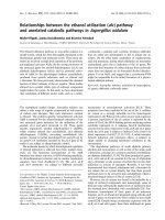
at 400× magnification. Only vessels exhibiting typical
morphology (lumen) were considered lymphatic micro-
vessels (Figure 1). The LMVD for each case was
expressed by the mean value (total number of vessels in
10 hot spot microscopic field s/10). The median value of
all the mean LMVD was the cutoff to divide tumors in
high or low LMVD, as suggested by Hall et al.[16].
Statistical analyses
The association between categorical variables and lymph
node status and surv ival were evaluated using Pearson’s
Chi-square test. Kaplan & Meier method was used to
produce time life tables and survival curves.
A p-value less than 0.05 was considered significant.
The software used was Epi Info
tm
, Version 3.5.1. [17].
Results
The tumors were classified as endometrioid (44 cases,
77.2%) and non-endometrioid classified as serous (6
cases , 10.5%), clear cell (4 cases, 7%), undifferentiated (2
cases, 3.5%), and mixed (1 case, 1.7%). The association
between all the variables with lymph node status and
patient death are summarized in Table 1. The mean
number of lymphatic vessels counted in each patient’s
tumor sample ranged from 0 to 4.7. The median value
was 0.5 vessels and defined the value above and below
which was considered high and low LMVD, respectively.
Low LMVD was found in 27 (47.4%) patients, and high
LMVD was found in 30 (52.6%) patients. We observed
that low intratumoral LMVD was associated with poor
outcome. Seventy-five percent of deaths occurred in
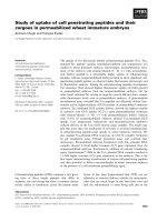
patients with low intratumoral LMVD (p = 0.031). The
mean survival time observed in the group with low
LMDV was 79 months (confidence interval [62 - 96
months]) and the mean survival time for high LMDV
group was 129 months (confidence interval [113 - 145
months]). The difference was significant at 5% level. The
survival curves for both LMDV groups are presented at
Figure 2.
Low LMVD was also associated with miometrial and
adnaexal infiltration, cervical and peritoneal involve-
ment. Patients below 70 years presented higher LMVD
(21/33, 63.3%) than older patients (9/24, 37.5%)
although the difference did not reach statistical signifi-
cance. No a ssociation was seen between LMVD and
FIGO staging, histological type, vascular invasion, or
lymph node involvement (Table 2).
Positive lymph nodes were significantly associated with
histological grade (p = 0.001), vascular invasion (p =
0.017), FIGO stage (p < 0.001), myometrial infiltration
(p < 0,001), cervical infiltration (p = 0,023), adnaexal
involvement (p = 0,023) and peritoneal inv olvem ent (p =
0,048). Besides LMVD, death was associated with histolo-
gical type (p = 0.010), vascular invasion (p = 0.040), and
lymph node involvement (p = 0.025). Mean survival time
was 110 months (range of 84 - 135 months) for patients
with negative lymph no de invo lvemen t, and 69 months
(range of 48 - 89 months) for pat ients with positive
lymph nodes.
Discussion
Lymphatic metastasis is one of the most important
prognostic factors for survival of patients with endome-
trial cancer as well as for other epithelial malignancies.
Many clinical and experimental data suggest that migra-
tion of tumor cells into the lymph nodes is facilitated by
lymphangiogenesis[18,19]. Studies evaluating lymphatic
neoformation and its role in tumoral migration are
facilitated by the recent identification of lymphangio-
genic factors and their receptors, and by lymphatic vas-
cular markers [5].
The most commonly used markers for immunohisto-
chemistry identification o f lymphatic endothelial tissue
Figure 1 Immu nohistochemistry staining of a histological
section of endometrioid carcinoma using D2-40 antibody
against podoplanin showing six lymphatic vessels. After
counting 10 high power fields, the mean was calculated for each
case. (Original magnification X400).
Kawamura et al. World Journal of Surgical Oncology 2010, 8:89
/>Page 3 of 7
include the vascular endothelial growth factor receptor 3
(VEGFR-3), PROX-1, LYVE-1, and podoplanin [5].
Among these, podoplanin, a 38 kDa mucin-type trans-
membrane glycoprotein which is recognized by the
monoclonal antibody D2-40, is considered the most reli-
able marker with a high specificity and sensitivity [5]. In
this study, we chose to evaluate podoplanin because it
was found to be expressed in lymphatic, but not blood
vascular endothelium [20], and is expressed in smaller
lymphatic vessels rather than larger ones [20], allowing
a more sensitive identification of lymphangiogenesis.
We then co nsidered which c ompartment (intra or
peritumoral) to evaluate for lymphangiogenesis in this
study, as this is a source of great controversy in the lit-
erature. Since our knowledge of lymphangiogenesis in
different types of cancer is still limited, we decided that
it would be worthwhile to begin accumulating data. We
believe that a better understanding of the role of lym-
phangiogenesis in tumoral dissemination will only be
achieved after contributions from different researchers
groups.
In this study, we evaluated LMVD in the intratumoral
compartment using podoplanin as the lymphatic marker.
Vascular density was determined by direct counting of
vessels in 10 high power microscope fields. Although
our study is limited by the fact that LMVD was assessed
only in TMA sections, the area of the tumor that was
investigated was rigorously chosen considering fixation
conditions of the specimen, morphology of the tumor,
and the peripheral localization of the sample. Besides,
our cores had 2.0 mm in diameter, bigger than the con-
ventional cores.
LMVD has been studied in several cancers by various
methods[21].TherearefewstudiesaboutLMVDin
gynecological cancer. Gombos et al. [9] studied intra-
and peritumo ral LMVD using D2-40 immunohisto-
chemistry in 111 cervical squamous cell carcinomas and
correlated them with vascular endothelial growth factor
Table 1 Clinicopathological variables and association with lymph node status and outcome
Variable Categories Lymph nodes p
1
Outcome p
1
negative
n
positive
n
alive
n
dead
n
Age 45-69 years 20 13 0.885 27 6 0.533
≥70 years 15 9 18 6
Histological type Endometrioid 30 14 0.053 38 6 0.010
Non-Endometrioid 5 8 7 6
Histological grade (Vancouver system) low 32 12 0.001 37 7 0.080
high 3 10 8 5
FIGO stage I 27 1 <0.001 24 4 0.303
II 4 0 4 0
III 3 19 15 7
IV 1 2 2 1
Vascular invasion no 12 3 0.017 14 1 0.040
yes 6 10 10 6
LMVD
2
Low 14 13 0.160 18 9 0.031
high 21 9 27 3
Myometrial infiltration <50% 25 5 <0.001 26 4 0.113
>50% 9 17 18 8
Cervical infiltration no 25 9 0.023 31 3 0.003
yes 9 12 12 9
Adnaexal involvement no 33 14 0.023 31 6 0.004
yes 2 8 4 6
Peritoneal involvement no 28 13 0.048 33 8 0.811
yes 1 5 4 2
Total 57 34 22 45 12
1
p value (Chi-square of Pearson);
2
Intratumoral lymphatic microvessel density.
Kawamura et al. World Journal of Surgical Oncology 2010, 8:89
/>Page 4 of 7
(VEGF)-C expression, clinicopathologic tumor features,
and outcome. They found only high peritumoral LMVD
to be associated with poor overall survival. Zhang et al. ,
using LYVE-1 (lymphatic vessel endothelial hyaluronan
receptor-1) as the lymphatic marker, studied intra- and
peritumoral lymphatic vascular density in early-stage
invasive cervical carcinomas. Both intravascular and
peritumoral high LMVD were associated with lymph
node metastasis [8]. In a conflicting study, Birn er et al.
studied 95 cases of stage pTIb cervical carcinomas and
observed a more favorable prognosis among tumors
with high LMVD [10], a result that is similar to ours.
LMVD studies in endometrial cancer are extremely
limited. To investigate whether increased peritumoral
and intratumoral LMVD was a good prognostic factor
for nodal metastasis, Gao et al. studied 102 patients
with endometrial carcinoma using LYVE-1 as the lym-
phatic marker [22]. They fo und peritumoral but not
intratumoral high LMVD to be associated with lymph
node metastases. Vandenput et al. studied 62 patients
with endometrial carcinoma and found that neither peri-
tumoral nor intratumoral lymphangiogenesis determined
by podoplanin expression were related to prognosis [11].
Donughue et al. evaluated lymphatic vessel density
(LVD) in 17 cases of endometrial carcinomas by count-
ing vessels expressing D2-40. Intratumoral LVD was sig-
nificantly increased when compared with endometrial
functionalis LVD, but not when compared with endo-
metrial basalis LVD. They also observed a tendency
towards increased intratumoral LVD in grade 3 tumors
compared with grade 1 tumours [23]. Other studies
have documented lymphatic metastases independent of
intratumoral lymphatic vessels suggesting that the intra-
tumoral lymphatic vasculature is usually not functioning
and only peritumoral lymphatic vessels conduct fluid
and cells [24].
In our study, low LMVD was associated with poor
outcome. Seventy-five percent of deaths occurred in
patients with low intratumoral LMVD compared with
only 25% of deaths in patients with high LMVD (p =
0.031). In contrast, high LMVD was associated with
favorable prognostic factors. High LMVD tumors were
associated with le ss myometrial infiltration > 50% (31%
vs. 63%, p = 0.034), less cervical involvement (24.1% vs.
53.8%, p = 0.023), and less peritoneal involvement (4%
vs. 29.4%, p = 0.055). The explanation for this finding is
not clear, but we can suppose that lymphangiogenesis is
an early event in tumor progression, when lymphatic
vessels are still not functional. Considering that higher
LMVD was present in early anatomic tumors with no
association with intrinsic tumors biological characteris-
tics, as histological type or grade, we can suppose that
this is an event related to the first steps of local interac-
tion between tumor and microenvironment. Besides,
high LMVD was associated with favorable outcome,
indicating that it might be clinically relevant. However,
it must be pointed that the vascular density results from
a more complex biologic pr ocess that involves a cr oss-
talk between tumor, vessels and host stroma. Our
results showed higher LMVD among tumor in the initial
Figure 2 Overall survival curve stratified for high and low intratumoral lymphatic microvessel density (LMVD).
Kawamura et al. World Journal of Surgical Oncology 2010, 8:89
/>Page 5 of 7
steps of invasion (no extrauterine extension) and better
prognosis. It is well known the role of normal lymphatic
vasculature in maintaining tissue homeostasis [25]. The
lymphatic network must be investigated not only by its
anatomic density, as we did, but also functionally. We
need intensive further investigation with more early
cases and adequate follow up, to explore the role of vas-
cular density in the tumoral progression. These future
results may help elucidate pathways of lymphan giogen-
esis leading to identification of potential drug targets for
endometrial carcinomas. Considering our preliminary
results, it is possible that the target would not be the
lymphatic vessel itsel f, but some ot her molecul es
involved with its function.
Conclusions
Our results show that high intratumoral LMVD is a
characteristic of endometrial carcinomas with lesser
extrauterine extension and is associated with bette r out-
come. However, further studies with larger series are
needed to elucidate t he biological meaning of these
intriguing findings.
Acknowledgements
We thank Antonio de Castro Bruni, M.S., for help with statistical analyses, and
Dr. Eloa Muniz de Freitas Alves and Prof. Joao Guidugli Neto for providing
pathological materials. This work was supported by grants of the Centro de
Estudos do Instituto Brasileiro de Controle do Cancer.
Author details
1
Department of Obstetrics and Gynaecology of Faculdade de Medicina da
Universidade de São Paulo, Sao Paulo (SP), Brazil.
2
Department of Pathology
of Faculdade de Medicina da Universidade de São Paulo, Sao Paulo (SP),
Brazil.
3
Consultoria em Patologia, Botucatu (SP), Brazil.
4
Instituto Brasileiro de
Controle do Cancer, Sao Paulo (SP) , Brazil.
Authors’ contributions
LK collected the data and participated in pathological analyses of the
samples and the drafting of the manuscript. FMC conceived of the study,
participated in its design and review the slides, and helped to the
coordination and drafting of the manuscript. BGLA reviewed all the
histological samples and performed the immunohistochemical analyses. CEB
carried out the immunohistochemical reactions and helped their
interpretation. JCSP, MAC and ECB participated in the design and the
coordination, and helped to draft the manuscript. JPC conceived of the
study and participated in its design, coordination and drafting. All authors
read and approved the final manuscript.
Competing interests
The authors declare that they have no competing interests.
Received: 13 June 2010 Accepted: 14 October 2010
Published: 14 October 2010
References
1. Sorosky JI: Endometrial cancer. Obstet Gynecol 2008, 111:436-447.
2. Mariani A, Webb MJ, Keeney GL, Aletti G, Podratz KC: Predictors of
lymphatic failure in endometrial cancer. Gynecol Oncol 2002, 84:437-442.
3. Prat J: Prognostic parameters of endometrial carcinoma. Hum Pathol
2004, 35:649-662.
4. Hoekstra AV, Kim RJ, Small W Jr, Rademaker AW, Helenowski IB, Singh DK,
Schink JC, Lurain JR: FIGO stage IIIC endometrial carcinoma: prognostic
factors and outcomes. Gynecol Oncol 2009, 114:273-278.
5. Van der Auwera I, Cao Y, Tille JC, Pepper MS, Jackson DG, Fox SB, Harris AL,
Dirix LY, Vermeulen PB: First international consensus on the methodology
of lymphangiogenesis quantification in solid human tumours. Br J Cancer
2006, 95:1611-1625.
6. Cao Y: Opinion: emerging mechanisms of tumour lymphangiogenesis
and lymphatic metastasis. Nat Rev Cancer 2005, 5:735-743.
7. Liersch R, Biermann C, Mesters RM, Berdel WE: Lymphangiogenesis in
cancer: current perspectives. Recent Results Cancer Res 2010, 180:115-135.
8. Zhang SQ, Yu H, Zhang LL: Clinical implications of increased lymph
vessel density in the lymphatic metastasis of early-stage invasive
cervical carcinoma: a clinical immunohistochemical method study. BMC
Cancer 2009, 9:64.
9. Gombos Z, Xu X, Chu CS, Zhang PJ, Acs G: Peritumoral lymphatic vessel
density and vascular endothelial growth factor C expression in early-
Table 2 Association between intratumoral lymphatic
microvessel density (LMVD) and clinicopathological
features
Variable categories LMVD p
1
low
n
high
n
Age 45-69 years 12 21 0.051
≥70 years 15 9
Histological type Endometrioid 19 25 0.396
Non-
Endometrioid
85
Histological grade (Vancouver
system)
low 20 24 0.594
high 7 6
FIGO stage I 9 19 0.145
II 2 2
III 14 8
IV 2 1
Vascular invasion no 6 9 0.576
yes 8 8
Myometrial infiltration <50% 10 20 0.034
>50% 17 9
Cervical infiltration no 12 22 0.047
yes 14 7
Adnaexal involvement no 19 28 0.023
yes 8 2
Peritoneal involvement no 17 24 0.055
yes 5 1
Lymph node involvement no 14 21 0.160
yes 13 9
Outcome alive 18 27 0.031
death 9 3
Total 57 27 30
1
p value (Chi-square of Pearson).
Kawamura et al. World Journal of Surgical Oncology 2010, 8:89
/>Page 6 of 7
stage squamous cell carcinoma of the uterine cervix. Clin Cancer Res
2005, 11:8364-8371.
10. Birner P, Schindl M, Obermair A, Breitenecker G, Kowalski H, Oberhuber G:
Lymphatic microvessel density as a novel prognostic factor in early-
stage invasive cervical cancer. Int J Cancer 2001, 95:29-33.
11. Vandenput I, Vanhove T, Calster BV, Gorp TV, Moerman P, Verbist G,
Vergote I, Amant F: The use of lymph vessel markers to predict
endometrial cancer outcome. Int J Gynecol Cancer 2010, 20 :363-367.
12. Stefansson IM, Salvesen HB, Akslen LA: Vascular proliferation is important
for clinical progress of endometrial cancer. Cancer Res 2006, 66:3303-3309.
13. Pecorelli S: Revised FIGO staging for carcinoma of the vulva, cervix, and
endometrium. Int J Gynaecol Obstet 2009, 105:103-104.
14. Tavassoli FA, Deville A: Pathology & Genetics Tumours of Breast and Female
Genital Organs Lion: International Agency for Research on Cancer - IARC
Press 2003.
15. Alkushi A, Abdul-Rahman ZH, Lim P, Schulzer M, Coldman A, Kalloger SE,
Miller D, Gilks CB: Description of a novel system for grading of
endometrial carcinoma and comparison with existing grading systems.
Am J Surg Pathol 2005, 29:295-304.
16. Hall FT, Freeman JL, Asa SL, Jackson DG, Beasley NJ: Intratumoral
lymphatics and lymph node metastases in papillary thyroid carcinoma.
Arch Otolaryngol Head Neck Surg 2003, 129:716-719.
17. Epi Info. Atlanta, GA: Centers for Disease Control and Prevention (CDC),
3.5.1 2008.
18. Alitalo K, Tammela T, Petrova TV: Lymphangiogenesis in development and
human disease. Nature 2005, 438:946-953.
19. He Y, Rajantie I, Pajusola K, Jeltsch M, Holopainen T, Yla-Herttuala S,
Harding T, Jooss K, Takahashi T, Alitalo K: Vascular endothelial cell growth
factor receptor 3-mediated activation of lymphatic endothelium is
crucial for tumor cell entry and spread via lymphatic vessels. Cancer Res
2005, 65:4739-4746.
20. Stacker SA, Achen MG, Jussila L, Baldwin ME, Alitalo K: Lymphangiogenesis
and cancer metastasis. Nat Rev Cancer 2002, 2:573-583.
21. Sundar SS, Ganesan TS: Role of lymphangiogenesis in cancer. J Clin Oncol
2007, 25:4298-4307.
22. Gao Y, Liu Z, Gao F, Meng XY: High density of peritumoral lymphatic
vessels is a potential prognostic marker of endometrial carcinoma: a
clinical immunohistochemical method study. BMC Cancer 2010, 10:131.
23. Donoghue JF, Lederman FL, Susil BJ, Rogers PA: Lymphangiogenesis of
normal endometrium and endometrial adenocarcinoma. Hum Reprod
2007, 22:1705-1713.
24. Padera TP, Kadambi A, di Tomaso E, Carreira CM, Brown EB, Boucher Y,
Choi NC, Mathisen D, Wain J, Mark EJ, Munn LL, Jain RK:
Lymphatic
metastasis in the absence of functional intratumor lymphatics. Science
2002, 296:1883-1886.
25. Szuba A, Skobe M, Karkkainen MJ, Shin WS, Beynet DP, Rockson NB,
Dakhil N, Spilman S, Goris ML, Strauss HW, Quertermous T, Alitalo K,
Rockson SG: Therapeutic lymphangiogenesis with human recombinant
VEGF-C. FASEB J 2002, 16:1985-1987.
doi:10.1186/1477-7819-8-89
Cite this article as: Kawamura et al.: Association between intratumoral
lymphatic microvessel density (LMVD) and clinicopathologic features in
endometrial cancer: a retrospective cohort study. World Journal of
Surgical Oncology 2010 8:89.
Submit your next manuscript to BioMed Central
and take full advantage of:
• Convenient online submission
• Thorough peer review
• No space constraints or color figure charges
• Immediate publication on acceptance
• Inclusion in PubMed, CAS, Scopus and Google Scholar
• Research which is freely available for redistribution
Submit your manuscript at
www.biomedcentral.com/submit
Kawamura et al. World Journal of Surgical Oncology 2010, 8:89
/>Page 7 of 7