Báo cáo khoa học: "Malignant gastrointestinal stromal tumor presenting with hemoperitoneum in puerperium: report of a case with review of the literature" ppt
Bạn đang xem bản rút gọn của tài liệu. Xem và tải ngay bản đầy đủ của tài liệu tại đây (664.38 KB, 7 trang )
CAS E REP O R T Open Access
Malignant gastrointestinal stromal tumor
presenting with hemoperitoneum in puerperium:
report of a case with review of the literature
Michail Varras
1*
, Nikolaos Vlachakos
2
, Christodoulos Akrivis
3
, Thivi Vasilakaki
4
, Evangelia Skafida
4
Abstract
Background: Gastrointestinal stromal tumors (GISTs) are mesenchymal tumors that develop in the wall of the
gastrointestinal tract and their diagnosis during pregnancy or puerperium is extremely rare.
Case: A 28-year old patient presented with acute abdomen due to hemoperitoneum from a large mass arising of
the small intestine with distended vessels on its top and a ruptured superficial vessel bleeding into the peritoneal
cavity. The patient was at the tenth postpartum day of her first pregnancy. The preoperative diagnosis was a
possible ovarian or uterine mass. After an emergency exploratory laparotomy a segmental bowel resection was
performed, removing the tumor with a part of 3-cm of the small intestine. Histology revealed GIST with maximum
diameter of 13 cm and mitotic rates more than 5 mitoses per 50 high power fields with some atypical forms,
indicating a high risk malignancy. Immunohistochemical staining of the tumor tissue demonstrated strongly
positive reactivity to CD 117 (c-kit) and CD34 in almost all the tumor cells. The patient was treated with oral
imatinib mesylate (Glee vec) 400 mg daily for one year. Three years after surgery, the patient was alive without
evidence of metastases or local recurrence.
Conclusion: Considering that only few patients with gastrointestinal stromal tumors have been reported in the
obstetrical and gynecological literature, the awareness of such an entity by the obstetricians-gyneco logists is
necessary in order to facilitate coordinated approach with the general surgeons and oncologists for the optimal
care of the patients.
Introduction
Gastrointestinal stromal tumors (GISTs) are uncommon
tumors that develop in the wall of the gastrointestinal
tract and usually present in the fifth to seventh decade
of life [1,2]. They account for approximately 0.1% to 3%
of all gastrointestinal neoplasms, with an incidence of
1-20 per million and up to 30% of these are considered
malignant [1,3,4]. The term gastrointestinal sromal
tumor, first used by Mazur and Clark in 1983, encom-
passes a heterogeneous group of nonepithelial neo-
plasms composed of spindle or epithelioid cells, which
display a range of differentiation [5]. Given the age dis-
tribution of occurrence, a diagnosis of gastrointestinal
stromal tumor during pregnancy [6-8] or puerperium is
very uncommon.
We hereby describe our experience of the case of a
GIST discovered during the puerperium, in a 28-year
old patient presented w ith acute abdomen due to spon-
taneous rupture of a superficial tumor vessel, an extre-
mely rare complication and review the current literature.
Case Report
A 28-year-old woman was brought to the emergency
department of our hospital with severe lower abdominal
pain, which became generalized and intolerable. The
patient was at the tenth postpartum day of her first
pregnancy and had no remarkable medical or surgical
history. Also, the patient had no history of an irregular
menstruation cycle. As the patient mentioned, during
her pregnancy the uterus was considered too large for
her gestational age and on routine ultrasounds a
* Correspondence:
1
Department of Obstetrics and Gynecology, ‘Tzaneio’ General State Hospital,
Piraeus, Greece
Full list of author information is available at the end of the article
Varras et al. World Journal of Surgical Oncology 2010, 8:95
/>WORLD JOURNAL OF
SURGICAL ONCOLOGY
© 2010 Varras et al; licensee BioMed Central Ltd. This is an Open Access article distributed under the terms of the Creative Commons
Attribution License ( which permits unrestricted use, distr ibution, and reprodu ction in
any medium, provided the origina l work is properly cited.
subserosal fibroid was suspected. The crown rump
length was in accordance with her last menstrual period
and the fetal growth was within the normal limits as
well, according to the patient’s information. She had a
normal delivery at term at a Private Maternity Hospital
of Athens. The patient denied any medical history of
gastrointestinal symptoms such as emesis, melaena,
abdominal pain or ileus during her pregnancy. At pre-
sentation, she was nauseous and had vomited a number
of times. Phy sical examination revealed a pale, moder-
ately obese young woman with a heart rate of 104 beats
per minute, blood pressure 130/70 mmHg and tempera-
ture 36°C. Her abdomen was extensively distended,
markedly tender with moderate spasm and rebound ten-
derness in both iliac fossae. Peristaltic sounds were
diminished. Her blood count demonstrated a haemoglo-
bin concentration of 9.4 g/dl, haematocrit 31%, white
blood count 18,000 cells/ml with 89.7% polymorphonuc-
lears and platelets 352,000/μl. Clotting time, bleeding
time, serum liver enzymes and kidney function tests
were within normal limits. L.D.H. was 297 U/l (normal
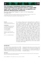
rates 100-240 U/l). An abdominal ultrasound examina-
tion revealed a large mass measuring 12.85 × 1 0.52-cm
with mixed echogenicity, occupying all the pelvis and
extending above the pubic symphysis and from the mid-
line to the left (Figures 1-2). Two cystic areas wit hin the
mass measuring approximately 4.30 × 4.97-cm and 3.54
× 3.66 -cm were found (Figures 2). The ovaries were not
visualized. Fre e fluid was present a t the Morisson’ s
space and the cul-de-sac with low levels of echogenic
debris. Chest X-ray examination was negative. Under
the diagnosis of hemoperitoneum from a possible ovar-
ian or uterine mass, an immediate exploratory laparot-
omy was performed. At laparotomy with a vertical,
midline infra-umbilical incision bloodstained fluid and
blood clots in the peritoneal cavity were found. Further
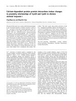
expl oration revealed a large mass arising from the small
intestine and growing exophytically out into the perito-
neal cavity (Figure 3). On the top of the mass, distended
vessels were observed and a ruptured superficial vessel
was actively bleeding into the abdominal cavity; no
other bleeding was indentified. Free fluid was obtained
for cytology. One liter of fluid and blood clots were
evacuated. A segmental bowel rese ction was perform ed,
removing the tumor with a part of 3-cm of the small
intestine (Figure 4). T he abdominal cavity was irrigated
and carefully inspected; the omentum, the ovaries and
the uterus had normal macroscopic appearance and no
visible findings suspicious of malignancy were found.
The patient made a good recovery post-operatively.
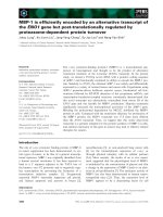
Grossly, the surgical specimen of the small intestine
showed a well-circumscribed tumor measuring 13 × 10
× 9-cm in size and located 3-cm from the nearest surgi-
cal martin (Figure 5). The external surface of the tumor
showed pronounced appearance of its vessels. Part of
the tumor was covered by the instinal musosa. The cut
surface demonstrated whitish-gray solid parenchyma,
with two cystic areas of degeneration with hemorrhagic
fluid; the largest cyst measured 5-cm in its maximum
diameter. The solid portion was soft in composition.
Microscopically, the neoplastic cells were mainly
Figure 1 Abdominal ultrasonography revealed a large mass
measuring 12.85 × 10.52-cm with mixed echogenicity,
occupying the pelvis and extending above the pubic
symphysis; two cystic areas within the mass measuring
approximately 4.30 × 4.97-cm and 3.54 × 3.66-cm are noted.
Figure 2 Abdominal ultrasonography revealed a large mass
measuring 12.85 × 10.52-cm with mixed echogenicity,
occupying the pelvis and extending above the pubic
symphysis; two cystic areas within the mass measuring
approximately 4.30 × 4.97-cm and 3.54 × 3.66-cm are noted.
Varras et al. World Journal of Surgical Oncology 2010, 8:95
/>Page 2 of 7
spindle-shaped or partly epithelioid (Figures 6, 7). The
mitotic rate was more than 5 mitoses per 50 HPFs (high
power fields) with some atypical forms. The neoplastic
stroma showed an important vascular component.
There were some areas with necrosis, hemorrhage, and
cystic degeneration. Immunohistochemical staining of
the tumor tissue demonstrated strongly positive reactiv-
ity to CD 117 (c-kit) (Figure 8) and CD34 (Figure 9) in
almost all the tumor cells, whereas a small percentage
of the neoplastic cells was positive for a-smoth muscle
actin. The immunostai ning was negative for desmin,
S-100 protein, and cytokeratins of high and low molecu-
lar weight. Cell proliferation by Ki-67 immunostaining
was low. The tumor was diagnosed as a primary malig-
nant gastrointestinal stromal tumor with high risk. The
surgical margins of the intestinal specimen were nega-
tive for tumor cells. Cytologic examination of the perito-
neal fluid obtained intraoperatively was negative for
malignancy.
The patient’s postoperative course was uneventful and
she was treated with oral imatinib mesylate (Gleevec)
400 mg daily for one year. Three years after surgery for
Figure 3 Exploration of the peritoneal cavity revealed a large
mass arising from the small intestine and growing
exophytically out into the peritoneal cavity. On the top of the
mass, distended vessels were observed and a ruptured superficial
vessel was actively bleeding into the abdominal cavity.
Figure 4 Demonstration of the resection of segmental bowel;
the tumor was removed with a part of 3-cm of the small
intestine.
Figure 5 The surgical specimen of the small intestine was
a well-circumscribed tumor measuring 13 × 10 × 9-cm in size
and located 3-cm from the nearest surgical margin. The cut
surface demonstrated whitish-gray solid parenchyma, with two
cystic areas of degeneration with hemorrhagic fluid; the solid
portion was soft in composition.
Figure 6 Microscopically, the neoplasticcells were mainly
spindle-shaped or partly epithelioid. Figures 13: H&E × 40;
Figure 14: H&E × 100.
Varras et al. World Journal of Surgical Oncology 2010, 8:95
/>Page 3 of 7
the primary gastrointestinal stromal tumor, the patient
is alive. A recent CT scan of the upper and lower abdo-
men was negative for local recurrences of the disease or
secondary metastases.
Discussion
GISTs occur anywhere in the intestine, with the most
common site being the stomach (50-60%), followed by
the small intestine (20-30%), large bowel (10%), t he
oesophagus (5%), and only 5% elsewhere in the abdom-
inal cavity such as in the mesentery, omentum or retro-
peritoneum [2,9].
GISTs may be detected during a gastroscopy as submu-
cosal tumors or occasionally as incidental radiologic find-
ings. The symptomatic GISTs of the esophagus typically
present with dysphagia. Gastric and small intestinal GISTs
often present with vague symptoms, but sometimes they
cause upper gastrointestinal bleeding. Colorectal GISTs
may manifest with lower gastrointestinal bleeding, colonic
perforation, pain, ob struction or combination [4,10,11].
Also, fever or liver metastasis have described as first symp-
toms [12]. Rarely, they can present with intraperitoneal
bleeding secondary to surface tumor ulteration [9,10]. Our
patient presented clinically with pelvic pain and a pelvic
mass diagnosed ultasonographically showing adequate
fluid in the abdominal and pelvic cavities. The preopera-
tive dia gnosis wa s consistent with acute ab domen from a
possible ovarian or uterine tumor. The ultrasonographic
findings of GISTs are non-characteristic and therefore a
preoperative presumptive diagnosis based on imaging is
virtually impossible [9]. In our patient, during the opera-
tion, on the to p of the mass a ruptured superficial vess el
was found to bleed actively into the abdominal cavity and
no other bleeding was indentified. It seems that the hemo-
peritoneum resulting from a solid mass is secondary to
passive blood congestion, subsequent rupture of a superfi-
cial tumoral vessel and spontaneous internal bleeding [13].
The postpartum hyperfibrinolysis might play a role for the
bleeding of the mass. A d iagnosis of GIST duri ng pr eg-
nancy is very uncommon [6-8]. Those few cases reported
were symptomatic and found in the second half of the
pregnancy, leading to an emergency cesarean section in
one case due to fetal distress during laparotomy [6,7]. The
delay in diagnosis of GISTs during pregnancy is normally
due to the clinicians’ reluctance to request diagnostic
examinations during pregnancy and to the non-specific
symptoms of the disease [8]. Our patient had a normal
delivery at term at a Private Maternity Hospital of Athens,
because no obstruction of labor had occurred obviously.
Possibly, the large mass arising from the small intestine
and growing exophytically out into the peritoneal cavity
Figure 7 Microscopically, the neoplasticcells were mainly
spindle-shaped or partly epithelioid. Figures 13: H&E × 40;
Figure 14: H&E × 100.
Figure 8 Immunohistochemical staining of the tumor tissue
demonstrated strongly positive reactivity to CD 117 (c-kit) in
almost all the tumor cells.
Figure 9 Immunohistochemical staining of the tumor tissue
demonstrated strongly positive reactivity to CD34 in almost all
the tumor cells.
Varras et al. World Journal of Surgical Oncology 2010, 8:95
/>Page 4 of 7
was removed outside the pelvis by the pregnant uterus.
For the same reason the patient might hav e tolerated the
high pressure during labor and the tumor did not stared
bleeding during labor.
Macroscopically, GISTs are usually grey-white in
appearance. They arise in the muscularis propria, and
can grow either exophytically out into the peritoneum,
or endophytically into the lumen of the gut [2]. Micro-
scopically, GISTs are well-circumscribed smooth lobu-
lated, uncapsulated tumours. They are composed of
spindle cells or epithelioid cells, or a mixture of both,
and may show areas of cystic degeneration, necrosis or
focal hemorrhage [Sanjay et al 2004]. The hypothe sis
that GISTs originate from the primitive stem cell that
can differentia te toward the interstitial cells of Cajal, has
been advocated. Interstitial cells of Cajal are autono-
mous nerve-related GI pacemaker cells that regulate
intestinal motility and are immunoreactive for a spe cific
immunostain (CD117) of a c-kit proto-oncogene protein
(KIT), which encodes for a transmembrane tyrosine-
kinase receptor [14-18]. Mutations of c-kit proto-
oncogen seem to produce overexpression of the KIT
protein, which is responsible for the pathogenesis of
GISTs [2]. Also, GISTs are often positive for CD34 and
variably positive for smooth muscle actin [19]. Mesench-
ymal tumors of the uterus and ovaries are thought to
rarely express c-kit, but if they do, the staining is usually
focal, with fewer than 5% of cells been positive [9,20,12].
The final diagnosis of our patient was established based
on the intestinal origin of the neoplasm and its histo-
pathologic and immunohistochemical findings. The
immunohistochemical analysis showed positive reactivity
to the c-kit gene, CD34, a-smooth muscle actin, but no
reactivity to S-100 protein.
Management depends on complete surgical resection,
incomplete resection being associated with a median
survival of less than 20 months. The wide margins of
resection are not necessary as there is minimal local
invasion and similarly lymphadenectomy is not routinely
necessary as local lymph nodes are not usually affected
[2]. Various systemic chemotherapeutic regimes, radia-
tion and intraperitoneal chemotherapy has been used
with little success [9]. However, Joensuu et al reported
the first use of STI-571 (imatinib mesylate, Gleevec) in a
case of recurrent metastatic GIST that failed extensive
surgical therapy and chemotherapy [21]. The dramatic
response in their case was documented both clinically
and histologically. They also documented a marked
decrease in tumor activity by 18FDG-PET scanning.
Shortly after reporting the case, confirmatory data were
published [22,23].
Prognostic factors indicating the possible malignant
potential of gastrointestinal tumors are mitotic activity
(>5 mitotic figures per 50 × high power field) and
tumor size (>5-cm). Tumors that have the c-kit exon
11 mutation are also at greater risk [2]. Tumor r upture
before or during surgery has also been linked to poor
prognosis [9,4,24]. Factors as mucosal invasion and
tumor necrosis have found to be related to increased
risk of aggressive behavior, but their clinical value
remains uncertain [24]. The spread pattern of gastroin-
testinal tumors with malignant potential shows predilec-
tion for liver metastasis and peritoneal dissemination.
Therefore, the presence of hepatic or peritoneal lesions
on presentation usually represents a sign of poor prog-
nosis [4,9]. In addition, incomplete surgical resection is
associated with a reduced survival [2]. Chemotherapy
with imatinib (Gleevec, Novartis, Switzerland) was per-
fomed in our patient due to the large tumor size
(>5-cm), and mitotic activity (>5/50 HPF). Reccurrence
or metastasis after complete surgical resec tion may
occur in more than two thirds of all gastrointestinal
stromal tumors. Recurrence is usually local or peritoneal
and often associated with liver metastases. Most recur-
rences occur within 2 years of the original tumor,
although intervals of up to 10 years have been reported
(2). In our case, three years after surgery, the patient
was alive and the recent CT scan of the upper and
lower abdomen showed no local recurrences of the dis-
ease or secondary metastases. Because of limited experi-
ence of GISTs diagnosed during pregnancy, no
reference is made to the possibility of metastatic disease
in the fetus or the developing of GIST in utero [6].
Conclusions
Since only few patients with gastrointestinal stromal
tumors have been reported in the obstetrical and gyne-
cological literature [6,7,12,8,25-31], the awareness of
such an entity by the obstetricians-gynecologists is
necessary in order to include this in t he differential
diagnosis of minor gastrointestinal discomfort during
pregnancy and in addition to facilitate coordinated
approach with the general surgeons and oncologists for
the optimal care of the patients. Complete surgical
resection and immediate therapy with imatinib are asso-
ciated with better survival of patients with such refrac-
tory tumors for radiotherapy and conventional
chemotherapy. For GISTs diagnosed during pregnancy
no reference is made to the possibility of metastatic dis-
ease in the fetus or the developing of GIST in utero [6].
For pregnant patients with suspicious tumor findings on
ultrasonography, the ultrasound examination should
determine the origin of the mass and its location, size
and internal structure. For ovarian masses, color
Dop pl e r imaging should also be p erformed. Pelvic MRI
with gadolinium injection can be performed after the first
trimester to remove any doubt or to provide additional
information if the ultrasound examination is not sufficient
Varras et al. World Journal of Surgical Oncology 2010, 8:95
/>Page 5 of 7
or as a tool for the assessment of cancer. Pelvic CT scan-
ning is not indicated during pregnancy. Surgery should be
immediate considered in cases of acute symptoms or sus-
picious tumors for malignancy [32]. The best predictor of
whether a uterine leiomyoma cause problem during preg-
nancy is its location. Submucosal leio myomas interfere
with the implantation of the placenta, subserosal feiomyo-
mas present with infarction, while leiomyomas in the cer-
vix or at lower uterine segment may cause obstruction of
the labor. Rarely, large submucosal nonpedunculated uter-
ine leiomyoma can interfere with puerpurium by obstruct-
ing the passage of lochia and leading to haematometra and
uterine atony [33].
Consent
Written informed consent was obtained from the patient
for publication of this case report and accompanying
images. A copy of the written consent is available for
review by the Editor-in-Chief of this journal.
Author details
1
Department of Obstetrics and Gynecology, ‘Tzaneio’ General State Hospital,
Piraeus, Greece.
2
Department of General Surgery, Tzaneio General State
Hospital, Piraeus, Greece.
3
Department of Obstetrics and Gynecology, ‘G.
Chatzikosta’ General State Hospital, Ioannina, Greece.
4
Department of
Pathology, ‘Tzaneio’ General State Hospital, Piraeus, Greece.
Authors’ contributions
MV was the principal investigator and responsible for the original
conception and design, has taken part in the operation, edited the
manuscript, supervised the whole attempt and was responsible as well for
images, correction, revision, and approval of the final version. NV has
operated, was clinically responsible for patient’s care and edited the
manuscript. ChA has edited the manuscript. ThV was responsible for the
histology consulting and pathology examination and has edited the
manuscript. ES has diagnosed and edited the manuscript. All authors read
and approved the final manuscript.
Competing interests
The authors declare that they have no competing interests.
Received: 31 March 2010 Accepted: 7 November 2010
Published: 7 November 2010
References
1. Crosby JA, Catton CN, Davis A, Couture J, O’Sullivan B, Kandel R,
Swallow CJ: Malignant gastrointestinal stromal tumors of the small
intestine: Review of 50 cases from a prospective database. Annals Surg
Oncol 2001, 8:50-59.
2. Towu E, Stanton M: Gastrointestinal stromal tumor presenting with
severe bleeding: a review of the molecular biology. Pediatr Surg Int 2006,
22:462-464.
3. Lewis JJ, Brennan MF: Soft tissue sarcomas. Curr Probl Surg 1996,
33:817-872.
4. Miettinen M, Virolainen M, Rikala MS: Gastrointestinal stromal tumors -
value of CD34 antigen in their identification and separation from true
leiomyomas and schwannomas. Am J Surg Pathol 1995, 19:207-216.
5. Mazur MT, Clark HB: Gastric stromal tumors: Reappraisal of histogenesis.
Am J Surg Pathol 1983, 7:507-519.
6. Scherjon S, Lam WF, Gelderblom H, Jansen FW: Gastrointestinal stomal
tumor in pregnancy: a case report. Case Rep Med 2009, article ID 456402.
7. Valente PT, Fine BA, Parra C, Schroeder B: Gastric stromal tumor with
peritoneal nodules in pregnancy: tumor spread or rare variant of diffuse
leiomyomatosis. Gynecol Oncol 1996, 63:392-397.
8. Lanzafame S, Mimutolo V, Caltabiano R, Minutolo O, Marino B, Gagliano G,
D’Asta S: About a case of GIST occuring during pregnancy with
immunohistochemical expression of epidermal growth factor receptor
and pregesterone receptor. Pathology Research Practice 2006, 202:119-123.
9. Zighelboim I, Gwendolyn H, Kunda A, Gutierrez C, Edwards C:
Gastrointestinal stromal tumor presenting as a pelvic mass. Gynecol
Oncol 2003, 91:630-635.
10. Cheon YK, Jung IS, Cho YD, Kim JO, Lee JS, Lee MS, Kim JH, Hur KY, Jin SY,
Shim CS: A spontaneously ruptured gastric stromal tumor with cystic
degeneration presenting with hemoperitoneum: a case report. J Korean
Med Sci 2003, 18:751-755.
11. Ueyama T, Guo KJ, Hashimoto H, Daimaru Y, Enjoji M: A clinicopathologic
and immunohistochemical study of gastrointestinal stromal tumors.
Cancer 1992, 69:947-955.
12. Wingen CBM, Pauwels PAA, Debiec-Rychter M, van Gemert WG, Vos MC:
Uterine gastrointestinal stromal tumor (GIST). Gynecol Oncol 2005,
97:970-972.
13. Varras M, Tsikini A, Polyzos D, Samara Ch, Akrivis Ch: Internal hemorrhage
caused by a twisted malignant ovarian dysgerminoma: ultrasonographic
findings of a rare case and review of the literature. Clin Exp Obstet
Gynecol 2004, 31:73-8.
14. Hirota S, Isozaki K, Moriyama Y, Hashimoto K, Nishida T, Ishiguro S,
Kawano K, Hanada M, Kurata A, Takeda M, Muhammad TG: Gain-of-
function mutations of c-kit in human gastrointestinal stromal tumors.
Science
1998, 279:577-580.
15. Thomsen L, Robinson TL, Lee JC, Farraway LA, Hughes MJ, Andrews DW,
Huizinga JD: Interstitial cells of Cajal generate a rhythmic pacemaker
current. Nat Med 1998, 4:848-851.
16. Kindblom LG, Remotti HE, Aldenborg F, Meis-Kindblom JM: Gastrointestitial
pacemaker cell tumor (GIPACT). Am J Pathol 1998, 152:1259-1269.
17. Kitabayashi K, Seki T, Kishimoto K, Saitoh H, Ueno K, Kita I, Takashima S,
Kurose N, Nojima T: A spontaneously ruptured gastric stromal tumor
presenting as generalized peritonitis: report of a case. Surg Today 2001,
31:350-354.
18. Maeda H, Yamagata A, Nishikawa S, Yoshinaga K, Kobayashi S, Nishi K,
Nishikawa S: Requirment of c-kit for development of intestinal
pacemaker system. Development 1992, 116:369-375.
19. Takano M, Saito K, Kita T, Furuya K, Aida S, Kikuchi Y: Preoperative needle
biopsy and immunohistochemical analysis for gastrointestinal stromal
tumor of the rectum mimicking vaginal leiomyoma. Inter J Gynecol
Cancer 2006, 16:884-943.
20. Klein WM, Kurman RJ: Lack of expression of c-kit protein (CD117) in
mesenchymal tumors of the uterus and ovary. Int J Gynaecol Pathol 2003,
22:181-184.
21. Joensuu H, Roberts PJ, Sarlomo-Rikala M, Andersson LC, Tervahartial P,
Tuveson D, Silberman S, Capdeville R, Dimitrijevic S, Druker B, Demetri GD:
Effect of the tyrosine kinase inhibitor STI571 in a patient with a
metastatic gastrointestinal stromal tumor. N Engl J Med 2001,
344:1052-1056.
22. Van Oosterom AT, Judson I, Verweij J, Stroobants S, Donato di Paola E,
Dimitrijevic S, Martens M, Webb A, Sciot R, Van Glabbekc M, Silberman S,
Nielsen OS: Safety and efficancy of Imatinib (STI571) in metastatic
gastrointestinal stromal tumours: A phase I study. Lancet 2001,
358:1421-1423.
23. Demetri GD, von Mehren M, Blanke CD, Van den Abbeele AD, Eisenberg B,
Roberts PJ, Heinrich MC, Tuveson DA, Singer S, Janicek M, Fletcher JA,
Silverman SG, Silberman SL, Capdeville R, Kiese B, Peng B, Dimitrijevic S,
Druker BJ, Corless C, Fletcher CD, Joensuu H: Efficacy and safety of
imatinib mesylate in advanced gastrointestinal stromal tumors. N Engl J
Med 2002, 347:462-463.
24. Nigri GR, Dente M, Valabrega S, Aurello P, D’Angelo F, Montrone G,
Ercolani G, Ramacciato G: Gastrointestinal stromal tumor of the anal
canal: an unusual presentation. World J Surg Oncol 2007, 5:20.
25. Hsu S, Chen SS: Gastrointestinal stromal tumors presenting as
gynecological tumors. Eur J Obstet Gynecol 2006, 125:139-145.
26. Nasu K, Ueda T, Kai S, Anai H, Kimura Y, Yokoyama S, Miyakawa :
Gastrointestinal stromal tumor arising in the rectovaginal septum. Int J
Gynecol Cancer 2004, 14(2):373-7.
27. Powell JL, Kotwall CA, Wright BD, Temple RH Jr, Ross SC, White WC:
Gastrointestinal stromal tumor mimicking ovarian neoplasia.
J Pelvic Surg
2002, 8:117-9.
Varras et al. World Journal of Surgical Oncology 2010, 8:95
/>Page 6 of 7
28. Erkanli S, Kayaselcuk F, Torer N, Bolat F, Tarim E, Simsek E, Kuscu E:
Gastrointestinal stromal tumors presenting as pelvic masses: report of
two cases. Eur J Gynaecol Oncol 2006, 27:101-103.
29. Belics Z, Csapo Z, Szabo I, Papay J, Szabo J, Papp Z: Large gastrointestinal
stromal tumor presenting as an ovarian tumor. A case report. J Reprod
Med 2003, 48:655-8.
30. Morimura Y, Yamashita N, Koyama N, Ohzeki T, Takayama T, Fujimori K,
Sato A: Gastrointestinal stromal tumor mimicking gynecological disease.
Fukushima J Med Sci 2006, 52:21-28.
31. Renaud MC, Plante M, Roy M: Metastatic gastrointestinal tract cancer
presenting as ovarian carcinoma. J Obstet Gynecol Can 2003, 25:819-824.
32. Marret H, Lhommé C, Lecuru F, Canis M, Lévèque J, Golfier F, Morice Ph:
Guidelines for the management of ovarian cancer during pregnancy. Eur
J Obstet Gynecol Reprod Biol 2010, 149:18-21.
33. Akrivis Ch, Varras M, Bellou A, Kitsiou E, Stefanaki S, Antoniou N: Primary
postpartum haemorrhage due to a large submocosal nonpedunculated
uterine leiomyoma: a case report and review of the literature. Clin Exp
Obstet Gynecol 2003, 30:156-158.
doi:10.1186/1477-7819-8-95
Cite this article as: Varras et al.: Malignant gastrointestinal stromal
tumor presenting with hemoperitoneum in puerperium: report of a
case with review of the literature. World Journal of Surgical Oncology 2010
8:95.
Submit your next manuscript to BioMed Central
and take full advantage of:
• Convenient online submission
• Thorough peer review
• No space constraints or color figure charges
• Immediate publication on acceptance
• Inclusion in PubMed, CAS, Scopus and Google Scholar
• Research which is freely available for redistribution
Submit your manuscript at
www.biomedcentral.com/submit
Varras et al. World Journal of Surgical Oncology 2010, 8:95
/>Page 7 of 7