Báo cáo y học: "Differential expression of the angiogenic Tie receptor family in arthritic and normal synovial tissue" doc
Bạn đang xem bản rút gọn của tài liệu. Xem và tải ngay bản đầy đủ của tài liệu tại đây (1.09 MB, 9 trang )
Research article
Differential expression of the angiogenic Tie receptor family in
arthritic and normal synovial tissue
Shiva Shahrara
1
, Michael V Volin
1
, Matthew A Connors
1
, G Kenneth Haines
2
and Alisa E Koch
1,3
1
Department of Medicine, Northwestern University Medical School, IL, USA
2
Department of Pathology, Northwestern University Medical School, IL, USA
3
Veterans Administration, Chicago Health Care System, Lakeside Division, Chicago, IL, USA
Correspondence: Alisa E Koch, MD, Northwestern University Medical School, Department of Medicine, Section of Rheumatology, 303 East Chicago
Avenue, Ward Building 3–315, Chicago, Illinois 60611, USA. Tel: +1 312 503 1963; fax: +1 312 503 0994; e-mail:
Introduction
Rheumatoid arthritis (RA) is characterized by synovial
tissue leukocyte infiltration and angiogenesis [1]. The neo-
vascularization process in RA is dependent on the balance
between angiogenic mediators and inhibitors. The angio-
genic mediators include growth factors, cytokines,
chemokines, adhesion receptors and proteolytic enzymes
[2]. These factors, which are released by endothelial cells
and macrophages, have been shown to play an important
role in the pathogenesis of RA [2].
Abstract
Angiopoietins (Ang) are vascular endothelial cell-specific growth factors that play important roles
principally during the later stages of angiogenesis. We have compared the distribution of the receptor
tyrosine kinase (Tie) and the Ang ligands in synovial tissues from normal subjects and those with
rheumatoid arthritis (RA) and osteoarthritis (OA).
Immunohistochemical analysis was used to determine the expression of Ang-1, Ang-2, Tie1 and Tie2 in
synovial tissue of normal subjects and those with RA and OA. Ang-1, Ang-2, Tie1 and Tie2 mRNA and
protein expression were quantified in synovial tissues and RA synovial tissue fibroblasts with real-time
reverse transcription polymerase chain reaction and western blot analysis.
In RA, Ang-1 positive immunostaining on lining cells, macrophages and endothelial cells was
significantly higher than in OA and normal synovial tissue. The expression pattern of Ang-2 in synovial
tissue was similar in RA and OA, whereas the Ang-2 expression was low in normal tissue. Synovial
tissue from subjects with RA and OA showed a significant upregulation of Tie1 on lining cells,
macrophages and endothelial cells compared to that from normal subjects. Tie2 was significantly
upregulated in the RA and OA synovial tissue lining cells, macrophages and smooth muscle cells
compared to normal synovial tissue. Generally Ang-1, Ang-2, Tie1 and Tie2 mRNA levels were higher
in RA synovial tissue compared to normal and OA synovial tissues, and RA synovial tissue fibroblasts.
Western blot analysis also demonstrated greater Tie1 and Tie2 protein expression in RA and OA
synovial tissue compared to RA synovial tissue fibroblasts. In conclusion, the dominance of Ang-1
mRNA and protein expression over Ang-2 is in agreement with an active neovascularization in RA
synovial tissue.
Keywords: angiopoietin, mRNA, rheumatoid arthritis, Tie receptor expression
Received: 6 March 2001
Revisions requested: 10 April 2001
Revisions received: 5 November 2001
Accepted: 27 November 2001
Published: 16 January 2002
Arthritis Res 2002, 4:201-208
This article may contain supplementary data which can only be found
online at />© 2002 Shahrara et al., licensee BioMed Central Ltd
(
Print ISSN 1465-9905; Online ISSN 1465-9913)
Ang = angiopoietin; BSA = bovine serum albumin; FBS = fetal bovine serum; HMVEC = human microvascular endothelial cell; OA = osteoarthritis;
PCR = polymerase chain reaction; RA = rheumatoid arthritis; RT-PCR = reverse transcription polymerase chain reaction, SE = standard error; Tie =
receptor tyrosine kinase; VEGF = vascular endothelial growth factor.
Available online />Arthritis Research Vol 4 No 3 Shahrara et al.
Tie receptors constitute a family of endothelial tyrosine
kinase receptors [3,4]. There are two members in this
receptor family, termed Tie1 and Tie2 (also known as Tek).
Tie1 is an orphan receptor, whereas the ligands of Tie2
receptor have been identified as angiopoietin (Ang)-1 and
Ang-2 [5,6]. Ang-1 has been shown to be responsible for
recruiting and sustaining periendothelial support cells [7].
It has been reported that Ang-2 disrupts blood vessel for-
mation in the developing embryo by antagonizing Ang-1-
induced autophosphorylation of Tie2 [6]. Transgenic
mouse models of Tie2/Ang-1 result in embryonic lethality
due to absence of remodeling and sprouting of blood
vessels [7,8]. The physiologic roles of the Tie2 receptor
and its ligands are limited to angiogenic processes that
occur subsequently to the earlier vasculogenic and angio-
genic actions of vascular endothelial growth factor (VEGF)
and its receptors [7]. VEGF and the Tie family orchestrate
optimal blood vessel formation [9,10].
Little is known about the role of Tie receptors and the
ligands Ang-1 and Ang-2 in RA synovial tissue. To deter-
mine which of these angiogenic factors may play a role in
RA, we investigated both the distribution and the levels of
mRNA for Tie1, Tie2, Ang-1 and Ang-2 in synovial tissue
obtained from RA patients, compared with that from sub-
jects with osteoarthritis (OA) and normal tissues.
Materials and methods
The patient population details and the methods used for
microscopic analysis and statistical analysis are described
in the supplementary material.
Antibodies and immunohistochemistry
Synovial tissues (4 µm) were fixed in cold acetone for
20 minutes. Endogenous peroxidase was quenched by
treatment with 3% H
2
O
2
for 5 minutes. Synovial tissues
were pretreated with either 3% horse or goat serum for
one hour at 37°C before application of primary antibody.
Indirect immunoperoxidase staining was performed at
37°C for one hour using Vector Elite ABC kits (Vector,
Burlingame, CA, USA) and diaminobenzidine (Kirkegaard
and Perry, Gaitherburg, MD, USA) as a chromogen
[11,12]. The polyclonal antibodies, goat antihuman Ang-1
and Ang-2, and rabbit antihuman Tie1 and Tie2, were pur-
chased from Santa Cruz Biotechnology (Santa Cruz, CA,
USA). Additionally, goat antihuman Tie1 and Tie2 antibod-
ies were purchased from R&D Systems (Minneapolis, MN,
USA). Antibodies did not cross-react with other members
of the Tie family.
Macrophage identity was confirmed by reactivity of cells
with the mouse antihuman mAb, CD11c (Becton Dickin-
son, Mountain View, CA, USA). Rabbit antihuman von
Willebrand factor (Dako, Carpinteria, CA, USA) was used
to identify endothelium. Isotype-specific IgG was used as
a negative control.
Cell culture, RNA quantification and western blot
analysis
Cell culture, RNA purification, reverse transcription, stan-
dard dilution preparation, real-time reverse transcription
polymerase chain reaction (RT-PCR) quantification
(TaqMan) and western blot analysis are detailed in the
supplementary material.
Results
Ang-1
Ang-1, was expressed on synovial lining cells in RA
patients (33%) to a significantly higher degree than in OA
(10%) and normal (1%) cells (P < 0.05) (Fig. 1). Ang-1
staining on macrophages was also significantly higher in
RA (39%) compared to OA (10%) and normal cells (0%)
(P < 0.05). The Ang-1 staining present on vascular
endothelium was upregulated in RA (86%) compared to
OA (8%) and normal synovial tissues (6%). The inflamma-
tory and vascularity scores were higher in RA synovial
tissue in comparison to OA and normal. In accordance
with the staining data, RA synovial tissue demonstrated
significantly higher Ang-1 mRNA expression compared to
OA and normal tissues. Ang-1 mRNA expression was not
detected in RA fibroblasts (Fig. 1).
Ang-2
The expression of Ang-2 was detected on synovial lining
cells, macrophages and vascular endothelium (Fig. 2). The
data demonstrate that Ang-2 was expressed on
macrophages (30%) and the vascular endothelium (78%)
in synovial tissue of RA patients more strongly than in the
tissue of OA patients (macrophages 11%, endothelium
66%) and normal subjects (macrophages 12%, endothe-
lium 15%). Additionally, Ang-2 was detected in the syn-
ovial vascular smooth muscle cells (13%) from RA
patients only.
The Ang-2 mRNA expression pattern was similar to Ang-2
immunostaining on vascular endothelium. The Ang-2
mRNA expression in RA synovial tissue was similar to that
from OA patients, and both were significantly higher than
normal. Interestingly, in RA synovial tissue fibroblasts, Ang-
2 mRNA was expressed at low levels compared to that
found in whole RA, OA or normal synovial tissue (Fig. 2).
Tie1
Tie1 expression was found to be significantly higher in RA
and OA synovial tissue lining cells (RA 79%, OA 81%)
and macrophages (RA 69%, OA 66%) compared to
normal synovial tissue (lining cells 24%, macrophages
22%) using antibodies from Santa Cruz Biotechnology
(Fig. 3). Tie1 immunostaining on endothelial cells was sig-
nificantly higher in RA synovial tissue (75%) in comparison
to OA (3%) and normal (9%) synovial tissue. The Tie1
positive immunostaining on vascular smooth muscle cells
was similar in all test groups (RA 74%, OA 78%, normal
71%). Additionally, all disease groups demonstrated
minimal percentages of Tie1 cell staining on synovial
tissue lymphocytes (RA 1%, OA 2%, normal 6%) and
fibroblasts (RA 18%, OA 18%, normal 30%).
Immunohistochemistry studies using the goat antihuman
Tie1 antibody purchased from R&D Systems showed pre-
dominantly endothelial cell staining, and minimal staining on
macrophages and fibroblasts. Tie1 mRNA expression was
significantly higher in RA synovial tissue compared to OA
synovial tissue (threefold), normal synovial tissue (9.5-fold)
and RA synovial tissue fibroblasts (sixfold). Western blot
analysis also confirmed the same pattern of expression.
Tie2
Tie2 was significantly upregulated in RA and OA synovial
tissue lining cells (RA 71%, OA 68%), macrophages
(RA 71%, OA 72%) and smooth muscle cells (RA 71%,
OA 66%) compared to normal synovial tissue (P < 0.05)
using the Santa Cruz antibodies (Fig. 4). Endothelial
cells showed relatively high Tie2 positive staining in all
disease groups (RA 71%, OA 66%, normal 49%). Only
synovial tissue from RA patients demonstrated positive
staining for Tie2 on lymphocytes. RA and OA patients
had similar patterns of Ang-2 (on cell lining and endothe-
lial cells), Tie1 (on lining cells, macrophages, smooth
muscle cells, lymphocytes and fibroblasts) and Tie2 (on
lining cells, macrophages, endothelial cells and vascular
smooth muscle cells) expression on various synovial
tissue components. The anti-Tie2 antibody from R&D
Systems demonstrated positive staining mainly on
endothelial cells and minimal staining on macrophages
and fibroblasts. Tie2 mRNA levels were significantly
higher in RA synovial tissue compared to OA synovial
tissue (1.9-fold), normal synovial tissue (fourfold) and RA
synovial tissue fibroblasts (9.6-fold). Tie2 protein expres-
sion by western blotting followed the same pattern as
the mRNA expression (Fig. 4).
Discussion
The Tie receptors and the ligands Ang-1 and Ang-2
appear to be involved in the later stages of vessel growth
and remodeling [8,13]. Transgenic mice with gene dele-
tions [7,8,13] or overexpression of Ang-2 [6] exhibit early
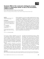
Available online />Figure 1
Expression pattern of angiopoietin-1 (Ang-1) in synovial tissues. The arrows denote the lining cell layer, the double arrowhead denotes subsynovial
macrophages and the single arrowhead denotes vascular endothelium. The immunohistochemistry was performed with a goat antihuman Ang-1
polyclonal antibody from Santa Cruz Biotechnology (Santa Cruz, CA, USA). (a) Synovial tissue from a patient with rheumatoid arthritis showing
positive staining for Ang-1. (b) Positive staining in osteoarthritis synovial tissue. (c) Ang-1 staining is absent in normal synovial tissue.
(a), (b) and (c) Original magnification × 226. (d) Immunohistochemistry of synovial tissues from normal (NL) subjects (n = 5) and those with
rheumatoid arthritis (RA) (n = 9) and osteoarthritis (OA) (n = 8). The bars represent mean ± SE. (e) Ang-1 mRNA levels in synovial tissue (ST)
and RA fibroblasts (Fib) were quantified using real-time reverse transcription-PCR and normalized to glyceraldehyde-3-phosphate dehydrogenase
(GAPDH). Bars represent the mean ± SE (n = 3). Lining, synovial tissue lining cell layer; Mac, subsynovial macrophages; Endo, vascular
endothelium. *P < 0.05.
embryonic lethality secondary to defects in the developing
vasculature.
Ang-1 was more highly expressed in synovial tissue from
RA patients than in either OA or normal synovial tissues;
expression in OA tissue was similar to normal. Positive
cells included lining cells, macrophages and endothelial
cells. In RA, Ang-1 was most upregulated on endothelial
cells. The results obtained from RT-PCR analysis con-
firmed that RA synovial tissue had significantly higher Ang-
1 mRNA expression than OA and normal synovial tissue.
Hence, no Ang-1 mRNA expression was detected in RA
fibroblasts (Fig. 1). The Tie receptor family is known to be
expressed on endothelial cells [5]. Otani et al [14] reported
Ang-1 and Ang-2 positive staining on macrophages and
endothelial cells in choroidal neovascular membranes.
Macrophages are key angiogenesis effector cells that
produce a number of growth factor stimulators and
inhibitors, proteolytic enzymes and cytokines that can acti-
vate one or more steps in the angiogenesis cascade
[15–17]. Recent studies have shown that angiopoietins
promote postnatal neovascularization by potentiating
angiogenic cytokines such as VEGF [9,10].
In situ hybridization and immunohistochemical analyses of
RA synovial biopsies revealed that VEGF mRNA and
protein localized to subsynovial macrophages, lining cells,
vascular smooth muscle cells and fibroblasts within the
pannus, which are the putative target of this cytokine
[18,19]. The localization of Ang-1 is similar to that of
VEGF, which further supports the crucial role of the inter-
action between these two pathways. Ang-1 modulates
VEGF-stimulated reorganization of endothelial cells and
promotes vascular network maturation [9,20]. Additionally,
both Ang-1 and VEGF are chemotactic and are involved in
recruiting endothelial cells to initiate and accelerate
endothelialization of blood vessels [21,22].
We detected Ang-2 immunopositive cells in synovial
lining, macrophages and vascular endothelium. Vascular
Arthritis Research Vol 4 No 3 Shahrara et al.
Figure 2
Expression pattern of angiopoietin-2 (Ang-2) mRNA and protein in synovial tissues. The double arrowhead denotes subsynovial macrophages, the
single arrowhead denotes vascular endothelium and the double headed arrows denote vascular smooth muscle cells. The immunohistochemistry
was performed with a goat antihuman Ang-2 polyclonal antibody from Santa Cruz Biotechnology (Santa Cruz, CA, USA). (a) Synovial tissue from a
patient with rheumatoid arthritis showing positive staining for Ang-2. (b) Positive staining for Ang-2 in osteoarthritis synovial tissue. (c) Ang-2
staining is absent in normal synovial tissue. (a), (b) and (c) Original magnification × 226. (d) Immunohistochemistry of synovial tissues from normal
(NL) subjects (n = 7) and those with rheumatoid arthritis (RA) (n = 11) and osteoarthritis (OA) (n = 12). The bars represent mean ± SE.
(e) Ang-2 mRNA levels in synovial tissue (ST) and RA fibroblasts (Fib) were quantified using real-time reverse transcription-PCR and
normalized to glyceraldehyde-3-phosphate dehydrogenase (GAPDH). Bars represent the mean ± SE (n = 3). Lining, synovial tissue lining
cell layer; Mac, subsynovial macrophages; Endo, vascular endothelium; Sm, vascular smooth muscle cells. *P < 0.05.
smooth muscle immunostaining was found only in RA. In
agreement with our finding, it has been reported that
primary cultured macrophages and precursor macrophage
cell lines mainly express Ang-2 mRNA [23]. In contrast to
Ang-1, Ang-2 was distributed in a similar pattern in RA
and OA compared to normal synovial tissues (Fig. 2). The
dominance of Ang-2 over Ang-1 mRNA and protein in OA
synovial tissue are in agreement with the inactive VEGF
receptor 1 pathway (angiogenic pathway) in OA [24]. The
Ang-2 mRNA expression pattern correlates with the Ang-2
immunostaining detected on endothelial cells. In RA syn-
ovial tissue, Ang-2 showed a lower percentage of staining
in all the cell types compared to Ang-1. Low Ang-2 mRNA
expression was also detected on RA fibroblasts. Interest-
ingly Ang-2 acts as an agonist for Tie2 receptors
expressed on fibroblasts [6]. Since the neovascularization
process is dependent on the dominance of angiogenic
mediators over inhibitors, the higher Ang-1 mRNA and
protein expression compared to Ang-2 in RA synovial
tissue is of importance. It has been shown that increasing
amounts of Ang-2 can block the chemotactic activity of
Ang-1 in endothelial cells [21]. Ang-2 alone did not stimu-
late corneal neovascularization, but the combination of
Ang-2 and VEGF resulted in longer vessels with greater
circumferential extent than those seen with VEGF alone
[9]. This suggests that Ang-2 expression in the absence of
VEGF leads to vessel regression, whereas expression of
Ang-2 in the presence of VEGF potentiates angiogenesis.
Using the Santa Cruz Biotechnology antibody, Tie1
expression was detected on all cell components exam-
ined, including a percentage of synovial lining cells,
macrophages and smooth muscle cells in RA and OA.
Synovial tissues from normal subjects, and those with OA,
had considerably lower positive immunostaining on
endothelial cells compared to RA tissues. Immunohisto-
chemical analysis performed with anti-Tie1 from R&D
Systems in synovial tissues supports its expression mainly
on endothelial cells. However, western blot analysis for
Tie1 also performed with an antibody from R&D Systems,
Available online />Figure 3
Expression pattern of Tie1 in synovial tissues. The double arrowhead denotes subsynovial macrophages, the single arrowhead denotes vascular
endothelium and the double headed arrows denote vascular smooth muscle cells. The immunohistochemistry was performed with a rabbit antihuman
Tie1 polyclonal antibody from Santa Cruz Biotechnology (Santa Cruz, CA, USA). (a) Synovial tissue from a patient with rheumatoid arthritis showing
positive staining for Tie1. (b) Positive staining for Tie1 in osteoarthritis synovial tissue. (c) Tie1 staining is absent in normal synovial tissue.
(a), (b) and (c) Original magnification × 212. (d) Immunohistochemistry of synovial tissues from normal (NL) subjects (n = 9) and those with
rheumatoid arthritis (RA) (n = 12) and osteoarthritis (OA) (n = 11). The bars represent mean ± SE. (e) Tie1 mRNA levels in synovial tissue (ST) and
RA fibroblasts (Fib) were quantified using real-time reverse transcription-PCR and normalized to glyceraldehyde-3-phosphate dehydrogenase
(GAPDH). Bars represent the mean ± SE (n = 3). (f) Western blot analysis of Tie1 in synovial tissues from RA, OA, RA fibroblasts and human
microvascular endothelial cells (Endo) using the goat antihuman antibody from R&D Systems (Minneapolis, MN, USA). Lining, synovial tissue lining
cell layer; Mac, subsynovial macrophages; Endo, vascular endothelium; Sm, vascular smooth muscle cells; Lymph, lymphocytes. *P < 0.05.
showed higher relative abundance of Tie1 in whole RA
and OA synovial tissue homogenates compared to RA
fibroblasts (Fig. 3). In accordance with the protein data,
the mRNA levels of Tie1 in RA synovial tissue were upreg-
ulated compared to OA synovial tissue, normal synovial
tissue and RA fibroblasts. Since the ligand for the Tie1
receptor is not known, it is difficult to speculate if there is
any correlation between this receptor and Tie2 and its
ligands. In agreement with previous studies, the Tie1
receptor was detected on vascular endothelial cells [4,25]
and recently on synovial lining cells [26]. It has been
shown that tumor necrosis factor-α and VEGF activate
membrane-associated metalloproteinases that release
soluble Tie1 from the cell surface of endothelial cells and
thereby decrease membrane-bound Tie1 expression [27].
The upregulation of Tie1 mRNA and protein expression in
RA is not consistent with these findings and this may be
due to the fact that the inflammatory milieu in RA synovial
tissue is far more complex than the in vitro system.
Using the Santa Cruz Biotechnology antibody we showed
that Tie2 expression was significantly upregulated both in
RA and OA synovial tissue lining cells, macrophages,
endothelial cells and vascular smooth muscle compared to
normal synovial tissue. In accordance with Otani et al. [14]
and Uchida et al. [26] we detected Tie2 immunostaining
on endothelial cells and fibroblasts in all disease groups.
Tie2 immunostaining using antibody from R&D Systems
confirmed the expression of this antigen mainly on
endothelial cells. Tie2 mRNA and protein expression fol-
lowed the same pattern of expression as Tie1. There are
some possible reasons why the immunohistochemistry
studies performed with Santa Cruz Biotechnology/R&D
Systems antibodies differ. The Santa Cruz Biotechnology
antibodies detected epitopes on the carboxy terminus
whereas the R&D Systems antibodies recognized the
extracelluar domain (N terminus or the ligand binding
domain) of Tie1 and Tie2. There is also some variability
between individual RA patient samples, however, we
Arthritis Research Vol 4 No 3 Shahrara et al.
Figure 4
Expression patterns of Tie2 mRNA and protein. The double arrowhead denotes subsynovial macrophages, the single arrowhead denotes vascular
endothelium and the double headed arrows denote vascular smooth muscle cells. The immunohistochemistry was performed with a rabbit
antihuman Tie2 polyclonal antibody from Santa Cruz Biotechnology (Santa Cruz, CA, USA). (a) Synovial tissue from a patient with rheumatoid
arthritis showing positive staining for Tie2. (b) Positive staining for Tie2 in osteoarthritis synovial tissue. (c) Tie2 staining is absent in normal
synovial tissue. (a), (b) and (c) Original magnification × 200. (d) Immunohistochemistry of synovial tissues from normal (NL) subjects (n = 11)
and those with rheumatoid arthritis (RA) (n = 15) and osteoarthritis (OA) (n = 11). The bars represent mean ± SE. (e) Tie2 mRNA levels in
synovial tissue (ST) and RA fibroblasts (Fib) were quantified using real-time reverse transcription-PCR and normalized to glyceraldehyde-3-
phosphate dehydrogenase (GAPDH). Bars represent the mean ± SE (n = 3). (f) Western blot analysis of Tie2 in synovial tissues from RA, OA,
RA fibroblasts and human microvascular endothelial cells (Endo) using the goat antihuman antibody from R&D Systems (Minneapolis, MN, USA).
Lining, synovial tissue lining cell layer; Mac, subsynovial macrophages; Endo, vascular endothelium; Sm, vascular smooth muscle cells; Lymph,
lymphocytes. *P < 0.05.
attempted to use the same RA patient samples in perform-
ing immunohistochemistry with the two different antibod-
ies. Additionally, the results obtained from the RT-PCR
and western blot analysis confirm our findings.
Conclusion
It is noteworthy that the activating ligand, Ang-1 is exclu-
sively higher than Ang-2 at the mRNA and protein levels in
RA synovial tissue compared to that from patients with OA
and normal subjects. Hypothetically, in RA synovial tissue
the dominating Ang-1 induces the Tie2 pathway, whereas
in normal synovial tissue higher Ang-2 presence blocks
Ang-1 binding to Tie2 and inhibits its autophosphorylation.
In conclusion, the dominance of Ang-1 over Ang-2 expres-
sion favors active neovascularization in RA synovial tissue.
Multiple pathways are probably required to regulate angio-
genesis. It may be that the inhibition of the VEGF and Tie2
pathways might be an effective therapeutic modality in RA.
Acknowledgements
This work was supported by NIH Grants HL58695, AI40987, the Gal-
lagher Professorship for Arthritis Research, and funds from the Veter-
an’s Administration Research Service.
References
1. Koch AE: Angiogenesis: implications for rheumatoid arthritis.
Arthritis Rheum 1998, 41:951-962.
2. Szekanecz Z, Szegedi G, Koch AE: Angiogenesis in rheumatoid
arthritis: pathogenic and clinical significance. J Invest Med
1998, 46:27-41.
3. Dumont DJ, Yamaguchi TP, Conlon RA, Rossant J, Breitman ML:
tek, a novel tyrosine kinase gene located on mouse chromo-
some 4, is expressed in endothelial cells and their presump-
tive precursors. Oncogene 1992, 7:1471-1480.
4. Partanen J, Armstrong E, Makela TP, Korhonen J, Sandberg M,
Renkonen R, Knuutila S, Huebner K, Alitalo K: A novel endothe-
lial cell surface receptor tyrosine kinase with extracellular epi-
dermal growth factor homology domains. Mol Cell Biol 1992,
12:1698-1707.
5. Davis S, Aldrich TH, Jones PF, Acheson A, Compton DL, Jain V,
Ryan TE, Bruno J, Radziejewski C, Maisonpierre PC, Yancopolous
GD: Isolation of Angiopoietin-1, a ligand for the TIE2 receptor,
by Secretion-Trap Expression Cloning. Cell 1996, 87:1161-1169.
6. Maisonpierre PC, Suri C, Jones PF, Bartunkova S, Wiegand SJ,
Radziejewski C, Compton D, McClain J, Aldrich TH, Papadopou-
los N, Daly TJ, Davis S, Sato TN, Yancopoulos GD: Angiopoietin-
2, a natural antagonist for Tie2 that disrupts in vivo
angiogenesis. Science 1997, 277:55-60.
7. Suri C, Jones PF, Patan S, Bartunkova S, Maisonpierre PC, Davis
S, Sato TN, Yancopoulos GD: Requisite role of angiopoietin-1,
a ligand for the Tie2 receptor, during embryonic
angiogenesis. Cell 1996, 87:1171-1180.
8. Sato TN, Tozawa Y, Deutsch U, Wolburg-Buchholz K, Fujiwara Y,
Gendron-Maguire M, Gridley T, Wolburg H, Risau W, Qin Y: Dis-
tinct roles of the receptor tyrosine kinases Tie-1 and Tie-2 in
blood vessel formation. Nature 1995, 376:70-74.
9. Asahara T, Chen D, Takahashi T, Fujikawa K, Kearney M, Magner
M, Yancopoulos GD, Isner JM: Tie2 receptor ligands, angiopoi-
etin-1 and angiopoietin-2, modulate VEGF-induced postnatal
neovascularization. Circ Res 1998, 83:233-240.
10. Hackett SF, Ozaki H, Strauss RW, Wahlin K, Suri C, Maisonpierre
P, Yancopoulos G, Campochiaro PA: Angiopoietin 2 expression
in the retina: upregulation during physiologic and pathologic
neovascularization. J Cell Physiol 2000, 184:275-284.
11. Szekanecz Z, Haines GK, Harlow LA, Shah MR, Fong TW, Fu R,
Lin SJ-W, Rayan G, Koch AE: Increased synovial expression of
TGF-
ββ
receptor endoglin and TGF-
ββ
1 in rheumatoid arthritis.
Clin Immunol Immunopathol 1995, 76:187-194.
12. Halloran MM, Szekanecz Z, Barquin N, Haines GK, Koch AE: Cel-
lular adhesion molecules in rat adjuvant arthritis. Arthritis
Rheum 1996, 39:810-819.
13. Dumont DJ, Gradwohl G, Fong GH, Puri MC, Gertsenstein M,
Auerbach A, Breitman ML: Dominant-negative and targeted null
mutations in the endothelial receptor tyrosine kinase, tek,
reveal a critical role in vasculogenesis of the embryo. Genes
Dev 1994, 8:1897-1909.
14. Otani A, Takagi H, Oh H, Koyama S, Matsumura M, Honda Y:
Expressions of angiopoietins and Tie2 in human choroidal
neovascular membranes. Invest Ophthalmol Vis Sci 1999, 40:
1912-1920.
15. Koch AE, Polverini PJ, Kunkel SL, Harlow LA, DiPietro LA, Elner
VM, Elner SG, Strieter RM: Interleukin-8 as a macrophage-
derived mediator of angiogenesis. Science 1992, 258:1798-
1801.
16. Koch AE, Kunkel SL, Harlow LA, Mazarakis DD, Haines GK,
Burdick MD, Pope RM, Walz A, Strieter RM: Epithelial neu-
trophil activating peptide-78: a novel chemotactic cytokine for
neutrophils in arthritis. J Clin Invest 1994, 94:1012-1018.
17. Polverini PJ: Role of the macrophage in angiogenesis-depen-
dent diseases. EXS 1997, 79:11-28.
18. Fava RA, Olsen NJ, Spencer-Green G, Yeo KT, Yeo TK, Berse B,
Jackman RW, Senger DR, Dvorak HF, Brown LF: Vascular per-
meability factor/endothelial growth factor (VPF/VEGF): accu-
mulation and expression in human synovial fluids and
rheumatoid synovial tissue. J Exp Med 1994, 180:341-346.
19. Koch AE, Harlow LA, Haines GK, Amento EP, Unemori EN, Wong
WL, Pope RM, Ferrara N: Vascular endothelial growth factor. A
cytokine modulating endothelial function in rheumatoid arthri-
tis. J Immunol 1994, 152:4149-4156.
20. Koblizek TI, Weiss C, Yancopoulos GD, Deutsch U, Risau W:
Angiopoietin-1 induces sprouting angiogenesis in vitro. Curr
Biol 1998, 8:529-532.
21. Witzenbichler B, Maisonpierre PC, Jones P, Yancopoulos GD,
Isner JM: Chemotactic properties of angiopoietin-1 and -2,
ligands for the endothelial-specific receptor tyrosine kinase
Tie2. J Biol Chem 1998, 273:18514-18521.
22. Tsurumi Y, Murohara T, Krasinski K, Chen D, Witzenbichler B,
Kearney M, Couffinhal T, Isner JM: Reciprocal relation between
VEGF and NO in the regulation of endothelial integrity. Nat
Med 1997, 3:879-886.
23. Kim I, Kim JH, Ryu YS, Jung SH, Nah JJ, Koh GY: Characteriza-
tion and expression of a novel alternatively spliced human
angiopoietin-2. J Biol Chem 2000, 275:18550-18556.
24. Giatromanolaki A, Sivridis E, Athanassou N, Zois E, Thorpe PE,
Brekken RA, Gatter KC, Harris AL, Koukourakis IM, Koukourakis
MI: The angiogenic pathway “vascular endothelial growth
factor/flk-1(KDR)-receptor” in rheumatoid arthritis and
osteoarthritis. J Pathol 2001, 194:101-108.
25. Sato TN, Qin Y, Kozak CA, Audus KL: Tie-1 and tie-2 define
another class of putative receptor tyrosine kinase genes
expressed in early embryonic vascular system. Proc Natl Acad
Sci USA 1993, 90:9355-9358.
26. Uchida T, Nakashima M, Hirota Y, Miyazaki Y, Tsukazaki T, Shindo
H: Immunohistochemical localisation of protein tyrosine
kinase receptors Tie-1 and Tie-2 in synovial tissue of rheuma-
toid arthritis: correlation with angiogenesis and synovial pro-
liferation. Ann Rheum Dis 2000, 59:607-614.
27. Yabkowitz R, Meyer S, Black T, Elliott G, Merewether LA, Yamane
HK: Inflammatory cytokines and vascular endothelial growth
factor stimulate the release of soluble tie receptor from
human endothelial cells via metalloprotease activation. Blood
1999, 93:1969-1979.
28. Altman R, Asch E, Bloch D, Bole G, Borenstein D, Brandt K,
Christy W, Cooke TD, Greenwald R, Hochberg M, Howell D,
Kaplan D, Koopman W, Longley S 3rd, Mankin H, McShane DJ,
Medsger TJ, Meenan R, Mikkelsen W, Moskowitz R, Murphy W,
Rothschild B, Segal M, Sokoloff L, Wolfe F: Development of cri-
teria for the classification and reporting of osteoarthritis.
Classification of osteoarthritis of the knee. Arthritis Rheum
1986, 29:1039-1049.
29. Altman R, Alarcon G, Appelrouth D, Bloch D, Borenstein D,
Brandt K, Brown C, Cooke TD, Daniel W, Feldman D, Greenwald
R, Hochberg M, Howell D, Ike R, Kapila P, Kaplan D, Koopman W,
Marino C, McDonald E, McShane DJ, Medsger TJ, Michel B,
Murphy WA, Osial T, Ramsey-Goldman R, Rothschild B, Wolfe F:
Available online />The American College of Rheumatology criteria for the classi-
fication and reporting of osteoarthritis of the hip. Arthritis
Rheum 1991, 34:505-514.
30. Koch AE, Polverini PJ, Leibovich SJ: Stimulation of neovascular-
ization by human rheumatoid synovial tissue macrophages.
Arthritis Rheum 1986, 29:471-479.
31. Chomczynski P, Sacchi N: Single-step method of RNA isolation
by acid guanidinium thiocyanate-phenol-chloroform extrac-
tion. Anal Biochem 1987, 162:156-159.
32. Endesfelder S, Krahn A, Kreuzer KA, Lass U, Schmidt CA,
Jahrmarkt C, von MA, Speer A: Elevated p21 mRNA level in
skeletal muscle of DMD patients and mdx mice indicates
either an exhausted satellite cell pool or a higher p21 expres-
sion in dystrophin-deficient cells per se. J Mol Med 2000, 78:
569-574.
33. Favy DA, Lafarge S, Rio P, Vissac C, Bignon YJ, Bernard-Gallon
D: Real-time PCR quantification of full-length and exon 11
spliced BRCA1 transcripts in human breast cancer cell lines.
Biochem Biophys Res Commun 2000, 274:73-78.
Supplementary material
Materials and methods
Patient population
Synovial tissues from patients with RA and OA, undergo-
ing arthroplasty or synovectomy, who fulfilled the criteria
set by the American College of Rheumatology were
obtained with Institutional Review Board (IRB) consent
[28,29]. Normal synovial tissues were obtained from fresh
autopsies or amputations. Synovial tissues were snap
frozen in Optimal Cutting Temperature (OCT) compound
(Miles, Elkhart, IN, USA).
Microscopic analysis
Synovial tissue components including lining cells,
macrophages, lymphocytes, fibroblasts, smooth muscle
cells and endothelial cells were graded for immunostain-
ing by a frequency of staining scale, scored 0–100%,
where 0% indicates no staining and 100% indicates that
all cells were immunoreactive. Five 400× fields were
examined per section by a single pathologist in a blinded
study. Selected sections were analyzed by two additional
observers.
Statistical analysis
Data were analyzed using a Kruskal Wallis test. There
were 5 to 15 patient samples per group. P values < 0.05
were considered to be significant.
Cell culture
Fibroblasts were isolated from synovial tissues that had
been minced and digested in a solution of dispase, colla-
genase and DNase. Synovial tissue fibroblasts were cul-
tured in Roswell Park Memorial Institute (RPMI)-1640
supplemented with 10% FBS and 1% penicillin/strepto-
mycin (P/S) and used at passage five or older, at which
time they were a homogeneous population of fibroblasts.
Human microvascular endothelial cells (HMVECs)
(BioWhittaker, CA, USA) were cultured in endothelial cell
growth medium-2 for microvascular cells (BioWhittaker,
Walkerville, MD, USA) and were used between passages
3 and 12. Upon confluence, the cells were passaged by
brief trypsinisation as previously described [30]. HMVECs
were used as positive controls for Tie1 and Tie2 protein
expression in western blot analysis.
RNA purification
Total RNA was prepared from synovial tissue by an acid
phenol method according to the procedure described by
Chomczynski and Sacchi [31]. RNA (20 µg) was digested
with five units of DNase in 1× reverse transcription buffer
(Gibco/Life Technologies, Grand Island, NY, USA) con-
taining eight units of RNase inhibitor (Gibco/Life Tech-
nologies, Grand Island, NY, USA) for 30 minutes at 37°C.
Sodium acetate (2 M, pH 4.0) was added (10% of the
total volume), and the solution was extracted with one
volume each of water-saturated phenol and chloroform
(Fisher, Itasca, IL, USA). The RNA was precipitated with
ethanol, washed with 75% ethanol, then air-dried. The
pellet was dissolved in water at 1 µg/µl.
Reverse transcription
Reverse transcription of RNA was performed using 5 µg of
RNA in a total volume of 20 µl. The RNA was reverse tran-
scribed by Superscript II RT (Gibco/Life Technologies,
Grand Island, NY, USA) according to manufacturer’s spec-
ification. After 10 minutes at 25°C, the enzyme was incu-
bated at 42°C for 50 minutes and thereafter inactivated at
70°C for 15 minutes. The solution was diluted to 40 µl. All
samples were reverse transcribed simultaneously.
Standard dilution preparation and PCR
All PCR reactions were performed on an ABI Prism 7700
Sequence Detection System (Perkin Elmer Applied Biosys-
tem, CA, USA). For each PCR run, a master mixture was
prepared on ice with 1× Platinum PCR buffer (Gibco/Life
Technologies, Grand Island, NY, USA), 1.5 mM MgCl
2
,
0.2 mM of each deoxynucleoside triphosphate, 0.2 µM
each primer, 2.5 units platinum Taq DNA polymerase
(Gibco/Life Technologies, Grand Island, NY, USA). The
diluted reverse transcription sample (3 µl) was added to 25
µl of the PCR mix. The thermal cycling conditions were 40
cycles of the denaturation step at 95°C for 30 seconds,
annealing at 60°C for one minute and an extention step at
72°C for one minute. PCR products were extracted from
agarose gels and purified. Serial dilutions of PCR products
were then prepared which ranged from 10
1
to 10
10
mole-
cules [32]. The sequence for the designed Tie receptors
and ligands are shown in Supplementary Table 1.
Real-time quantitative RT-PCR using the TaqMan system
The PCR primer and the TaqMan fluorogenic probe were
designed using the Primer Express program v 1.01 (Perkin
Arthritis Research Vol 4 No 3 Shahrara et al.
Elmer Applied Biosystem, CA, USA). The TaqMan probe
carries a 5′ FAM (6-carboxy-fluorescein) reporter dye and a
3′ TAMRA (6-carboxy-tetramethyl-rhodamine) quencher
dye (Mega Bases, Chicago, IL, USA) The quantity of cDNA
of the gene of interest was directly related to the fluores-
cence detection of FAM after 40 cycles. The amount of
cDNA was calculated using a comparative C
T
method and
the standard curve method [33] according to Perkin Elmer
ABI PRISM 7700 User Bulletin No. 2, 1997. The calibra-
tion curves showed a strong linear correlation, with correla-
tion coefficients between 0.96 and 0.99. In both methods,
the estimated amount of the gene of interest was normal-
ized by the amount of glyceraldehyde-3-phosphate dehy-
drogenase to compensate for variations in quantity and for
differences in reverse transcription efficiency.
Briefly, 3 µl of the cDNA was in a reaction of 25 µl that
contained final concentrations of 1× Platinum PCR buffer
(Gibco/Life Technologies, Grand Island, NY, USA),
3.5 mM MgCl
2
, 200 µM dNTP, 500 nM each primer
(Mega Bases, Chicago, IL, USA), 200 nM FAM-TAMRA
probe (Mega Bases, IL, USA), 100 nM Blue 636 (BD
636), 0.05 units platinum Taq DNA polymerase
(Gibco/Life Technologies, Grand Island, NY, USA). The
thermal cycling conditions included 94°C for five minutes,
followed by 40 cycles of amplification at 94°C for
30 seconds and 60°C for one minute for denaturing and
annealing-extension, respectively. All samples were ampli-
fied in triplicate.
Western blot analysis
Synovial tissues were homogenized in a 50 ml conical
centrifuge tube containing 3 ml of Complete Mini protease
inhibitor cocktail homogenization buffer (Roche, Indi-
anapolis, IN, USA). Synovial tissue homogenization was
completed on ice using a motorized homogenizer, fol-
lowed by sonication for 30 seconds. Homogenates were
centrifuged at 2000 × g for 10 minutes, filtered through a
0.45 µm pore size filter (Bedpore, Bedford, MA, USA) and
stored at –80°C until use. Complete Mini protease
inhibitor cocktail (1 ml) was added to one million RA
fibroblasts and HMVECs for lysis. The concentration of
protein in each synovial tissue and cell lysate was deter-
mined using a bicinchoninic acid assay (Pierce, Rockford,
IL, USA), using BSA as the standard. Protein extracts
(25 µg) were mixed with an equal volume of 2× Laemmli’s
sample buffer. Equal amounts of each sample were loaded
and run on a 10% SDS-PAGE gel and transferred to nitro-
cellulose membranes using a semi-dry transblotting appa-
ratus (Bio-Rad, Richmond, CA, USA). Nitrocellulose
membranes were blocked with 5% nonfat milk in Tris-
buffered saline Tween (20 mM Tris, 137 mM NaCl,
pH 7.6, with 0.1% Tween 20) for 60 minutes at room tem-
perature. Blots were incubated overnight with anti-Tie1
and anti-Tie2 antibodies (R&D Systems) at 1: 5000, in
Tris-buffered saline Tween containing 5% nonfat milk.
Blots were washed three times and then incubated in
horseradish-peroxidase-conjugated antibody (1:10,000
dilution) for one hour at room temperature. All blots were
developed using the enhanced chemiluminesence
reagents (Amersham, Piscataway, NJ, USA) as per the
manufacturer’s instructions.
Available online />Supplementary Table 1
The sequence for the designed Tie receptors and ligands
Names Forward primers TaqMan probes Reverse primers
Ang-1 GCC ATT ACC AGT CAG AGG CAG T CAT GCT AAG AAT TGA GTT AAT AAT AGG CTC GGT TCC CTT CC
Ang-2 CGC TCG AAT ACG ATG ACT CG TGC AGA GGC TGC AAG TGC TGG AGA A CCA CTG AGT GTT GTT TTC CAT GAT
Tie1 GCC ACG TTC TGG CTG GAT TCA GGC CTC CTC AGC TGT GGC AT ACT TCA CTT ACG CGG GCA TT
Tie2 GGC AAC TTG ACT TCG GTG CT ACT TAC ATC CCA GGG AGC AGT ACG TGG TC GGC CTT GGT GTT GAC TCT AGC T