Báo cáo y học: "The potential of human regulatory T cells generated ex vivo as a treatment for lupus and other chronic inflammatory diseases" pot
Bạn đang xem bản rút gọn của tài liệu. Xem và tải ngay bản đầy đủ của tài liệu tại đây (294.03 KB, 6 trang )
241
CTLA-4 = cytotoxic T-lymphocyte-associated antigen-4; TGF-β = transforming growth factor-β; IL = interleukin; MHC = major histocompatibility
complex; SLE = systemic lupus erythematosus; TCR = T-cell receptor; Th = T helper cells; Tr1 = Treg 1 regulatory CD4
+
cells.
Available online />Introduction
It has become evident that self-reactive T cells with the
potential to cause autoimmune disease comprise a part of
the normal T-cell repertoire, but their activation is prevented
by suppressor cells [1–3]. Although originally described in
the 1970s [4], significant progress in characterizing sup-
pressor T-cell subsets has been made only recently, where
they have been renamed ‘regulatory’ T cells.
A subset of thymus-derived CD4
+
cells that constitutively
expresses CD25, the α-chain of the IL-2 receptor, protect
their host from spontaneous organ-specific autoimmune
diseases. These CD4
+
CD25
+
cells have been called
‘professional’ suppressor cells and have a contact-depen-
dent mechanism of action, at least in vitro [5]. Other
subsets of CD4
+
and CD8
+
cells, natural killer T cells, and
cells displaying γδ TCRs also have downregulatory (sup-
pressor) activity. In the periphery, suppressor T cells gen-
erated in response to environmental antigens protect their
hosts from immune-mediated tissue injury by producing
immunosuppressive cytokines.
The mechanisms responsible for the generation of sup-
pressor T cells were poorly understood until recently. Our
group has accumulated evidence that the multifunctional
cytokine transforming growth factor-β (TGF-β) plays an
essential role in the expansion of thymus-derived, profes-
sional, CD4
+
CD25
+
precursors that circulate in the
blood. TGF-β also plays a key role in the generation of
peripherally induced CD4
+
and CD8
+
cytokine-producing
suppressor cell subsets.
This article will briefly review the evidence for contact-
mediated and cytokine-producing suppressor cells, espe-
cially in humans, and the role of TGF-β in the generation of
these cells. This knowledge can be used to generate sup-
pressor T cells ex vivo in large numbers, and raises the
possibility that the transfer of these cells back to the donor
Commentary
The potential of human regulatory T cells generated
ex vivo
as a
treatment for lupus and other chronic inflammatory diseases
David A Horwitz, J Dixon Gray and Song Guo Zheng
The Division of Rheumatology and Immunology, Department of Medicine, Keck School of Medicine, University of Southern California, Los Angeles,
California, USA
Corresponding author: David A Horwitz (e-mail: )
Received: 5 December 2001 Revisions received: 1 February 2002 Accepted: 7 February 2002 Published: 12 March 2002
Arthritis Res 2002, 4:241-246
© 2002 BioMed Central Ltd (
Print ISSN 1465-9905; Online ISSN 1465-9913)
Abstract
Regulatory T cells prevent autoimmunity by suppressing the reactivity of potentially aggressive self-
reactive T cells. Contact-dependent CD4
+
CD25
+
‘professional’ suppressor cells and other cytokine-
producing CD4
+
and CD8
+
T-cell subsets mediate this protective function. Evidence will be reviewed
that T cells primed with transforming growth factor (TGF)-β expand rapidly following restimulation.
Certain CD4
+
T cells become contact-dependent suppressor cells and other CD4
+
and CD8
+
cells
become cytokine-producing regulatory cells. This effect is dependent upon a sufficient amount of IL-2
in the microenvironment to overcome the suppressive effects of TGF-β. The adoptive transfer of these
suppressor cells generated ex vivo can protect mice from developing chronic graft versus host disease
with a lupus-like syndrome and alter the course of established disease. These data suggest that
autologous T cells primed and expanded with TGF-β have the potential to be used as a therapy for
patients with systemic lupus erythematosus and other chronic inflammatory diseases. This novel
adoptive immunotherapy also has the potential to prevent the rejection of allogeneic transplants.
Keywords: autoimmunity, IL-2, regulatory T cells, systemic lupus erythematosus, transforming growth factor-β
242
Arthritis Research Vol 4 No 4 Horwitz et al.
can serve as a therapy for autoimmune diseases such as
systemic lupus erythematosus (SLE). This T-cell-based
therapy could also be used to prevent graft rejection.
Thymus-dependent, ‘professional’, contact-
dependent, regulatory T cells
The existence of thymus-derived suppressor cells was sug-
gested by studies in mice where neonatal thymectomy on
day 3 led to the development of a multiorgan autoimmune
disease [6]. This disease is due to the loss of CD4
+
CD25
+
suppressor cells that do not appear until the first
week after birth [7,8]. Mature CD4
+
CD25
+
cells are found
in the CD45RB
low
activated/memory fraction mouse T
cells. Because potentially aggressive, self-reactive T cells
are found in the CD45RB
hi
naive fraction of mouse T cells,
the injection of CD45RB
hi
cells from nonautoimmune,
normal mice into immunodeficient mice results in general-
ized, multiorgan inflammatory disease. Similar to neonatal
thymectomy, this disease is prevented by supplementing
the injected cells with purified CD4
+
CD25
+
cells [9,10].
Because these thymus-derived CD4
+
CD25
+
T cells
appear to be crucial for the prevention of spontaneous
autoimmune diseases, they have been called ‘professional’
suppressor cells [5,8].
In general, the properties of rodent and human CD4
+
CD25
+
T cells appear to be very similar. In humans, 6–18%
of CD4
+
T cells constitutively express CD25 [11–17]. Puri-
fied CD4
+
CD25
+
cells do not proliferate in response to
cross-linking of their TCRs. They inhibit the activation of
other T cells by a contact-dependent mechanism [5–17]. A
large percentage constitutively express intracellular cyto-
toxic T-lymphocyte-associated antigen 4 (CTLA-4 or
CD152), the IL-2 receptor β-chain (CD122), transferrin
receptors (CD71) and class II MHC markers [17].
Almost all the CD4
+
CD25
+
are in the ‘activated’ state
(CD45RB
low
in the mouse, CD45RA
–
RO
+
in the human).
This suggests they may be continuously stimulated by their
internal environment. Although activation of CD4
+
CD25
+
cells is antigen specific, once these cells are activated they
not only suppress T cells stimulated by the same antigen,
but they also inhibit T cells stimulated by other antigens;
so-called bystander effects [18]. Although CD4
+
CD25
+
cells are nonresponsive to cross-linking their TCRs, they do
proliferate when costimulated with IL-2 or anti-CD28.
Cytokine production by CD4
+
CD25
+
cells is controver-
sial. While some groups claim that these cells do not
produce cytokines [8,17], other groups have found that
they can produce IL-10 [12,13,19], TGF-β [15,19], IL-4
[12] and low amounts of interferon-γ [15]. All groups
agree that these cells do not produce IL-2 and that they
have a contact-dependent mechanism of action. Their
suppressive activities are not abolished by neutralizing
antibodies to IL-10, and all groups agree that suppression
is not abolished by anti-TGF-β, but for one exception [19].
Nakamura et al. reported that immunosuppression by
CD4
+
CD25
+
regulatory T cells is mediated by cell
surface-bound TGF-β [19]. Many of these differences can
possibly be explained by the heterogeneity of CD4
+
CD25
+
T cells. One group separated human CD4
+
CD25
+
cells into high- and low-intensity fractions by cell
sorting, and they found that the suppressive effects were
only displayed by the high-intensity fraction [17]. This
subset did not produce cytokines.
Cytokine-dependent regulatory T cells
CD8
+
and CD4
+
T cells that produce immunosuppressive
cytokines have been described. Those that produce pre-
dominantly TGF-β and variable amounts of IL-10 and IL-4
have been called Th3-type cells, and they have been gen-
erated in vivo by immunization through an oral or other
mucosal route [2,20]. This route of antigen administration,
however, does not only result in Th3 cells. Both Th2 cells
and CD4
+
CD25
+
cells can also be generated by this pro-
cedure [20–22]. The conditions needed for the generation
of Th3 cells are poorly understood.
Other workers have produced regulatory CD4
+
cells by
repeatedly stimulating with the antigen in the presence of
IL-10 [23–26], or using immature antigen-presenting cells
that lack potent costimulatory activity [27]. These regula-
tory CD4
+
cells have been called Treg 1 (Tr1) cells and
they produce significant quantities of IL-10. They do not
proliferate in response to antigen and do not produce IL-2.
Therefore, they are anergic.
Th3 and Tr1-like cells have been described in humans.
One group has reported the appearance of Th3 cells in
patients with multiple sclerosis following oral administra-
tion of myelin basic protein [28]. Human Tr1 cells sup-
pressed an alloantigen-induced proliferative response
[29]. Th3 or Tr1 cells mediate antigen-specific cellular
hyporesponsiveness in patients with chronic helminth
infections [30].
The fact that some regulatory T cells produce predomi-
nantly TGF-β and others IL-10 is not fortuitous. The com-
bination of TGF-β and IL-10 is more immunosuppressive
than either of the cytokines by themselves [31]. Signifi-
cantly, shortly after antigen activation, T cells downregu-
late their signal transducing type II receptor (TGF-βRII)
and become refractory to the effects of TGF-β [32]. These
cells then become mature effector cells. IL-10 appears as
a feedback regulator later in the response and induces the
re-expression of TGF-βRII. The synergistic inhibitory
effects of TGF-β and IL-10 then terminate the response.
Whether Th3 cells and Tr1 cells come from similar precur-
sors or comprise different subsets of regulatory T cells is
not known. Many variables determine the differentiation
243
pathway that a naive T cell will take following activation.
These include the antigen concentration and route of
administration, the cytokine milieu, and the pattern of co-
stimulatory signals. Self-MHC-reactive T cells in humans
can either provide B-cell helper function or suppress anti-
body production, depending on how they are activated. In
each case, regulatory function depends on the cytokines
produced [33]. In determining the T-cell response to
myelin basic protein, another group found that TCR usage
was similar whether the T cells became Th1 encephalito-
genic cells or regulatory Th3 cells [34]. These studies
suggest common precursors for T cells that take different
differentiation pathways.
TGF-
ββ
induces CD4
+
and CD8
+
T cells to
become suppressor cells
While TGF-β has well-known inhibitory effects on lympho-
cyte cytokine production and functional properties [35],
our laboratory has accumulated data that these effects
can be overcome by IL-2 and can be superceded by co-
stimulatory activities. The net effect is that TGF-β induces
IL-2-activated CD8
+
and CD4
+
T cells to develop potent
suppressive activities. In parallel, other groups have
observed that TGF-β inhibits the differentiation of T cells
to Th1 or Th2 subsets [36,37].
The initial observation that TGF-β is an IL-2-dependent
differentiation factor for regulatory T cells was made in a
study designed to determine the conditions required for
human CD8
+
T cells to become suppressors of antibody
production. Using a model where we could induce T-cell-
dependent antibody production without accessory cells,
we found that CD4
+
T cells, by themselves, lacked sup-
pressor-inducing activity. The CD4
+
cells produced IL-2
but, notwithstanding previous reports [38,39], this
cytokine could not induce suppressor cells by itself. We
learned that the interaction of IL-2-activated natural killer
cells with CD8
+
cells leads to the production of active
TGF-β, and that the presence of this cytokine was critical
for CD8
+
cells to suppress antibody production (Figure 1)
[40,41]. Moreover, the suppression was cytokine depen-
dent and was abolished by a neutralizing anti-TGF-β
monoclonal antibody (JD Gray and DA Horwitz, unpub-
lished observation, 2000). Both IL-2 and TGF-β were thus
critical for CD8
+
cells to become Th3-like regulatory cells.
We have also induced CD4
+
T cells to become Th3 cells.
We used the superantigen, staphylococcal enterotoxin B,
as the T-cell activating agent. Low-dose staphylococcal
enterotoxin B can induce T-cell-dependent antibody pro-
duction without additional accessory cells [42]. Briefly
exposing CD4
+
cells to TGF-β downregulated B-cell
helper activity and induced certain CD4
+
cells to develop
suppressive activity that was neutralized by anti-TGF-β.
Activating both CD4
+
and CD8
+
cells in the presence of
TGF-β thus induced them to develop cytokine-dependent
suppressive activity [43]. Other workers have also reported
similar effects of TGF-β on CD8
+
T cells [44]. One group
found that IL-4 and TGF-β are involved in the differentiation
of naive CD4
+
cells to cytokine-producing Th3-type cells
[45]. Another group reported that in vitro differentiation of
Th3-type cells from Th0 precursors from TCR transgenic
mice is enhanced by culture with TGF-β [20].
We next focused our attention on the induction of naive
(CD45RA
+
RO
–
) CD4
+
T cells to become suppressor
cells. Using the alloantigens as the T-cell activating agent,
we found that TGF-β induced naive CD4
+
T cells to
develop extremely potent suppressive activity. These
CD4
+
cells had the phenotype and functional characteris-
tics of ‘professional’ regulatory T cells. Using the genera-
tion cytotoxic T-cell activity and T-cell proliferation to
assess suppressive activity, we learned that the suppres-
sor cells were CD25
+
, and that a large percentage
expressed CTLA-4. Their suppressive effects were
contact dependent and were not neutralized by anti-TGF-β
or IL-10. Adding less than 1% of these cells to T cells
strongly inhibited the generation of cytotoxic T-lymphocyte
activity by preventing the activation of CD8
+
cells [14].
Other workers have also reported that CD4
+
CD25
+
cells
have potent suppressive effects on CD8
+
cells. Rodent
CD4
+
CD25
+
regulatory cells cause CD8
+
cells to enter
cycle arrest [46].
The precursors of the human CD4
+
CD25
+
T cells induced
by IL-2 and TGF-β appear to be the small number of CD25
+
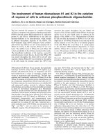
Available online />Figure 1
The role of transforming growth factor-β (TGF-β) in the differentiation
pathway of CD8
+
regulatory T cells. In response to antigen stimulation,
the combination of IL-2 produced by CD4
+
cells and the active form of
TGF-β produced by natural killer (NK) cells or macrophages (not
shown) induce CD8
+
cells to lose their cytotoxic potential and become
regulatory, TGF-β-producing, Th3-like cells. IL-2 also enhances the
extracellular conversion of TGF-β from the latent to the biologically
active form.
244
cells in the naive fraction. Although <1% of these cells
express CD25, depletion of these cells abrogated the gen-
eration of suppressive activity in some experiments [14].
The principal difference between the cytokine-induced
CD4
+
CD25
+
cells and the murine and human positively
selected CD4
+
CD25
+
cells that are predominantly found in
the CD45RO
+
‘memory’ fraction is their capacity for expan-
sion. The positively selected cells are anergic while the
CD4
+
CD25
+
cells generated from naive cells can be
expanded in IL-2 and retain their suppressive activity [14].
Studies on the mechanism of action of TGF-β have
revealed that this cytokine has potent costimulatory effects
on IL-2-activated T cells. These effects include upregula-
tion of CD25, CTLA-4 and CD40 ligand expression on
CD4
+
cells [14,47], and increased tumor necrosis factor-α
production by both CD4
+
and CD8
+
cells [47]. The TGF-β
costimulated human CD4
+
T cells are resistant to activa-
tion-induced apoptosis. They took up less annexin and
expanded fivefold greater in primary cultures than control,
alloactivated CD4
+
T cells [14] (SG Zheng and DA
Horwitz, unpublished observations, 2001). Some workers
have reported that TGF-β can accelerate activation-
induced cell death of some T cells [48,49], while others
observed that this cytokine protected T cells from apopto-
sis [50,51]. We favor the hypothesis that TGF-β promotes
the death of mature Th1 and Th2 cells while protecting
newly generated regulatory T cells from undergoing apop-
tosis. This view is consistent with a report indicating posi-
tive effects of TGF-β on naive T cells [52].
In summary, using several different stimuli to activate T
cells, we have found that the combination of IL-2 and
TGF-β can induce CD4
+
and CD8
+
T cells to become
either cytokine-producing Th3-like or contact-dependent
professional suppressor cells. In our studies with CD8
+
cells, the cultures were always supplemented with IL-2.
When human CD4
+
cells are activated in the presence of
TGF-β by irradiated allogeneic stimulator cells or with
superantigens, however, sufficient IL-2 is produced for the
costimulatory effects of TGF-β and suppressor cell differ-
entiation. By contrast, cultures with mouse lymphocytes
must generally be supplemented with IL-2.
As shown in Figure 2, we propose that TGF-β induces
thymic-derived CD25 precursors in the naive fraction of
CD4
+
cells to expand and to become contact-dependent
‘professional’ regulatory T cells. TGF-β also induces CD4
+
and CD8
+
cells that are CD25
–
to become Th3-like cells.
Although almost all naive CD4
+
cells are CD25
–
, why the
predominant TGF-β effect on T cells in this fraction is the
generation of ‘professional’ regulatory T cells remains to
be determined. Our finding that both IL-2 and TGF-β are
critical in the generation of regulatory T cells is of particu-
lar importance in patients with SLE since production of
IL-2 and the active form of TGF-β is decreased [53].
In vivo
effects of Treg
Cloned Th3 cells protect mice from several autoimmune
diseases that include experimental allergic encephalitis,
diabetes mellitus, colitis, and uveitis [20,29,54–56].
Cytokine-producing CD8
+
cells were described initially
[55], but reports of CD4
+
cells with this characteristic have
become predominant. Cloned Tr1 cells protect rodents
from an experimental colitis [29]. Small numbers of adop-
tively transferred noncloned CD4
+
CD25
+
cells protect
lymphopenic mice from developing spontaneous organ-
specific autoimmune diseases and also protect animals
from developing graft-versus-host disease [8–10,57].
We have begun to learn whether regulatory T cells gener-
ated ex vivo with TGF-β can have protective effects in
vivo. For this purpose, we selected a mouse model of SLE
that has a rapid onset. The transfer of parental T cells to
F1 mice can result in acute or chronic graft-versus-host
disease depending on the precursor frequency of CD8
+
parental cells reactive against the allogeneic MHC anti-
gens [58,59]. The transfer of DBA/2 T cells into DBA/2 x
C57BL/6 F1 mice results in a lupus-like syndrome with
high titers of anti-DNA antibodies and an immune complex
glomerulonephritis. While alloactivated DBA/2 T cells
accelerated the disease, alloactivation of splenic T cells or
CD4
+
cells in the presence of TGF-β markedly sup-
pressed and even prevented the development of the
lupus-like syndrome. Both anti-DNA antibody production
and proteinuria were significantly suppressed [60]. Recent
studies have revealed that these suppressor T cells can
also alter the course of established disease. A single
transfer of 5 million T cells conditioned with TGF-β
markedly improved survival of these mice (SG Zheng and
DA Horwitz, unpublished observations, 2001).
Arthritis Research Vol 4 No 4 Horwitz et al.
Figure 2
The role of transforming growth factor-β (TGF-β) in the differentiation
pathway of CD4
+
regulatory T cells. Following T-cell activation where a
sufficient amount of IL-2 is produced to overcome the inhibitory effects
of TGF-β, the costimulatory effects of this cytokine induce the
precursors of CD4
+
CD25
+
T cells to become contact-dependent
‘professional’ suppressor cells or induces CD4
+
CD25
–
cells to
produce immunosuppressive quantities of TGF-β. IFN, interferon;
Tr-1, Treg 1 regulatory CD4
+
cells.
245
Since it has been possible to significantly expand regula-
tory T cells generated with TGF-β, it should be possible to
generate sufficient numbers in humans for clinical trials.
Although this will be carried out initially with mitogens as
the T-cell activating agent, the ultimate goal is to induce
autoantigen-specific regulatory T cells. This should be pos-
sible based on the progress being made in characterizing
the pathogenic peptides that trigger autoimmune diseases.
It may even be possible to induce potentially aggressive
naive self-reactive cells to become protective suppressor
cells by activating them with TGF-β. An adoptive
immunotherapy using the patients own T cells that have
regained a protective function they had lost should lack the
serious toxic effects associated with the agents now in use.
This treatment is especially promising in autoimmune dis-
eases characterized by a relapsing and remitting course
such as SLE, inflammatory bowel disease or certain forms
of multiple sclerosis. The adoptive transfer of regulatory T
cells generated ex vivo also has the potential to prevent
the rejection of allogeneic organ transplants.
Acknowledgements
This research was supported in part by National Institutes of Health grant
AI 41768, The Nora Eccles Treadwell Foundation, and the Arthritis
Foundation-Southern California Chapter.
References
1. Fowell D, Mason D: Evidence that the T cell repertoire of
normal rats contains cells with the potential to cause dia-
betes. Characterization of the CD4+ T cell subset that inhibits
this autoimmune potential. J Exp Med 1993, 177:627-636.
2. Hafler DA, Weiner HL: Immunologic mechanisms and therapy
in multiple sclerosis. Immunol Rev 1995, 144:75-107.
3. Sakaguchi S, Sakaguchi N, Asano M, Itoh M, Toda M: Immuno-
logic self-tolerance maintained by activated T cells express-
ing IL-2 receptor-chains (CD25). Breakdown of a single
mechanism of self-tolerance causes various autoimmune dis-
eases. J Immunol 1995, 155:1151-1164.
4. Gershon RKA: A disquisition on suppressor T cells. Transplant
Rev 1975, 26:170-185.
5. Shevach EM: Certified professionals: CD4+ CD25+ suppres-
sor T cells. J Exp Med 2001, 193:41-46.
6. Sakaguchi S, Fukuma K, Kuribayashi K, Masuda T: Organ-specific
autoimmune diseases induced in mice by elimination of T cell
subset. I. Evidence for the active participation of T cells in
natural self-tolerance; deficit of a T cell subset as a possible
cause of autoimmune disease. J Exp Med 1985, 161:72-87.
7. Kuniyasu Y, Takahashi T, Itoh M, Shimizu J, Toda G, Sakaguchi S:
Naturally anergic and suppressive CD25+ CD4+ T cells as a
functionally and phenotypically distinct immunoregulatory T
cell subpopulation. Int Immunol 2000, 12:1145-1155.
8. Shevach EM: Regulatory T cells in autoimmmunity. Annu Rev
Immunol 2000, 18:423-449.
9. Mason D, Powrie F: Control of immune pathology by regulatory
T cells. Curr Opin Immunol 1998, 10:649-655.
10. Maloy KJ, Powrie F: Regulatory T cells in the control of immune
pathology. Nat Immunol 2001, 2:816-822.
11. Taams LS, Smith J, Rustin MH, Salmon M, Poulter LW, Akbar AN:
Human anergic/suppressive CD4+ CD25+ T cells: a highly
differentiated and apoptosis-prone population. Eur J Immunol
2001, 31:1122-1131.
12. Stephens LA, Mottet C, Mason D, Powrie F: Human CD4+
CD25+ thymocytes and peripheral T cells have immune sup-
pressive activity in vitro. Eur J Immunol 2001, 31:1247-1254.
13. Dieckmann D, Plottner H, Berchtold S, Berger T, Schuler G: Ex
vivo isolation and characterization of CD4+CD25+ T cells with
regulatory properties from human blood. J Exp Med 2001,
193:1303-1310.
14. Yamagiwa S, Gray JD, Hashimoto S, Horwitz DA: A role for
TGF-beta in the generation and expansion of CD4+CD25+
regulatory T cells from human peripheral blood. J Immunol
2001, 166:7282-7289.
15. Levings MK, Sangregorio R, Roncarolo MG: Human CD25+
CD4+ T regulatory cells suppress naive and memory T cell
proliferation and can be expanded in vitro without loss of
function. J Exp Med 2001, 193:1295-1302.
16. Jonuleit H, Schmitt E, Stassen M, Tuettenberg A, Knop J, Enk AH.
Identification and functional characterization of human
CD4+CD25+ T cells with regulatory properties isolated from
peripheral blood. J Exp Med 2001, 193:1285-1294.
17. Baecher-Allan C, Brown JA, Freeman GJ, Hafler DA:
CD4+CD25high regulatory cells in human peripheral blood. J
Immunol 2001, 167:1245-1253.
18. Thornton AM, Shevach EM: Suppressor effector function of
CD4+CD25+ immunoregulatory T cells is antigen nonspecific.
J Immunol 2000, 164:183-190.
19. Nakamura K, Kitani A, Strober W: Cell contact-dependent
immunosuppression by CD4+CD25+ regulatory T cells is
mediated by cell surface-bound transforming growth factor
beta. J Exp Med 2001, 194:629-644.
20. Weiner HL: Oral tolerance: immune mechanisms and the gen-
eration of Th3-type TGF-beta-secreting regulatory cells.
Microbes Infect 2001, 3:947-954.
21. Zhang X, Izikson L, Liu L, Weiner HL: Activation of CD25+ CD4+
regulatory T cells by oral antigen administration. J Immunol
2001, 167:4245-4253.
22. Thorstenson KM, Khoruts A: Generation of anergic and poten-
tially immunoregulatory CD25+ CD4 + T cells in vivo after
induction of peripheral tolerance with intravenous or oral
antigen. J Immunol 2001, 167:188-195.
23. Asseman C, Powrie F: Interleukin 10 is a growth factor for a
population of regulatory T cells. Gut 1998, 42:157-158.
24. Cottrez F, Hurst SD, Coffman RL, Groux H: T regulatory cells 1
inhibit a Th2-specific response in vivo. J Immunol 2000, 165:
4848-4853.
25. Levings MK, Roncarolo MG: T-regulatory 1 cells: a novel subset
of CD4 T cells with immunoregulatory properties. J Allergy
Clin Immunol 2000, 106:S109-S112.
26. Levings MK, Sangregorio R, Galbiati F, Squadrone S, de Waal
Malefyt R, Roncarolo MG: IFN-alpha and IL-10 induce the dif-
ferentiation of human type 1 T regulatory cells. J Immunol
2001, 166:5530-5539.
27. Jonuleit H, Schmitt E, Schuler G, Knop J, Enk AH: Induction of
interleukin 10-producing, nonproliferating CD4+ T cells with
regulatory properties by repetitive stimulation with allogeneic
immature human dendritic cells. J Exp Med 2000, 192:
1213-1222.
28. Fukaura H, Kent SC, Pietrusewicz MJ, Khoury SJ, Weiner HL,
Hafler DA: Induction of circulating myelin basic protein and
proteolipid protein-specific transforming growth factor-beta1-
secreting Th3 T cells by oral administration of myelin in multi-
ple sclerosis patients. J Clin Invest 1996, 98:70-77.
29. Groux H, O’Garra A, Bigler M, Rouleau M, Antonenko S, de Vries
JE, Roncarolo MG: A CD4+ T-cell subset inhibits antigen-spe-
cific T-cell responses and prevents colitis. Nature 1997, 389:
737-742.
30. Doetze A, Satoguina J, Burchard G, Rau T, Loliger C, Fleischer B,
Hoerauf A: Antigen-specific cellular hyporesponsiveness in a
chronic human helminth infection is mediated by Th3/
Tr1-type cytokines IL-10 and transforming growth factor-beta
but not by a Th1 to Th2 shift. Int Immunol 2000, 12:623-630.
31. Zeller JC, Panoskaltsis-Mortari A, Murphy WJ, Ruscetti FW,
Narula S, Roncarolo MG, Blazar BR: Induction of CD4+ T cell
alloantigen-specific hyporesponsiveness by IL-10 and
TGF-beta. J Immunol 1999, 163:3684-3691.
32. Cottrez F, Groux H: Regulation of TGF-beta response during T cell
activation is modulated by IL-10. J Immunol 2001, 167:773-778.
33. Kitani A, Chua K, Nakamura K, Strober W: Activated
self-MHC-reactive T cells have the cytokine phenotype of
Th3/T regulatory cell 1 T cells. J Immunol 2000, 165:691-702.
34. Chen Y, Kuchroo VK, Inobe J, Hafler DA, Weiner HL: Regulatory
T cell clones induced by oral tolerance: suppression of auto-
immune encephalomyelitis. Science 1994, 265:1237-1240.
35. Letterio JJ, Roberts AB: Regulation of immune responses by
TGF-beta. Annu Rev Immunol 1998, 16:137-161.
Available online />246
36. Heath VL, Murphy EE, Crain C, Tomlinson MG, O’Garra A: TGF-
beta1 down-regulates Th2 development and results in
decreased IL-4-induced STAT6 activation and GATA-3 expres-
sion. Eur J Immunol 2000, 30:2639-2649.
37. Ludviksson BR, Seegers D, Resnick AS, Strober W: The effect of
TGF-beta1 on immune responses of naive versus memory
CD4+ Th1/Th2 T cells. Eur J Immunol 2000, 30:2101-2111.
38. Ting CC, Yang SS, Hargrove ME: Induction of suppressor T
cells by interleukin 2. J Immunol 1984, 133:261-266.
39. Yamamoto H, Hirayama M, Genyea C, Kaplan J: TGF-beta medi-
ates natural suppressor activity of IL-2-activated lymphocytes.
J Immunol 1994, 152:3842-3847.
40. Gray JD, Hirokawa M, Horwitz DA: The role of transforming
growth factor beta in the generation of suppression: an inter-
action between CD8+ T and NK cells. J Exp Med 1994, 180:
1937-1942.
41. Gray JD, Hirokawa M, Ohtsuka K, Horwitz DA: Generation of an
inhibitory circuit involving CD8+ T cells, IL-2, and NK cell-
derived TGF-beta: contrasting effects of anti-CD2 and anti-
CD3. J Immunol 1998, 160:2248-2254.
42. Stohl W, Elliott JE, Linsley PS: Human T cell-dependent B cell
differentiation induced by staphylococcal superantigens.
J Immunol 1994, 153:117-127.
43. Zheng SG, Yamagiwa S, Ohtsuka K, Gray JD, Horwitz DA:
Inhibitory effects of TGF-
ββ
on the generation of T cell help for
B cells [abstract]. Arthritis Rheum 2001, 44:S97.
44. Rich S, Seelig M, Lee HM, Lin J: Transforming growth factor
beta 1 costimulated growth and regulatory function of
staphylococcal enterotoxin B-responsive CD8+ T cells.
J Immunol 1995, 155:609-618.
45. Seder RA, Marth T, Sieve MC, Strober W, Letterio JJ, Roberts AB,
Kelsall B: Factors involved in the differentiation of TGF-beta-
producing cells from naive CD4+ T cells: IL-4 and IFN-gamma
have opposing effects, while TGF-beta positively regulates its
own production. J Immunol 1998, 160:5719-5728.
46. Piccirillo CA, Shevach EM: Cutting edge: control of CD8+ T cell
activation by CD4+CD25+ immunoregulatory cells. J Immunol
2001, 167:1137-1140.
47. Gray JD, Liu T, Huynh N, Horwitz DA: Transforming growth
factor beta enhances the expression of CD154 (CD40L) and
production of tumor necrosis factor alpha by human T lym-
phocytes. Immunol Lett 2002, 78:83-88.
48. Chung EJ, Choi SH, Shim YH, Bang YJ, Hur KC, Kim CW: Trans-
forming growth factor-beta induces apoptosis in activated
murine T cells through the activation of caspase 1-like pro-
tease. Cell Immunol 2000, 204:46-54.
49. Sillett HK, Cruickshank SM, Southgate J, Trejdosiewicz LK:
Transforming growth factor-beta promotes ‘death by neglect’
in post-activated human T cells. Immunology 2001, 102:310-
316.
50. Genestier L, Kasibhatla S, Brunner T, Green DR: Transforming
growth factor beta1 inhibits Fas ligand expression and subse-
quent activation-induced cell death in T cells via downregula-
tion of c-myc. J Exp Med 1999, 189:231-239.
51. Chen W, Jin W, Tian H, Sicurello P, Frank M, Orenstein JM, Wahl
SM: Requirement for transforming growth factor beta1 in con-
trolling T cell apoptosis. J Exp Med 2001, 194:439-453.
52. de Jong R, van Lier RA, Ruscetti FW, Schmitt C, Debre P,
Mossalayi MD: Differential effect of transforming growth
factor-beta 1 on the activation of human naive and memory
CD4+ T lymphocytes. Int Immunol 1994, 6:631-638.
53. Ohtsuka K, Gray JD, Stimmler MM, Toro B, Horwitz DA:
Decreased production of TGF-beta by lymphocytes from
patients with systemic lupus erythematosus. J Immunol 1998,
160:2539-2545.
54. Han HS, Jun HS, Utsugi T, Yoon JW: Molecular role of TGF-
beta, secreted from a new type of CD4+ suppressor T cell,
NY4.2, in the prevention of autoimmune IDDM in NOD mice.
J Autoimmun 1997, 10:299-307.
55. Pankewycz OG, Guan JX, Benedict JF: A protective NOD islet-
infiltrating CD8+ T cell clone, I.S. 2.15, has in vitro immuno-
suppressive properties. Eur J Immunol 1992, 22:2017-2023.
56. Keino H, Takeuchi M, Suzuki J, Kojo S, Sakai J, Nishioka K,
Sumida T, Usui M: Identification of Th2-type suppressor T cells
among in vivo expanded ocular T cells in mice with experi-
mental autoimmune uveoretinitis. Clin Exp Immunol 2001,
124:1-8.
57. Gao Q, Rouse TM, Kazmerzak K, Field EH: CD4+CD25+ cells
regulate CD8 cell anergy in neonatal tolerant mice. Transplan-
tation 1999, 68:1891-1897.
58. Pals ST, Radaszkiewicz T, Gleichmann E: Induction of either
acute or chronic graft-versus-host disease due to genetic dif-
ferences among donor T cells. Adv Exp Med Biol 1982, 149:
537-544.
59. Shustov A, Nguyen P, Finkelman F, Elkon KB, Via CS: Differential
expression of Fas and Fas ligand in acute and chronic graft-
versus-host disease: up-regulation of Fas and Fas ligand
requires CD8+ T cell activation and IFN-gamma production.
J Immunol 1998, 161:2848-2855.
60. Zheng SG, Koss MN, Quismorio FP, Horwitz DA: Suppression of
a lupus-like syndrome with regulatory T cells generated ex-
vivo with TGF-
ββ
[abstract]. Arthritis Rheum 2001, 44:S283.
Correspondence
David A Horwitz, MD, The Division of Rheumatology and Immunology,
Department of Medicine, Keck School of Medicine, University of Southern
California, Los Angeles, California, USA. Tel: +1 323 442 1946;
fax: +1 323 442 2874; e-mail:
Arthritis Research Vol 4 No 4 Horwitz et al.