Báo cáo y học: "Successful immunotherapy with matrix metalloproteinasederived peptides in adjuvant arthritis depends on the timing of peptide administration" pdf
Bạn đang xem bản rút gọn của tài liệu. Xem và tải ngay bản đầy đủ của tài liệu tại đây (355.37 KB, 8 trang )
Introduction
Rheumatoid arthritis (RA) is an autoimmune disease, in
which autoreactive T cells are considered to play a central
role in the autodestructive process [1,2]. For the develop-
ment of long-term acting, antigen-specific immunothera-
pies, considerable attention has been devoted to
intervening in the autoimmune process via modulation of
the autoaggressive T-cell response. Several studies in
experimental autoimmune models have shown that it is
possible to induce antigen-specific tolerance, leading to
disease resistance [3–9]. This tolerance induction is
dependent on several factors such as adjuvant use,
antigen dose and the route of antigen administration (as
reviewed in [10]). Nasal antigen administration appears to
be an especially efficient way to induce mucosal tolerance
in experimental arthritis models [9,11–13].
We recently published a computer search profile to predict
T-cell self-epitopes recognized in adjuvant arthritis (AA)
[14]. This search profile led to the identification of matrix
metalloproteinases (MMPs) as targets for T-cell recognition
in experimental arthritis. Interestingly, active arthritis could
be induced by immunization with synthetic peptides con-
taining the predicted MMP-3, MMP-10 or MMP-16 T-cell
epitope. Moreover, CD4
+
T cells isolated from rats immu-
nized with MMP-3 peptide, MMP-10 peptide or MMP-16
peptide could transfer arthritis into naïve Lewis rats.
As well as being a target for T cells, we have demon-
strated that MMPs also become a target for B cells during
the course of AA. This is shown by the appearance of
MMP-3-specific IgM and IgG titers after AA induction
(JHM van Bilsen et al., unpublished observations).
In the present study, we investigate the usefulness of the
recently identified MMP epitopes to modulate MMP-
specific T-cell responses, thereby interfering in the course
of AA in Lewis rats.
Materials and methods
See Supplementary material for full details of the peptides,
the animals and the arthritis model.
AA = adjuvant arthritis; DTH = delayed-type hypersensitivity; IC
50
= relative MHC binding affinity; IFN = interferon; IL = interleukin; hsp65 = 65 kDa
heat-shock protein; MHC = major histocompatibility complex; MMP = matrix metalloproteinase; OVA
323–339
= ovalbumin peptide; PBS = phos-
phate-buffered saline; PLN = popliteal lymph node; RA = rheumatoid arthritis; RT1.B
L
= Lewis rat MHC class II molecule.
Available online />Research article
Successful immunotherapy with matrix metalloproteinase-
derived peptides in adjuvant arthritis depends on the timing of
peptide administration
Jolanda HM van Bilsen, Josée PA Wagenaar-Hilbers, Maarten JF van der Cammen,
Mariska EA van Dijk, Willem van Eden and Marca HM Wauben
Department of Infectious Diseases and Immunology, Faculty of Veterinary Medicine, Utrecht University, The Netherlands
Corresponding author: Marca HM Wauben (e-mail: )
Received: 20 November 2001 Revisions received: 13 March 2002 Accepted: 10 April 2002 Published: 7 May 2002
Arthritis Res 2002, 4:R2
© 2002 van Bilsen et al., licensee BioMed Central Ltd (Print ISSN 1465-9905; Online ISSN 1465-9913)
Abstract
We have recently found that matrix metalloproteinases
(MMPs) are targets for T-cell and B-cell reactivity in
experimental arthritis. In the present article, we investigate
whether modulation of MMP-specific T-cell responses could
influence the course of adjuvant arthritis (AA). Lewis rats were
treated nasally with MMP peptides prior to or after AA
induction. Administration of the MMP-10 or the MMP-16
peptide prior to AA induction reduced the arthritic symptoms.
In contrast, administration of the MMP-10 peptide after AA
induction aggravated the arthritic symptoms. The present
study shows the possible usefulness of MMP peptides for
immunotherapy. However, a clear understanding of proper
timing of peptide administration is crucial for the development
of such therapies.
Keywords: adjuvant arthritis, immunotherapy, matrix metalloproteinase, nasal treatment, peptides
Page 1 of 8
(page number not for citation purposes)
Page 2 of 8
(page number not for citation purposes)
Arthritis Research Vol 4 No 4 van Bilsen et al.
Nasal peptide treatment
Rats were treated nasally with 10 µl of 10 µg/µl peptide in
PBS using a micropipette. In the pretreatment protocol,
rats were treated four times on day –14, day –11, day –7
and day –4, preceding AA induction. In the treatment pro-
tocol, treatment was started after AA induction when more
than 50% of the animals showed weight loss (i.e. at the
onset of clinical arthritis). Animals were equally divided
over different treatment groups based on their weight loss
and the AA score. Nasal peptide administration was
repeated four times with an interval of 3–4 days.
Delayed-type hypersensitivity assay
Peptides were dissolved in PBS (2 mg/ml) and 50 µl was
injected in one ear. PBS was injected in the contra-lateral
ear, and served as the control. The delayed-type hypersen-
sitivity (DTH) reaction was determined by measuring the
ear thickness 48 hours after injection with a pressure sen-
sitive micrometer. Data are expressed as the mean differ-
ence in ear swelling between the peptide-treated and the
PBS-treated ears (mm/100) ± standard error of the mean.
MHC class II–peptide binding assay
See Supplementary material for full details of the MHC
class II–peptide binding assay.
Ex vivo
proliferation assays
See Supplementary material for full details of the ex vivo
proliferation assays.
Results
Natural occurrence of MMP peptide-specific T cells
during AA
We have recently identified T-cell epitopes in MMP-3,
MMP-10 and MMP-16 recognized during AA (JHM van
Bilsen et al., unpublished observations). We monitored
T-cell responses to these MMP epitopes during the
course of AA in the popliteal lymph nodes (PLNs) to repre-
sent the local immune response in the arthritic joints.
Although only very low proliferative responses were
detected, they tend to rise during the course of the
disease (see Supplementary material).
To gain more insight into these MMP-specific T-cell
responses, we also performed DTH assays. As presented
in Table 1, MMP-3
444–458
, MMP-10
329–343
and
MMP-16
539–553
induced a significant DTH reaction at
day 20 and day 27 after arthritis induction. Naïve rats did
not show DTH responses to any of the tested peptides,
and no DTH reaction was observed with the control oval-
bumin peptide OVA
323–339
.
We next evaluated the MMP peptides for binding to the
Lewis rat MHC class II molecule, RT1.B
L
, which was used
as a prerequisite for the selection of the peptides [14].
Although all three peptides (MMP-3
444–458
, MMP-10
329–343
and MMP-16
539–553
) bound to RT1.B
L
, as was expected
from the search profile, no correlation was observed
between their actual MHC binding affinity and the
observed T-cell responses (see Supplementary material).
Altogether, these results indicate that the selected MMP epi-
topes are naturally processed and presented during arthritis,
and that they are able to activate MMP-specific T cells.
Nasal peptide administration prior to AA induction
We then investigated whether we could modulate MMP-
specific T-cell responses, thereby interfering in the develop-
ment of AA. To this end, rats were treated nasally with MMP
peptides or OVA
323–339
(negative control) before AA induc-
tion. As shown in Figure 1, nasal administration of MMP-
3
444–458
had no influence on AA development as compared
with treatment with OVA
323–339
. In contrast, nasal adminis-
tration of MMP-10
329–343
and MMP-16
539–553
significantly
Table 1
Delayed-type hypersensitivity responses of naïve and
Mycobacterium tuberculosis
/incomplete Freund’s adjuvant (IFA)-immunized
rats
Ear swelling (mm/100) ± SEM
a
M. tuberculosis/IFA-immunized M. tuberculosis/IFA-immunized
Tested peptide Naïve (n = 3) day 20 (n = 5) day 27 (n = 3–4)
OVA
323–339
3.3 ± 1.3 3.4 ± 2.4 ND
MMP-3
444–458
3.3 ± 4.6 30.0 ± 1.3***
, †
14.0 ± 1.7*
, ‡
MMP-10
329–343
3 ± 1.5 17.2 ± 3.6**
, †
12.0 ± 2.3*
MMP-16
539–553
–0.7 ± 0.9 11.4 ± 1.6**
, †
16.3 ± 3.2*
MMP, matrix metalloproteinase; OVA
323–339
, ovalbumin peptide.
a
Mean ± standard error of the mean (SEM) difference in ear swelling between the
PBS-treated ear and the peptide-treated ear. * P < 0.05, ** P < 0.01, *** P < 0.001 difference between PBS-treated ear and the peptide-treated
ear (Student’s t test);
†
P < 0.05 difference between the M. tuberculosis/IFA-immunized group and the naïve group (Mann–Whitney test);
‡
P <
0.05 difference between the M. tuberculosis/IFA-immunized group at day 20 and at day 27 (Mann–Whitney test).
ND, not determined.
Page 3 of 8
(page number not for citation purposes)
reduced the clinical signs of AA. The clinical findings were
consistent with the observed changes in bodyweight (see
Supplementary material).
Nasal peptide administration after AA induction
Furthermore, we investigated whether it was possible to
intervene in ongoing AA. We treated rats nasally with
MMP peptides and OVA
323–339
at the onset of clinical
arthritis. As shown in Figure 2, treatment with
MMP-3
444–458
or MMP-16
539–553
did not influence the
course of the disease compared with OVA
323–339
-treated
rats. In contrast, MMP-10
329–343
-treated rats showed an
aggravation of AA. These findings were consistent with
the higher weight loss in the MMP-10 peptide-treated
group (see Supplementary material).
Lymphocyte proliferation of MMP peptide-treated rats
To analyze whether the interference in AA after nasal
administration of MMP-10
329–343
or MMP-16
539–553
was
accompanied by tolerance induction, PLN cells of
peptide-treated rats were tested for proliferative
responses to the dominant mycobacterial 65 kDa heat-
shock protein epitope 178–186 (hsp65
178–186
), recog-
nized by arthritogenic T cells in AA [9].
Although very low proliferative responses were detected,
MMP-10 peptide treatment before AA induction resulted
in a decreased proliferation to mycobacterial hsp65
178–186
as compared with the untreated group. In contrast, PLN
cells from rats that received MMP-10 peptide treatment
after AA induction showed an increased proliferative
response to mycobacterial hsp65
178–186
(Fig. 3).
Both these findings are consistent with the respectively
lower and higher AA scores observed after MMP-10
peptide pretreatment or treatment after AA induction. In
accordance with the marginal disease modulation after
MMP-16
539–553
therapy, no alteration of the proliferative
mycobacterial hsp65
178–186
response was observed.
Discussion
In the present study, we explored the possibility of using
recently identified MMP T-cell epitopes for immunotherapy
in AA. First, we monitored T-cell responses to the MMP
epitopes during the course of AA. In general, low prolifera-
tive responses to these epitopes were detected, which
were accompanied by specific DTH reactions. We have
previously shown that the proliferative response to
mycobacterial 65 kDa heat-shock protein (hsp65)
178–186, which is the dominant epitope recognized by
arthritogenic T cells in AA [9], is also very low when tested
in a polyclonal lymph node cell population [9] (Fig. 3).
To analyze whether the low MMP-specific proliferative
responses are due to, for example, low precursor fre-
quency or tolerance, it would be necessary to isolate and
further characterize the MMP-specific T cells. We are cur-
rently developing a specific T-cell capture assay based on
liposomal-bound MHC–peptide complexes to isolate such
cells [15]. Interestingly, although the MMP epitopes
greatly differed in MHC class II RT1.B
L
binding affinity, no
differences in DTH reaction and/or proliferation were
observed, indicating that these epitopes become a target
for T-cell recognition irrespective of their MHC binding
affinity. It was previously suggested that immunotherapy is
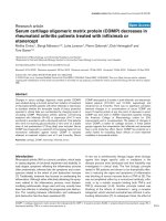
Available online />Figure 1
Modulation of adjuvant arthritis development after nasal administration of matrix metalloproteinase (MMP) peptides prior to adjuvant arthritis. The
nasal treatment was repeated four times (arrows). Data shown are mean arthritis scores ± standard error of the mean of two experiments (n = 10
rats per group). * P < 0.05 (two-tailed Mann–Whitney test) compared with the ovalbumin peptide (OVA
323–339
)-treated group.
most successful with high-affinity MHC binders [16].
However, the present study shows that the strongest
immunomodulatory peptide, MMP-10
329–343
, was a weak
MHC class II RT1.B
L
binder.
The upregulation of MMP-3 and its pathogenic role in
arthritis has been shown in numerous reports, while only
a few reports describe the presence of MMP-10 and
MMP-16 in the synovium of RA patients [17,18].
Although MMP-10 and MMP-16 have been suggested to
be involved in connective tissue/bone remodeling around
prostheses [19,20], their role in arthritis is less clear.
Surprisingly, peptides derived from MMP-10 and MMP-
16, but not from MMP-3, can alter the course of AA after
nasal administration. The observed opposite effect of
nasal therapy using MMP-10 peptide furthermore illus-
trates that we seem to target the proper cell population
to interfere in arthritis, but that the desired disease
Arthritis Research Vol 4 No 4 van Bilsen et al.
Page 4 of 8
(page number not for citation purposes)
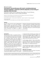
Figure 2
Modulation of adjuvant arthritis (AA) development after nasal treatment with matrix metalloproteinase (MMP) peptides after AA induction. The nasal
treatment was started at day +11, after immunization with Mycobacterium tuberculosis, when more than 50% of the animals showed weight loss
(i.e. at the onset of clinical arthritis). The nasal treatment was repeated four times (arrows). Data shown are mean arthritis scores ± standard error
of the mean of five rats (MMP-10 and MMP-16 peptide groups) or 10 rats (MMP-3 peptide group) per group. OVA
323–339
, ovalbumin peptide.
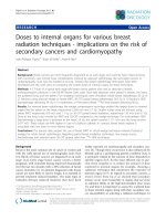
Figure 3
Popliteal lymph node cell proliferative responses after nasal matrix metalloproteinase (MMP) peptide administration prior to (left) or after (right)
adjuvant arthritis (AA) induction. Responses were measured 35 days after AA induction. The irrelevant peptides used were MBP
72–85
or ovalbumin
peptide, OVA
323–339
. Data are expressed as the mean stimulation index (SI) of three or four organs ± standard error of the mean. * P < 0.05.
inhibitory effect strongly depends on the timing of T-cell
modulation.
Other studies have also shown that mucosal therapy
might in some cases induce or even exacerbate T helper 1
cell autoimmunity [21–23]. Moreover, the critical aspect of
proper timing has also been described for cytokine thera-
pies as shown by both disease inhibition and exacerbation
after in vivo administration of IFN-γ, IL-2 or IL-12 in experi-
mental arthritis models [24–26].
In the present study, we stated that the improvement of
arthritis symptoms after MMP-10 peptide pretreatment
coincided with a decreased proliferative response to the
critical T-cell epitope for the induction of AA, mycobacterial
hsp65
178–186
. Although the proliferative response to this
epitope is difficult to measure in a polyclonal lymph node
cell population, our results suggest that MMP-10 peptide
pretreatment inhibits the response to the arthritogenic
epitope via bystander suppression. Immunotherapy using
agents that induce T-cell-mediated bystander suppression
makes it unnecessary to identify the self-antigens involved
in the initiation of the arthritis process, but makes it possi-
ble to exploit spreading epitopes or other self-antigens that
become available during the arthritis process for disease
intervention.
Conclusion
The present study shows the possible usefulness of MMP
peptide immunotherapy in arthritis, based on modulation
of the T-cell response. However, a clear understanding of
proper timing of MMP peptide-based immunotherapy and
the consequences for T-cell modulation will be crucial for
the development of such therapies.
Acknowledgements
The authors thank MC Grosfeld for the peptide–MHC binding assays.
The research by Dr MHM Wauben was made possible by a fellowship
of the Royal Netherlands Academy of Arts and Sciences.
References
1. Breedveld FC: New insights in the pathogenesis of rheuma-
toid arthritis. J Rheumatol 1998, 53(suppl):S3-S7.
2. Panayi GS, Lanchbury S, Kingsley GH: The importance of the T
cell in initiating and maintaining the chronic synovitis of
rheumatoid arthritis. Arthritis Rheum 1992, 35:729-735.
3. Barchan D, Souroujon MC, Im SH, Antozzi C, Fuchs S: Antigen-
specific modulation of experimental myasthenia gravis: nasal
tolerization with recombinant fragments of the human acetyl-
choline receptor alpha-subunit. Proc Natl Acad Sci USA 1999,
96:8086-8091.
4. Karachunski PI, Ostlie NS, Okita DK, Conti-Fine BM: Prevention
of experimental myasthenia gravis by nasal administration of
synthetic acetylcholine receptor T epitope sequences. J Clin
Invest 1997, 100:3027-3035.
5. Tian J, Atkinson MA, Clare-Salzler M, Herschenfeld A, Forsthuber
T, Lehmann PV, Kaufman DL: Nasal administration of glutamate
decarboxylase (GAD65) peptides induces Th2 responses and
prevents murine insulin-dependent diabetes. J Exp Med 1996,
183:1561-1567.
6. Dick AD, Cheng YF, Liversidge J, Forrester JV: Intranasal admin-
istration of retinal antigens suppresses retinal antigen-
induced experimental autoimmune uveoretinitis. Immunology
1994, 82:625-631.
7. Metzler B, Wraith DC: Inhibition of experimental autoimmune
encephalomyelitis by inhalation but not oral administration of
the encephalitogenic peptide: influence of MHC binding affin-
ity. Int Immunol 1993, 5:1159-1165.
8. Laliotou B, Liversidge J, Forrester JV, Dick AD: Interphotorecep-
tor retinoid binding protein is a potent tolerogen in Lewis rat:
suppression of experimental autoimmune uveoretinitis is
retinal antigen specific. Br J Ophthalmol 1997, 81:61-67.
9. Prakken BJ, van der Zee R, Anderton SM, van Kooten PJ, Kuis W,
van Eden W: Peptide-induced nasal tolerance for a mycobac-
terial heat shock protein 60 T cell epitope in rats suppresses
both adjuvant arthritis and nonmicrobially induced experi-
mental arthritis. Proc Natl Acad Sci USA 1997, 94:3284-3289.
10. Wauben MH: Immunological mechanisms involved in experi-
mental peptide immunotherapy of T-cell-mediated diseases.
Crit Rev Immunol 2000, 20:451-469.
11. Myers LK, Seyer JM, Stuart JM, Kang AH: Suppression of
murine collagen-induced arthritis by nasal administration of
collagen. Immunology 1997, 90:161-164.
12. Joosten LA, Coenen-de Roo CJ, Helsen MM, Lubberts E, Boots
AM, van den Berg WB, Miltenburg AM: Induction of tolerance
with intranasal administration of human cartilage gp-39 in
DBA/1 mice: amelioration of clinical, histologic, and radiologic
signs of type II collagen-induced arthritis. Arthritis Rheum
2000, 43:645-655.
13. Staines NA, Harper N, Ward FJ, Malmström V, Holmdahl R,
Bansal S: Mucosal tolerance and suppression of collagen-
induced arthritis (CIA) induced by nasal inhalation of syn-
thetic peptide 184-198 of bovine type II collagen (CII)
expressing a dominant T cell epitope. Clin Exp Immunol 1996,
103:368-375.
14. van Bilsen JHM, Wagenaar-Hilbers JPA, Boot EPJ, van Eden W,
Wauben MHM: Searching for the cartilage-associated mimicry
epitope in adjuvant arthritis. Autoimmunity 2002, in press.
15. Prakken B, Wauben M, Genini D, Samodal R, Barnett J, Mendivil
A, Leoni L, Albani S: Artificial antigen-presenting cells as tool
to exploit the immune ‘synapse’. Nat Med 2000, 6:406-410.
16. Anderton S, Burkhart C, Metzler B, Wraith D: Mechanisms of
central and peripheral T-cell tolerance: lessons from experi-
mental models of multiple sclerosis. Immunol Rev 1999, 169:
123-137.
17. Konttinen YT, Ainola M, Valleala H, Ma J, Ida H, Mandelin J, Kinne
RW, Santavirta S, Sorsa T, Lopez-Otin C, Takagi M: Analysis of
16 different matrix metalloproteinases (MMP-1 to MMP-20) in
the synovial membrane: different profiles in trauma and
rheumatoid arthritis. Ann Rheum Dis 1999, 58:691-697.
18. Pap T, Shigeyama Y, Kuchen S, Fernihough JK, Simmen B, Gay
RE, Billingham M, Gay S: Differential expression pattern of
membrane-type matrix metalloproteinases in rheumatoid
arthritis. Arthritis Rheum 2000, 43:1226-1232.
19. Takei I, Takagi M, Santavirta S, Ida H, Ishii M, Ogino T, Ainola M,
Konttinen YT: Messenger ribonucleic acid expression of 16
matrix metalloproteinases in bone-implant interface tissues
of loose artificial hip joints. J Biomed Mater Res 2000, 52:613-
620.
20. Ishiguro N, Ito T, Kurokouchi K, Iwahori Y, Nagaya I, Hasegawa Y,
Iwata H: mRNA expression of matrix metalloproteinases and
tissue inhibitors of metalloproteinase in interface tissue
around implants in loosening total hip arthroplasty. J Biomed
Mater Res 1996, 32:611-617.
21. Blanas E, Carbone FR, Allison J, Miller JFAP, Heath WR: Induc-
tion of autoimmune diabetes by oral administration of
autoantigen. Science 1996, 274:1707-1709.
22. Lens JW, van den Berg WB, van de Putte LB, van den Bersselaar
L: Flare-up of antigen-induced arthritis in mice after challenge
with oral antigen. Clin Exp Immunol 1984, 58:364-371.
23. Weinberg AD, Whitham R, Swain SL, Morrison WJ, Wyrick G,
Hoy C, Vandenbark AA, Offner H: Transforming growth factor-b
enhances the in vivo effector function and memory phenotype
of antigen-specific T helper cells in experimental autoimmune
encephalomyelitis. J Immunol 1992, 148:2109-2117.
24. Jacob CO, Holoshitz J, van der Meide P, Strober S, McDevitt HO:
Heterogeneous effects of IFN-gamma in adjuvant arthritis.
J Immunol 1989, 142:1500-1505.
25. Joosten LA, Lubberts E, Helsen MM, van den Berg WB: Dual role
of IL-12 in early and late stages of murine collagen type II
arthritis. J Immunol 1997, 159:4094-4102.
Available online />Page 5 of 8
(page number not for citation purposes)
26. Thornton S, Boivin GP, Kim KN, Finkelman FD, Hirsch R: Hetero-
geneous effects of IL-2 on collagen-induced arthritis.
J Immunol 2000, 165:1557-1563.
Correspondence
Marca HM Wauben, Department of Infectious Diseases and Immunology,
Faculty of Veterinary Medicine, Utrecht University, P.O. Box 80.165,
3508 TD Utrecht, The Netherlands. Tel: +31 30 253 1872; fax: +31
30 253 355; e-mail:
Supplementary material
Supplementary Materials and methods
Peptides
Guinea pig myelin basic protein (MBP)
72–85
(QKSQRSQ-
DENPV), Mycobacterium tuberculosis hsp65
178–186
(SNTFGLQLE) and chicken OVA
323–339
(ISQAVHAA-
HAEINEAGR) peptides were obtained from Isogen Bio-
science (Maarn, The Netherlands). Rat MMP-3
444–458
(FLYFFSGSSQLEFDP), rat MMP-10
329–343
(SAFWP-
SLPSGLDAAY) and human MMP-16
539–553
(VKEGHSP-
PDDVDIVI) were synthesized via automated simultaneous
multiple peptide synthesis [S1]. Peptides were obtained
as C-terminal amides after cleavage with 90–95% trifluo-
roacetic acid/scavenger cocktails. Peptides were analyzed
by reversed-phase high-performance liquid chromatogra-
phy, and checked via electrospray ionization mass spec-
trometry on an ion-trap mass spectrometer (LCQ;
Thermoquest, Breda, The Netherlands). Peptide MBP
72–85
was biotinylated during synthesis and used as the marker
peptide in the RT1.B
L
MHC–peptide binding assays.
Animals
Male inbred Lewis rats were obtained from Maastricht
University (Maastricht, The Netherlands). Rats were
6–8 weeks old at the start of each experiment. This study
was approved by the Dutch Committee of Animal
Experiments.
Induction and clinical evaluation of AA
AA was induced by intradermal injection at the base of the
tail with 0.1 ml of 5 mg/ml heat-killed M. tuberculosis
H37RA in incomplete Freund’s adjuvant (DIFCO Labora-
tories, Detroit, MI, USA). Rats were examined for clinical
signs of arthritis in a blinded set-up. The severity of arthri-
tis was scored by grading each paw from 0 to 4 based on
erythema, swelling and deformity of the joints, resulting in
a maximum score of 16.
MHC class II–peptide binding assay
Lewis rat MHC class II molecules, RT1.B
L
, were purified
from the Z1a T cell clone through affinity chromatography
using the OX6 monoclonal antibody, as described previ-
ously [S2]. Affinity-purified RT1.B
L
MHC molecules were
solubilized in 1% n-β-octyl glucopyranoside (Sigma-
Aldrich Chemie BV, Zwijndrecht, The Netherlands). The
MHC class II–peptide binding studies were performed
using the competitive binding assay as described previ-
ously [S2]. Briefly, 3 µM RT1.B
L
was incubated with
400 nM biotinylated marker peptide and a concentration
range (0–256 µM) of unlabeled competitor peptide at
room temperature for 40 hours at pH 5 in the presence of
a protease inhibitor mix.
The MHC–peptide mixtures were analyzed via nonreducing
SDS-PAGE, followed by western blotting (Hybond-ECL;
Amersham, Pharmacia Biotech Benelux, Roosendaal, The
Netherlands). Biotinylated peptides were visualized on
hyperfilm (Amersham, Pharmacia Biotech Benelux,
Roosendaal, The Netherlands), through enhanced chemi-
luminescence (Western Blot ECL kit; Amersham, Pharma-
cia Biotech Benelux). Spots on the films were quantified
by Molecular Analyst Software version 1.5 (Bio-Rad Labo-
ratories B.V., Veenendaal, The Netherlands). The relative
MHC binding affinity (IC
50
value) is expressed as the con-
centration range of competitor peptide (µM) resulting in
50% inhibition of the MHC binding of the marker peptide.
Ex vivo
proliferation assays
At different time points after AA induction, PLNs were iso-
lated and single cell suspensions were cultured in 96-well
flat-bottomed microtiter plates (2 × 10
5
cells/well in tripli-
cate) in the absence or presence of peptide (1 µg/ml or
10 µg/ml). After 3 days of culturing, proliferation was
determined by overnight incorporation of [
3
H]thymidine
(0.4 µCi/well; specific activity, 1 Ci/mmol; Amersham
Pharmacia Biotech Benelux). Proliferation was expressed
as the stimulation index (counts per minute of cells cul-
tured with peptide divided by the counts per minute of
cells cultured in medium only).
Statistics
Statistical evaluation was performed using GraphPad
Prism version 3.0 (GraphPad Software, San Diego, CA,
USA). To evaluate the effect of each peptide in the DTH
reaction, the paired, two-tailed Student’s t test was per-
formed on the mean difference in ear swelling between
the peptide-treated ear and the PBS-treated ear. The two-
tailed Mann–Whitney U test was performed to compare
the mean differences in DTH reactions between naïve and
arthritic rats. For the comparison of arthritis scores and
proliferative responses between the different groups, a
two-tailed Mann–Whitney U test was used. P < 0.05 was
considered statistically significant.
Supplementary Results
Natural occurrence of MMP peptide-specific T cells
during AA
The PLN responses were analyzed in lymphocyte prolifera-
tion assays at day 0 (naïve animals), day 10 (before clinical
onset of disease), day 14 (onset of disease), day 21 (overt
Arthritis Research Vol 4 No 4 van Bilsen et al.
Page 6 of 8
(page number not for citation purposes)
disease) and day 35 (no or limited clinical disease) after
disease induction with M. tuberculosis/incomplete
Freund’s adjuvant. Supplementary Figure 1 shows the pro-
liferative responses to the MMP peptides in a representa-
tive experiment. As can be seen, only very low proliferative
responses were detectable, which tend to rise during the
course of the disease.
MHC binding affinity of MMP peptides
We previously selected the MMP peptides based on their
putative binding to Lewis rat MHC class II RT1.B
L
[14]. In
the present study, we determined the binding affinity of
the MMP peptides for RT1.B
L
in a competitive
peptide–MHC binding assay.
The relative binding affinities of the MMP epitopes are
depicted in Supplementary Figure 2. The MMP epitopes
could be ranked into three broadly defined categories:
MMP-3
244–258
is a good RT1.B
L
binder (IC
50
≤ 8 µM),
MMP-16
539–553
is an intermediate RT1.B
L
binder (IC
50
=
64–128 µM), and MMP-10
329–343
is a poor RT1.B
L
binder
(IC
50
= 128–256 µM).
Nasal peptide administration prior to AA induction
Nasal administration of MMP-10
329–343
and MMP-16
539–553
significantly reduced the severity of AA. Weight loss (a sen-
sitive objective measure of physical well-being) was ana-
lyzed as an addition parameter (Supplementary Figure 3).
The mean body weight demonstrated that MMP-3
444–458
-
treated rats showed the same weight loss as the
OVA
323–339
-treated group, which is consistent with the com-
parable AA severity. In contrast, MMP-10
329–343
-treated rats
showed a clear reduction in weight loss as compared with
the OVA
323–339
-treated group, consistent with the lower AA
scores. MMP-16
539–553
-treated rats showed no difference in
weight loss as compared with the OVA
323–339
control group.
Nasal peptide administration after AA induction
MMP-10 peptide treatment after AA induction aggravated
the arthritic symptoms of the Lewis rats. These findings
were consistent with the lower mean body weight in the
MMP-10 peptide-treated group compared with the
OVA
323–339
control group (Supplementary Figure 4).
MMP-3 peptide-treated rats and MMP-16 peptide-treated
rats showed no differences in body weight and clinical
scores as compared with the OVA
323–339
-treated group.
Available online />Page 7 of 8
(page number not for citation purposes)
Supplementary Figure 1
Monitoring of proliferative responses to matrix metalloproteinase (MMP) peptides during adjuvant arthritis in popliteal lymph node cells (PLNC) of
Lewis rats. All organs were tested separately. Data are expressed as the mean of the stimulation index (SI) of three to four rats ± standard error of
the mean. At day 0, PLNC and inguinal lymph node cells of n = 4 rats were pooled and subsequently tested.
Supplementary Figure 2
Binding affinity of matrix metalloproteinase (MMP) peptides to purified
RT1.B
L
. The binding affinity of the competitor peptides (MMP
peptides) was tested in a competitive MHC class II peptide binding
assay in a dose range of 0–256 µM. * IC
50
, concentration of
competitor peptide (µM) resulting in 50% inhibition of the binding of
marker peptide to RT1.B
L
, as evaluated by Molecular Analyst Software
(see Supplementary Materials and methods).
Supplementary References
S1. van der Zee R, Anderton SM, Buskens CAF, Alonso de Velasco E,
van Eden W: Heat shock protein T-cell epitopes as immuno-
genic carriers in subunit vaccines. In Peptides 1994. Proceed-
ings of the Twenty-Third European Peptide Symposium,
September 4–10, 1994, Braga, Portugal. Edited by Maia HLS.
Leiden, The Netherlands: ESCOM; 1994:841-842.
S2. Joosten I, Wauben MHM, Holewijn MC, Reske K, Pedersen LO,
Roosenboom CF, Hensen EJ, van Eden W, Buus S: Direct
binding of autoimmune disease related T cell epitopes to
purified Lewis rat MHC class II molecules. Int Immunol 1994,
6:751-759.
Arthritis Research Vol 4 No 4 van Bilsen et al.
Page 8 of 8
(page number not for citation purposes)
Supplementary Figure 3
Mean body weight of rats treated nasally with matrix metalloproteinase (MMP) peptide prior to adjuvant arthritis (AA) induction. The mean body
weight is depicted as the percentage of the body weight at the time of AA induction. Data shown are the percentage of the body weight of two
experiments (n = 10 rats per group) ± standard error of the mean. OVA
323–339
, ovalbumin peptide.
Supplementary Figure 4
Mean body weight of rats treated nasally with matrix metalloproteinase (MMP) peptides after induction of adjuvant arthritis (AA). The mean body
weight is depicted as the percentage of the body weight at the time of AA induction. Data shown are percentage body weight ± standard error of
the mean of two experiments with five rats (MMP-10 and MMP-16 peptide groups) or 10 rats (MMP-3 peptide group) per group. OVA
323–339
,
ovalbumin peptide.