Báo cáo khoa học: "Hepatic splenosis mimicking HCC in a patient with hepatitis C liver cirrhosis and mildly raised alpha feto protein; the important role of explorative laparoscopy" potx
Bạn đang xem bản rút gọn của tài liệu. Xem và tải ngay bản đầy đủ của tài liệu tại đây (392.03 KB, 4 trang )
BioMed Central
Page 1 of 4
(page number not for citation purposes)
World Journal of Surgical Oncology
Open Access
Case report
Hepatic splenosis mimicking HCC in a patient with hepatitis C liver
cirrhosis and mildly raised alpha feto protein; the important role of
explorative laparoscopy
M Abu Hilal*, A Harb, B Zeidan, B Steadman, JN Primrose and NW Pearce
Address: Hepatobiliary-Pancreatic and Laparoscopic Surgical Unit, Southampton University Hospital, Southampton, SO16 6YD, UK
Email: M Abu Hilal* - ; A Harb - ; B Zeidan - ;
B Steadman - ; JN Primrose - ; NW Pearce -
* Corresponding author
Abstract
Background: Splenosis is a heterotropic implantation of splenic fragments onto exposed
vascularised peritoneal and intrathoracic surfaces, following splenic injury or elective splenectomy.
Case presentation: A 60 year old cirrhotic patient was referred to us with a hepatic mass,
suspected to be HCC in a cirrhotic liver. A computerized tomography scan (CT) demonstrated a
cirrhotic liver with a 2 × 2.7 cm focal hypervascular nodule, lying peripherally at the junction of
segment 7 and 8. Diagnostic laparoscopy demonstrated a 3 cm exofitic dark brown splenunculus
attached to the diaphragm and indenting the surface of segment 7 of the liver. The lesion was easily
resected laparoscopically and shaved from the live surface with no need for a liver resection. The
histopathological assessment confirmed the diagnosis of splenunculus, with no evidence of
neoplasia.
Conclusion: Hepatic splenosis is not a rare event and should be suspected in patients with a
history of splenic trauma or splenectomy. Correct diagnosis is essential and will determine
subsequent management plans. In doubtful cases laparoscopic investigation can offere essential
information and should be part of the standard protocol for investigating suspected splenosis.
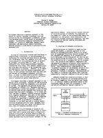
Background
Splenosis is a heterotropic implantation of splenic frag-
ments onto exposed vascularised peritoneal and intratho-
racic surfaces, following splenic injury or elective
splenectomy.
There are a few reported cases of hepatic splenosis in the
English literature, which has usually a challenging and
difficult differential diagnosis with hepatic adenoma, hae-
mangioma, focal nodulal hyperplasia, lymphoma and
hepatocellular carcinoma [1-4]. Excluding a diagnosis of
Hepato cellular carcinoma (HCC) has proven difficult
despite different suggested radiological investigation
methods [5,6], particularly in patients with chronic liver
disease [7-11], when HCC is an expected development
and a more likely underlying pathology than is intrahe-
patic splenosis.
Laparoscopic exploration provides a port of minimally
invasive entry for the visualisation of suspect masses, and
allows access for potential subsequent biopsy or resection.
To our knowledge, this is the first case were explorative
Published: 5 January 2009
World Journal of Surgical Oncology 2009, 7:1 doi:10.1186/1477-7819-7-1
Received: 28 May 2008
Accepted: 5 January 2009
This article is available from: />© 2009 Abu Hilal et al; licensee BioMed Central Ltd.
This is an Open Access article distributed under the terms of the Creative Commons Attribution License ( />),
which permits unrestricted use, distribution, and reproduction in any medium, provided the original work is properly cited.
World Journal of Surgical Oncology 2009, 7:1 />Page 2 of 4
(page number not for citation purposes)
laparoscopy has been an essential tool to confirm the
diagnosis of splenosis and rule out the doubt of malig-
nancy. This had a significant positive impact on this
patient's management, avoiding unneccassary laparot-
omy or'/and surgical resection in a high-risk patient.
Hepatic splenosis is not a rare event and should be sus-
pected in patients with a history of splenic trauma or
splenectomy. Correct diagnosis is essential and will deter-
mine subsequent management plans. In doubtful cases;
laparoscopic investigation can offer essential information
and should be part of the standard protocol for investigat-
ing suspected splenosis.
Case presentation
A 60 year old cirrhotic patient was referred to us with a
hepatic mass, suspected to be HCC in a cirrhotic liver. The
patient was diagnosed with liver cirrhosis in December
2003, secondary to Hepatitis C infection after receiving
blood transfusions at splenectomy for a ruptured spleen
46 years ago.
Serial blood tests showed sudden derangement in his liver
function, and a mild rise in alpha-feto protein levels. Clin-
ically, he complained of non-specific flu-like symptoms
and also reported recent weight loss and reduced appetite.
On examination he appeared jaundiced, there were signs
of clubbing and on examination of his abdomen, there
was a mild degree of ascites and the liver edge was palpa-
ble.
He was a persistent alcoholic and was prone to frequent
binging. A computerized tomography scan (CT) demon-
strated a cirrhotic liver with a 2 × 2.7 cm focal hypervascu-
lar nodule, lying peripherally at the junction of segment 7
and 8 (figure 1). There was an increased enhancement in
the venous phase scans, and the picture was very suspi-
cious for a focal hepatoma.
A double contrast MR study, using Gadolinium and reso-
vist contrasts, confirmed the presence of a solitary 2 × 2.5
cm mass with features suggestive of hepatoma, lying
within segment 7 in a subcapsular position (figures 2 &3).
Although a similar characteristics 4.5 cm mass was also
noted in the left upper quadrant; malignancy couldn't be
excluded. The multi-disciplinary meeting advised an
explorative laparoscopy for further investigation of this
lesion, and better assessment of the extent of the disease.
Diagnostic laparoscopy demonstrated a 3 cm exofitic dark
brown splenunculus attached to the diaphragm and
indenting the surface of segment 7 of the liver. Multiple
other typical looking splenunculi were found. Intraopera-
tive ultrasound was performed and excluded any other
lesions within the liver or surrounding tissues.
The lesion was easily resected laparoscopically and shaved
from the live surface with no need for a liver resection. The
histopathological assessment confirmed the diagnosis of
splenunculus, with no evidence of neoplasia.
The patient finished a 24 weeks course of pegylated inter-
feron α and ribavirin as part of his hepatitis C treatment
regime post operatively. At two years his follow up
CT scanFigure 1
CT scan. (A) Axial IV contrast enhanced CT. Arterial phase image showing a 2 × 2.7 cm hypervascular, subcapsular nodule in
segment VII of the liver. (B) Portal venous phase image.
World Journal of Surgical Oncology 2009, 7:1 />Page 3 of 4
(page number not for citation purposes)
showed no radiologic evidence of HCC, and his LFTs and
AFP were within normal limits.
Discussion
Splenosis is the heterotropic implantation of splenic frag-
ments onto exposed vascularised peritoneal and intratho-
racic surfaces, following splenic injury or elective
splenectomy. This can occur anywhere within the abdom-
inal cavity and the resultant splenunculus will receive its
blood by parasitizing the surrounding tissue.
There are few previous reports of hepatic splenosis mim-
icking hepatocellular carcinoma [10,12,13]. In most
cases, correct diagnosis was only possible on histological
examination after a laparotomy and open liver resection
[10,14]. A missed diagnosis of hepatic splenosis can have
a significant negative impact on patient's management
[15]. Interestingly, in all cases a history of post-traumatic
splenectomy was reported and all patients were known to
have an underlying chronic liver disease [10,16,17]. There
are no typical radiological features of intrahepatic spleno-
sis and it is usually difficult to distinguish this condition
from other liver tumors. In the presence of chronic liver
disease, although mild but raised tumoral markers and
strong suspicion of HCC on clinical ground, establishing
the correct diagnosis can prove to be difficult.
Distinguishing the nature of a hepatic mass is important
because it significantly alters patient management. In this
case, if a diagnosis of HCC was confirmed, this patient
would be suitable for resection (Child-Pugh class B) or for
a liver transplant, satisfying the Milan criteria. Liver cir-
rhosis, having recent LFT derangement and with the above
radiological picture made HCC strongly suspected. How-
ever, the pervious history of traumatic splenic rupture and
the presence of multiple splenunculi within the abdomi-
Arterial (A) and portal venous phase (B) of IV Gadlinium enhanced axial MRI images demonstrating a solitary 2 cm hypervascu-lar nodule in segment VII (Arrow)Figure 2
Arterial (A) and portal venous phase (B) of IV Gadlinium enhanced axial MRI images demonstrating a solitary
2 cm hypervascular nodule in segment VII (Arrow). A 4.5 cm nodule with similar enhancement characteristics is also
noted in the left upper quadrant (Arrow).
Post Resovet Axial MRI imagesFigure 3
Post Resovet Axial MRI images. The segment VII lesion
demonstrates higher signal than the background, reflecting a
relative lack of functioning hepatocytes.
World Journal of Surgical Oncology 2009, 7:1 />Page 4 of 4
(page number not for citation purposes)
nal cavity suggested that the best way to proceed would be
for a laparoscopic exploration and a secure diffinition of
the lesions nature before planning for future manage-
ment.
Laparoscopy was sufficient in confirming diagnosis of
splenosis, as well as excluding coexistent malignancy. This
had a significant impact of clinical plans and patients
management. It is already recognized that laparoscopy
provides a port of minimally invasive entry for the visual-
isation of suspect masses, and allows access for potential
subsequent biopsy or resection.
The abnormal liver function behavior in this case can be
explained by an active hepatitis C process, which have
improved following further anti viral treatment. Yet, a low
threshold for HCC is a must with any similar scenario of
suspicious liver function tests and radiological findings.
Laparoscopic resection of symptomatic or suspicious sple-
nosis is a minimally invasive and feasible procedure. This
was reported to be a successful diagnostic and interven-
tional tool even in laparoscopically challenging scenarios
involving the pancreas [18,19].
To the best of our knowledge, this is the first case where
laparoscopy has been the main tool in difining the correct
diagnosis in a case of splenosis, suspected to be an HCC
on radiological investingations and strong clinical bases.
We therefore propose that laparoscopic investigation
should be part of a new approach for investigating suspect
intrahepatic masses.
Conclusion
Hepatic splenosis is not a rare event and should be consid-
ered with the differential diagnosis in case of suspected
lesions especially in patients who had previous splenec-
tomy. Correct diagnosis is essential and can significantly
influence patient management. We propose that laparo-
scopic investigation should be part of a new protocol for
confirming the diagnosis of suspected intrahepatic
masses.
Consent
Written informed consent was obtained from the patient
for publication of this case report and accompanying
images. A copy of the written consent is available for
review by the Editor-in-Chief of this journal.
Competing interests
The authors declare that they have no competing interests.
Authors' contributions
MAH wrote the paper. AH was responsible for literature
review, medline search and wrote the first draft. BZ wrote
the case history and collected all clinical information. JNP
reviewed the article and made suggestions. NWP was the
surgeon, reviewed the paper and made suggestions. BS
was the radiologist who selected the images and com-
mented on the manuscript.
References
1. Di Costanzo GG, Picciotto FP, Marsilia GM, Ascione A: Hepatic
splenosis misinterpreted as hepatocellular carcinoma in cir-
rhotic patients referred for liver transplantation: report of
two cases. Liver Transpl 2004, 10:706-709.
2. Nakata Y, Yoshida H, Shiono T, Asai S, Araki T: [Intrahepatic sple-
nosis: a case report]. Nippon Igaku Hoshasen Gakkai Zasshi 2003,
63:111-113.
3. Mathurin J, Lallemand D: Splenosis simulating an abdominal
lymphoma. Pediatr Radiol 1990, 21:69-70.
4. Gruen DR, Gollub MJ: Intrahepatic splenosis mimicking hepatic
adenoma. AJR Am J Roentgenol 1997, 168:725-726.
5. De VS, Van SW, Aerts R, Van HH, Van BD, Van HL: Intrahepatic
splenosis: imaging features. Abdom Imaging 2000, 25:187-189.
6. Vento JA, Peng F, Spencer RP, Ramsey WH: Massive and widely
distributed splenosis. Clin Nucl Med 1999, 24:845-846.
7. Kondo M, Okazaki H, Takai K, Nishikawa J, Ohta H, Uekusa T, Yosh-
ida H, Tanaka K: Intrahepatic splenosis in a patient with
chronic hepatitis C. J Gastroenterol 2004, 39:1013-1015.
8. Di Costanzo GG, Picciotto FP, Marsilia GM, Ascione A: Hepatic
splenosis misinterpreted as hepatocellular carcinoma in cir-
rhotic patients referred for liver transplantation: report of
two cases. Liver Transpl 2004, 10:706-709.
9. Kim KA, Park CM, Kim CH, Choi SY, Park SW, Kang EY, Seol HY,
Cha IH: An interesting hepatic mass: splenosis mimicking a
hepatocellular carcinoma (2003:9b). Eur Radiol 2003,
13:2713-2715.
10. Lee JB, Ryu KW, Song TJ, Suh SO, Kim YC, Koo BH, Choi SY:
Hepatic splenosis diagnosed as hepatocellular carcinoma:
report of a case. Surg Today 2002, 32:180-182.
11. Galloro P, Marsilia GM, Nappi O: [Hepatic splenosis diagnosed
by fine-needle cytology]. Pathologica
2003, 95:57-59.
12. Galloro P, Marsilia GM, Nappi O: [Hepatic splenosis diagnosed
by fine-needle cytology]. Pathologica 2003, 95:57-59.
13. Di Costanzo GG, Picciotto FP, Marsilia GM, Ascione A: Hepatic
splenosis misinterpreted as hepatocellular carcinoma in cir-
rhotic patients referred for liver transplantation: report of
two cases. Liver Transpl 2004, 10:706-709.
14. Khosravi MR, Margulies DR, Alsabeh R, Nissen N, Phillips EH, Mor-
genstern L: Consider the diagnosis of splenosis for soft tissue
masses long after any splenic injury. Am Surg 2004, 70:967-970.
15. Di Costanzo GG, Picciotto FP, Marsilia GM, Ascione A: Hepatic
splenosis misinterpreted as hepatocellular carcinoma in cir-
rhotic patients referred for liver transplantation: report of
two cases. Liver Transpl 2004, 10:706-709.
16. Di Costanzo GG, Picciotto FP, Marsilia GM, Ascione A: Hepatic
splenosis misinterpreted as hepatocellular carcinoma in cir-
rhotic patients referred for liver transplantation: report of
two cases. Liver Transpl 2004, 10:706-709.
17. Khosravi MR, Margulies DR, Alsabeh R, Nissen N, Phillips EH, Mor-
genstern L: Consider the diagnosis of splenosis for soft tissue
masses long after any splenic injury. Am Surg 2004, 70:967-970.
18. Fiamingo P, Veroux M, Da RA, Guerriero S, Pariset S, Buffone A,
Tedeschi U: A rare diagnosis for a pancreatic mass: splenosis.
J Gastrointest Surg 2004, 8:915-916.
19. Barbaros U, Dinccag A, Kabul E: Minimally invasive surgery in the
treatment of splenosis. Surg Laparosc Endosc Percutan Tech 2006,
16:187-189.