Báo cáo khoa học: "Interstitial pneumonitis following intrapleural chemotherapy" docx
Bạn đang xem bản rút gọn của tài liệu. Xem và tải ngay bản đầy đủ của tài liệu tại đây (952.07 KB, 5 trang )
BioMed Central
Page 1 of 5
(page number not for citation purposes)
World Journal of Surgical Oncology
Open Access
Case report
Interstitial pneumonitis following intrapleural chemotherapy
Luis Zappa
1
, Renaldo Savady
1
, Gary N Humphries
2
and Paul H Sugarbaker*
1
Address:
1
Program in Peritoneal Surface Malignancy, Washington Cancer Institute, Washington, DC, USA and
2
Grand Valley Medical Specialists,
Grand Rapids, MI, USA
Email: Luis Zappa - ; Renaldo Savady - ; Gary N Humphries - ;
Paul H Sugarbaker* -
* Corresponding author
Abstract
Background: Mucinous neoplasms within the abdomen may disseminate by direct extension
through the diaphragm to involve the pleural space. Treatment of this condition is by parietal and
visceral pleurectomy followed by hyperthermic intrapleural chemotherapy.
Case presentation: In this case report a patient developed persistent right upper lobe interstitial
pneumonitis and progressive parenchymal fibrosis following intrapleural chemotherapy treatment
with mitomycin C and doxrubicin. The condition persisted until death 28 months later. Death was
from progressive intraabdominal disease with intestinal obstruction and sepsis associated with
progressive pulmonary parenchymal disease. The right pleural space disease did not recur.
Conclusion: This manuscript is the first case report describing interstitial pneumonitis and lung
fibrosis following intrapleural chemotherapy. Since pulmonary toxicity from chemotherapy is a
dose-dependent phenomenon, dose reduction of intrapleural as compared to intraperitoneal
hyperthermic chemotherapy may be necessary.
Background
Pseudomyxoma peritonei is the clinical syndrome of
mucinous ascites associated with diffuse peritoneal
implants of mucin-producing neoplasms [1,2]. It shows
varying amounts of invasiveness and histologically the
neoplasms producing pseudomyxoma peritonei can be
classified as low grade and high grade mucinous carcino-
mas [3,4]. Appendiceal epithelial cancers represent 1% of
colorectal cancers in the USA [5]. Almost all patients with
appendiceal mucinous neoplasms have no lymphatic or
haematogenous dissemination. Intrathoracic dissemina-
tion that occurs from penetration of the diaphragm by the
intraabdominal mucinous tumor is rare but does occur in
patients with pseudomyxoma peritonei [6]. In the
absence of progressive disease in the abdomen, pleurec-
tomy and intrapleural chemotherapy have been used to
treat pleural dissemination with excellent clinical results
and low morbidity [7]. In this report we present a patient
with appendiceal mucinous neoplasm with right pleural
dissemination treated by pleurectomy and intrapleural
hyperthermic chemotherapy. Acute interstitial pneumoni-
tis was followed by a debilitating long-term fibrosis.
Case presentation
A 48-year-old woman in early 2004 developed increasing
abdominal girth associated with discomfort and pain. A
CT scan was performed and a right pelvic mass was visu-
alized. In April 2004, she underwent a debulking proce-
dure which included a right hemicolectomy and
omentectomy. Her pathology showed a high grade muci-
Published: 12 February 2009
World Journal of Surgical Oncology 2009, 7:17 doi:10.1186/1477-7819-7-17
Received: 21 October 2008
Accepted: 12 February 2009
This article is available from: />© 2009 Zappa et al; licensee BioMed Central Ltd.
This is an Open Access article distributed under the terms of the Creative Commons Attribution License ( />),
which permits unrestricted use, distribution, and reproduction in any medium, provided the original work is properly cited.
World Journal of Surgical Oncology 2009, 7:17 />Page 2 of 5
(page number not for citation purposes)
nous epithelial malignancy of the appendix with perito-
neal metastases. Because of the extent of disease the
patient was referred to the Washington Cancer Institute.
In November 2004 she had a 12-hour cytoreductive pro-
cedure with hyperthermic (41.5°C) intraoperative intra-
peritoneal mitomycin C. Early postoperative
intraperitoneal chemotherapy with 5-fluorouracil was
also used. An end ileostomy was created at the time of her
procedure to protect a low colorectal anastomosis and
reversed in 2005.
At the time of this operation at our institution, the mid-
portions of both right and left hemidiaphragms were
resected because of cancer invasion. Visual inspection
through the open hemidiaphragm of the pleural space on
the left side was clear and remained clear by CT scan. On
the right side there were approximately 50 mucinous
tumor nodules visualized within the pleural space which
were positive by histology for mucinous adenocarcinoma.
The patient remained asymptomatic.
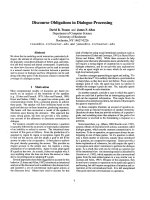
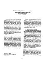
In March 2006 chest CT showed obvious progression of
disease in the right pleural space (Figure 1). She had a
right thoracotomy with complete stripping of the parietal
pleura, partial removal of the visceral pleura and hyper-
thermic (41.5°) intrapleural chemotherapy with doxoru-
bicin and mitomycin C plus intravenous 5-fluorouracil
and leucovorin for 90 minutes. Postoperatively, the
patient had an air leak requiring prolonged chest tube
drainage. Pathology reported the findings as adenomuci-
nosis [3].
In April 2006 she began to develop progressive dyspnea at
rest and increased with exertion. She was unable to take a
deep breath without coughing. Her first postoperative CT
scan in August 2006 showed minimal visceral cortical
scarring on the right with a vague peripheral right mid-
lung field opacity.
A CT-guided core needle biopsy of the right upper lobe
was performed in April 2007 which demonstrated benign
densely fibrotic pulmonary parenchyma with chronic
inflammation and entrapped alveolar spaces without evi-
dence of malignancy. The patient was placed on daily
prednisone with improvement of her respiratory symp-
toms. The symptoms remained steroid-dependent with
exacerbation on attempted steroid reduction.
In early 2008 the patient was functionally performing the
normal activities of daily living but had limited respira-
tory reserve. A persistent cough was present without asso-
ciated sputum production or fever. A chest radiograph
showed contraction and fibrosis of the right upper lobe
(Figure 2). A CT scan demonstrated a persistent interstitial
fibrosis without evidence of progression (Figure 3).
Abdominal CT showed a 1.9 cm × 2.6 cm mass anterior to
Chest CT obtained February 2006 showing pleural accumula-tion of mucinous adenocarcinomaFigure 1
Chest CT obtained February 2006 showing pleural
accumulation of mucinous adenocarcinoma.
Chest radiograph obtained April 2008, approximately 2 years after pleurectomy with hyperthermic intraoperative intrap-leural chemotherapyFigure 2
Chest radiograph obtained April 2008, approxi-
mately 2 years after pleurectomy with hyperthermic
intraoperative intrapleural chemotherapy. A persist-
ent contraction from severe fibrosis of the right upper lobe
is seen.
World Journal of Surgical Oncology 2009, 7:17 />Page 3 of 5
(page number not for citation purposes)
the superior mesenteric vein associated with an increased
CA 19-9 level compatible with progressive intraabdomi-
nal disease causing gastric outlet obstruction. No recur-
rence of disease in the right pleural space was seen. The
patient died 28 months after the pleurectomy with pro-
gressive fibrosis and infection of the right lung causing
sepsis unresponsive to intensive systemic antibiotics.
Intestinal obstruction had caused severe nutritional depri-
vation.
Discussion
It is known that chemotherapy affects lung function, but
it is still unclear to what extent hyperthermic intraopera-
tive intrapleural chemotherapy may cause postoperative
complications. Prior to this patient, intrapleural chemo-
therapy had not been associated with pneumonitis or
interstitial fibrosis [7]. An adverse effect on lung function
has been documented for many cytotoxic drugs, but the
chemotherapy-induced pathologic mechanisms of pul-
monary impairment remain unknown [8].
Verweij and colleagues prospectively studied pulmonary
interstitial pneumonitis induced by mitomycin C admin-
istered by an intravenous route [9]. These authors con-
cluded that mitomycin C-related lung toxicity is a dose-
dependent adverse side effect, occurring at cumulative
dose levels of 20 mg/m
2
or more. The incidence was esti-
mated at less than 10%. Gonzalez-Moreno and coworkers
reported a single patient with severe pulmonary intersti-
tial pneumonitis that occurred 37 days after intraperito-
neal hyperthermic mitomycin C given at a dose of 50 mg
[10]. Oxygen and prolonged oral prednisone led to a full
resolution of the symptoms and CT abnormalities after
two months of treatment. Gonzalez-Moreno commented
that the interstitial pneumonitis they documented was the
only case encountered in more than 150 similarly treated
patients. They suggested that an idiosyncratic susceptibil-
ity may occur with this unusual condition.
When systemic chemotherapy results in lung disease, radi-
ologically the pulmonary damage is always diffuse
[11,12]. A normal radiologic study does not exclude the
presence of cytotoxic injury, however, it is usually seen.
The presence of pleural effusion is unusual with chemo-
therapy-induced lung disease as a whole but it does not
exclude drug toxicity. Purely lobar or segmental density
should prompt an alternate explanation. The pulmonary
toxicity in our patient was different from prior descrip-
tions in the oncology literature in that intrapleural chem-
otherapy had caused the damage rather than systemic
chemotherapy. Also, the pulmonary damage was local-
ized to the right upper lobe rather than being diffused. We
hypothesize that more extensive surgical trauma from vis-
ceral pleurectomy at this anatomic site allowed greater
localized parenchymal exposure to the sclerotic effects of
mitomycin C and doxorubicin.
The second local-regional chemotherapy agent used in the
pleural space of this patient was low-dose doxorubicin.
This drug diluted in 3 liters of chemotherapy solution has
been shown to be well tolerated in the peritoneal cavity
and has been used in more than 400 patients [13]. How-
ever, for both intrapleural mitomycin C and for intrapleu-
ral doxorubicin, an increased risk of local-regional toxicity
may exist. As the hyperthermia lavage of the lung
progresses over 90 minutes, the intrapleural drugs are
slowly cleared into the systemic circulation. If even
minute amounts of chemotherapy enter the alveolar
spaces of the lung, this may greatly amplify the toxicities
caused by drugs present in the blood. The case report of
Gonzalez-Moreno confirmed that the systemic effects of
intraperitoneal chemotherapy can cause pulmonary inter-
stitial pneumonitis [10].
Diffuse pulmonary disease has been seen in patients
receiving chemotherapy. There may be a wide variety of
etiologies which include pulmonary infection, complica-
tions of the underlying disease and direct toxicity from
cytotoxic drugs. The clinical syndromes of pneumonitis
and fibrosis have a biphasic pathological course and are
dependent upon the dose and volume of lung exposed.
Chest CT obtained July 2007, approximately 1 year after pleurectomy with hyperthermic intraoperative intrapleural chemotherapyFigure 3
Chest CT obtained July 2007, approximately 1 year
after pleurectomy with hyperthermic intraoperative
intrapleural chemotherapy. An interstitial fibrosis is evi-
dent. Subsequent CT are largely unchanged but the patient's
symptoms persist.
World Journal of Surgical Oncology 2009, 7:17 />Page 4 of 5
(page number not for citation purposes)
The pulmonary complications following systemic chemo-
therapy can be acute in onset or may develop insidiously
months or years after treatment. Usually the clinical syn-
drome occurs 1–3 months after completion of the drug
therapy. The severity of symptoms of the acute pneumo-
nitis syndrome is dependent on the degree of pulmonary
involvement. It progresses eventually to a fibrotic phase
[12]. The mainstay of treatment has been steroids.
Although unusual, thoracic involvement by pseu-
domyxoma peritonei of appendiceal origin has been well
documented as pleural effusions or pulmonary metastasis
[7,14]. The time frame between initial diagnosis of pseu-
domyxoma peritonei and the discovery of thoracic disease
related to this syndrome ranged from less than 1 year to
nearly 15 years. The average period of time, however, is
between 2 and 6 years [7]. There are several mechanisms
which have been proposed in order to explain how neo-
plastic cells can spread to the pleural space from the peri-
toneal cavity: (1) iatrogenic perforation of the diaphragm
while a subdiaphragmatic peritonectomy is performed;
(2) incidental finding, when the pleural space is entered
as in the case presented; (3) dissemination via direct inva-
sion or through lymphovascular spaces of the diaphragm;
(4) and in a small proportion of cases, congenital or
acquired pleuroperitoneal communications which allow
neoplastic cells to reach the pleural spaces [15].
The cytologic and histologic features of the metastasis
resemble the primary appendiceal neoplasm [1,3]. As the
diagnosis of pseudomyxoma peritonei is usually made
early in the patient's history, the differential diagnosis
from the thorax should not pose a great challenge. The
presence of intracytoplasmic and extracellular mucin
should lead to the correct diagnosis. Mucinous material is
often abundant in the background of very few cancer cells.
An aspiration cytology of tumor in the pleural space may
show acellular mucin or the tumor cells may be sur-
rounded by large volumes of mucin and difficult to posi-
tively identify.
Conclusion
From our review of this patient's clinical course and our
experience with other patients who had the same proce-
dure, the causation of this interstitial fibrosis is not appar-
ent. Although parenchymal damage to the lung is well
described from chemotherapy, a localized and persistent
fibrosis of lung parenchyma as seen in this patient given
intrapleural chemotherapy has not been previously
reported. This localized pneumonitis which progressed to
fibrosis and then systemic sepsis was likely related to com-
bined systemic and local-regional toxic effects of chemo-
therapy. It is likely that visceral pleurectomy prior to the
hyperthermic intrapleural chemotherapy treatment could
result in access of small amounts of drug solution to lung
parenchyma. The multi-agent chemotherapy used for this
pleural extension of adenocarcinoma contains vesicant
drugs that could cause fibrosis to the delicate lung paren-
chyma. Theoretically, surface treatment should have no
parenchymal toxicity; however, damage to lung paren-
chyma by visceral pleurectomy may allow tissue penetra-
tion by chemotherapy. Progressive lung inflammation
and fibrosis may result in a long-term disability and con-
tribute to the demise of the patient.
Abbreviations
CT: computed tomography; CA 19-9: cancer antigen 19-9
Competing interests
The authors declare that they have no competing interests.
Consent
Written informed consent was obtained from the patient's
next of kin for publication of this case report and accom-
panying images. A copy of the written consent is available
for review by the Editor-in-Chief of this journal.
Authors' contributions
All authors made substantial contributions to the concept,
design, acquisition of data, analysis and interpretation of
data, drafting and revising the intellectual content of the
manuscript. All authors read and approved the final man-
uscript.
References
1. Sugarbaker PH, Ronnett BM, Archer A, Averbach AM, Bland R, Chang
D, Dalton RR, Ettinghausen SE, Jacquet P, Jelinek J, Koslowe P, Kur-
man RJ, Shmookler B, Stephens AD, Steves MA, Stuart OA, White S,
Zahn CM, Zoetmulder FA: Pseudomyxoma peritonei syn-
drome. Adv Surg 1996, 30:233-280.
2. Sugarbaker PH: New standard of care for appendiceal epithe-
lial neoplasms and pseudomyxoma peritonei syndrome? Lan-
cet Oncol 2006, 7:69-76.
3. Ronnett BM, Zahn CM, Kurman RJ, Kass ME, Sugarbaker PH,
Shmookler BM: Disseminated peritoneal adenomucinosis and
peritoneal mucinous carcinomatosis: A clinicopathologic
analysis of 109 cases with emphasis on distinguishing patho-
logic features, site of origin, prognosis, and relationship to
"pseudomyxoma peritonei". Am J Surg Pathol 1995,
19:1390-1408.
4. Yan H, Pestieau SR, Shmookler BM, Sugarbaker PH: Histopatho-
logic analysis in 46 patients with pseudomyxoma peritonei
syndrome: Failure vs. success with a second-look operation.
Mod Pathol 2001, 14:164-171.
5. Fann JI, Vierra M, Fisher D, Oberhelman HA Jr, Cobb L: Pseu-
domyxoma peritonei. Surg Gynecol Obstet 1993, 177:441-447.
6. Geisinger KR, Levine EA, Shen P, Bradley RF: Pleuropulmonary
involvement in pseudomyxoma peritonei: Morphologic
assessment and literature review. Am J Clin Pathol 2007,
127:135-143.
7. Pestieau SR, Esquivel J, Sugarbaker PH: Pleural extension of muci-
nous tumor in patients with pseudomyxoma peritonei syn-
drome. Ann Surg Oncol 1999, 7:199-203.
8. Abid SH, Malhotra V, Perry MC: Radiation-induced and chemo-
therapy-induced pulmonary injury. Curr Opin Oncol 2001,
13:242-248.
9. Verweij J, van Zanten T, Souren T, Golding R, Pinedo HM: Prospec-
tive study on the dose relationship of mitomycin C-induced
interstitial pneumonitis. Cancer 1987, 60:756-761.
Publish with BioMed Central and every
scientist can read your work free of charge
"BioMed Central will be the most significant development for
disseminating the results of biomedical research in our lifetime."
Sir Paul Nurse, Cancer Research UK
Your research papers will be:
available free of charge to the entire biomedical community
peer reviewed and published immediately upon acceptance
cited in PubMed and archived on PubMed Central
yours — you keep the copyright
Submit your manuscript here:
/>BioMedcentral
World Journal of Surgical Oncology 2009, 7:17 />Page 5 of 5
(page number not for citation purposes)
10. Gonzalez-Moreno S, Lambert LA, Mansfield PF: Interstitial pneu-
monitis: An exceptional toxicity of hyperthermic intraperi-
toneal mitomycin C. Eur J Surg Oncol 2008, 34:482-484.
11. Sostman HD, Putman CE, Gamsu G: Diagnosis of chemotherapy
lung. AJR 1981, 136:233-240.
12. McDonald S, Rubin P, Phillips TL, Marks LB: Injury to the lung from
cancer therapy: Clinical syndromes, measurable endpoints,
and potential scoring systems. Int J Radiation Oncology Biol Phys
1995, 31:1187-1203.
13. Sugarbaker PH: Early postoperative intraperitoneal adriamy-
cin as an adjuvant treatment for visceral and retroperitoneal
sarcoma. In Peritoneal Carcinomatosis: Drugs and Diseases Edited by:
Sugarbaker PH. Boston: Kluwer; 1996:7-14.
14. Mortman KD, Sugarbaker PA, Shmookler BM, DeGuzman VC, Sober-
man MS: Pulmonary metastases in pseudomyxoma peritonei.
Ann Thorac Surg 1997, 64:1434-1436.
15. Pestieau SR, Wolk R, Sugarbaker PH: Congenital pleuroperito-
neal communication in a patient with pseudomyxoma peri-
tonei. J Surg Oncol 2000, 73:174-178.