Báo cáo khoa học: "Synchronous infiltrating ductal carcinoma and primary extramedullary plasmacytoma of the breast" pot
Bạn đang xem bản rút gọn của tài liệu. Xem và tải ngay bản đầy đủ của tài liệu tại đây (1.43 MB, 3 trang )
BioMed Central
Page 1 of 3
(page number not for citation purposes)
World Journal of Surgical Oncology
Open Access
Case report
Synchronous infiltrating ductal carcinoma and primary
extramedullary plasmacytoma of the breast
Shui Cao
1
, Hong-Gang Kang
1
, Yan-Xue Liu
2
and Xiu-Bao Ren*
1
Address:
1
Department of Biotherapy, Tianjin Cancer Hospital & Institute, Tianjin Medical University, Tianjin, PR China and
2
Department of
Pathology, Tianjin Cancer Hospital & Institute, Tianjin Medical University, Tianjin, PR China
Email: Shui Cao - ; Hong-Gang Kang - ; Yan-Xue Liu - ; Xiu-
Bao Ren* -
* Corresponding author
Abstract
Background: Extramedullary plasmacytomas are seldom solitary and usually progress to diffuse
myelomatosis. Plasmacytomas of the breast are rare, especially when not associated multiple
myeloma. Synchronous infiltrating ductal carcinoma and primary extramedullary plasmacytoma of
the breast have not previously reported.
Case presentation: A 27-years-old woman with an untreated upper outer quadrant breast mass
for 1-year was referred to our cancer hospital for surgical evaluation of increasing breast pain.
Postoperatively, microscopic examination revealed an infiltrating ductal carcinoma complicated by
an extramedullary plasmacytoma divided by fibrous tissue in one section. Following surgery, the
patient received chemotherapy for the carcinoma and radiotherapy for the plasmacytoma.
Conclusion: In this case, careful histopathology examination was essential to make the correct
diagnosis and therapy for these synchronous lesions. The patient finished chemotherapy and
radiotherapy without significant adverse effects.
Background
Extramedullary plasmacytoma is described most fre-
quently in the upper respiratory tract but it may also be
found in the oral cavity, gastrointestinal tract, lung, lymph
nodes, skin, and subcutaneous tissue [1,2]. Involvement
of the breast is rare. While infiltrating ductal breast cancer
is very common throughout the world, synchronous pri-
mary extramedullary plasmacytoma and breast cancer
have not previously been reported.
Case presentation
A 27-years-old woman with an untreated upper outer
quadrant breast mass for 1-year was referred to our cancer
hospital for surgical evaluation of increasing breast pain.
She had no history of bone pain, weight loss, fatigue, fever
or other systemic complaints, and no family history of
breast cancer. On physical examination there were no skin
changes or nipple discharges, and the mass was firm,
freely moveable, and nontender. Mammography con-
firmed a well-defined 5.2 cm mass in upper outer quad-
rant of the right breast. There were no satellite lesions.
Laboratory tests including complete blood count, total
protein, glucose, hepatic and renal function panels were
normal. Because primary breast carcinoma was suspected
patient agreed to a modified radical mastectomy. There
was no extension from the capsulated masses to pectoral
muscles or chest wall, and no axillary lymph node
involvement.
Published: 24 April 2009
World Journal of Surgical Oncology 2009, 7:43 doi:10.1186/1477-7819-7-43
Received: 9 January 2009
Accepted: 24 April 2009
This article is available from: />© 2009 Cao et al; licensee BioMed Central Ltd.
This is an Open Access article distributed under the terms of the Creative Commons Attribution License ( />),
which permits unrestricted use, distribution, and reproduction in any medium, provided the original work is properly cited.
World Journal of Surgical Oncology 2009, 7:43 />Page 2 of 3
(page number not for citation purposes)
Gross pathology examination revealed a soft, pale, encap-
sulated gritty mass surrounded by normal breast tissue,
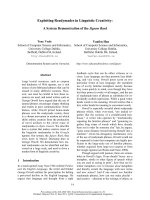
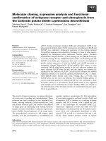
measuring 5.0 cm × 4.0 cm × 2.5 cm. Histopathological
examination showed an infiltrating ductal carcinoma
complicated by an extramedullary plasmacytoma divided
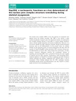
by fibrous tissue in one section (Figure 1). Immunohisto-
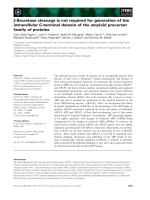
chemical stains were negative for estrogen, P170, and pro-
gesterone receptors. Her-2 was negative (Figure 2).
Nuclear prognostic marker (Ki-67) showed 25% to 50%
nuclear expression. Topoisomerase-IIα (+<5%), CylinD-
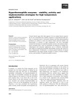
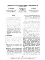
1, Cytokeratin, and S-100 were negative. The tumor cells
were strongly positive for light kappa chains (Figure 3),
and negative for light lambda, delta, and my chains.
After the surgery and pathologic findings, serum immu-
noglobulins were measured and found to be: IgG 10.03 g/
l (8.0–16 g/l), IgA 1.34 g/l (0.7–3.3 g/l), IgM 1.44 g/l
(0.5–2.2 g/l). No Bence Jones or other M components
were detected in the urine. Serum calcium and phospho-
rus were normal. Posterior iliac crest bone marrow biopsy
was negative for plasma cells. PET and CT scans, except for
absence of the right breast, did not detect other lesions.
The patient was offered and accepted chemotherapy and
radiotherapy based on previous research results about
infiltrating ductal carcinoma and case reports about
extramedullary plasmacytoma.
Discussion
Primary soft tissue extrameullary plasmacytoma (SEP) is
uncommon and is defined as a malignant tumor of
plasma cells arising in the soft tissue in the absence of
bone involvement. It can occur in any organ as a solitary
form of plasma cell neoplasm. [1] Some authors consider
SEP to be unrelated to multiple myeloma. Although SEP
can arise throughout the body, almost 80% to 90% of the
cases arise in the head and neck areas. [2]. Approximately
70% occur in patients with multiple myeloma. Since Vis-
alia reported the first case in 1928 [3], approximately
sixty-three cases have been described in published litera-
ture [1-13]. More than two-thirds of the lesions were uni-
lateral in breast [2,4]. Extramedullary plasmacytomas are
seldom solitary and usually progress to diffuse myeloma-
tosis as a first manifestation of multiple myeloma
[7,14,15], or recurrence of multiple myeloma [4]. Plasma-
Extramedullary plasmacytoma and breast cancer were divided by fibrous tissueFigure 1
Extramedullary plasmacytoma and breast cancer
were divided by fibrous tissue. (Hematoxylin &
Eosin×40).
Immunohistochemical stain for Her-2 was negative ×40Figure 2
Immunohistochemical stain for Her-2 was negative
×40.
Plasma cells are diffusely and strongly positive for light kappa chains ×200Figure 3
Plasma cells are diffusely and strongly positive for
light kappa chains ×200.
World Journal of Surgical Oncology 2009, 7:43 />Page 3 of 3
(page number not for citation purposes)
cytomas of the breast are rare, especially those not associ-
ated with multiple myeloma.
The circulating paraproteins vary in type and include
kappa and lambda light-chain patterns, IgA and IgG [4].
In this patient paraproteins were absent in serum and
urine, but strongly positive for kappa light chains in the
lesion.
In our case the tumor was not more than 5 cm in greatest
dimension, and tests for estrogen receptor, progesterone
receptor, and Her-2 were negative. As recommended for
primary breast cancer in "Practice Guidelines in Oncology
– 2008", post surgery the patient received chemotherapy.
In this case it was three cycles of AC→T (doxorubicin/
cyclophosphamide followed by paclitaxel) and no hor-
monal therapy. It is generally accepted that plasmacyto-
mas are radiosensitive, and excellent long-term results
have been reported with local control rates following radi-
otherapy of 79% to 90%, and 10-years survival rates from
50% to 100% [15-17]. Mendenhall reported a threshold
dose of 40 Gy for local control [10]. Local recurrence
develops most frequently in the first five years of follow-
up but maybe found many years later [8]. Based on these
previous reports, after chemotherapy this patient received
a dose of 50 Gy radiation therapy.
This case emphasizes the importance of excision biopsy
and immunohistochemical panel in the differential diag-
nosis and correct therapy of breast masses. The infiltrating
ductal carcinoma and extramedullary plasmacytoma were
divided by fibrous tissue so it is possible they were two
independent diseases, juxtaposed by accident in one clin-
ical lesion. As this is the first case reported, a causal rela-
tionship between synchronous extramedullary
plasmacytoma and infiltrating ductal carcinoma of the
breast must remain speculative.
Consent
Patient's permission was obtained for publishing her case
records.
Competing interests
The authors declare that they have no competing interests.
Authors' contributions
SC was involved in treatment of the patient, collected case
details, literature search and prepared the article. HGK
was involved in treatment of the patient, collected case
details, literature search and helped in preparation of
manuscript. YXL was involved in pathological diagnosis
and figures, wrote the pathological part of the manuscript.
XBR was involved in treatment planning of the patient
and manuscript preparation. All authors read and
approved the final manuscript.
Acknowledgements
Authors also wish to thanks Song-Yan GAO, Department of pathology,
Tianjin Cancer Institute and Hospital, Tianjin Medical University, Tianjin,
China.
References
1. Van Nieuwkoop C, Giard RW, Veen HF, Dees RW: Extramedul-
lary plasmacytoma of the breast simulating breast cancer.
Neth J Med 2001, 58:174-176.
2. Kaviani A, Djamali-Zavareie M, Noparast M, Keyhani-Rofagha S:
Recurrence of primary extra medullary plasmacytoma in
breast both simulating breast carcinoma. World J Sur Oncol
2004, 2(1):29.
3. Wasiliu T, Popa R: Forme gastrointestinale destumeurs dites
plasmacytomas. Comptes Rendus de la Societe Roumaine de Biologie
1928, 98:738-740.
4. Lamy O, Von Bremen K, Burckhardt P: Breast plasmacytoma.
Leuk Lymphoma 2000, 37:611-615.
5. Cangiarella J, Waisman J, Cohen JM, Chhieng D, Symmans WF, Gold-
enberg A: Plasmacytoma of the breast: A Report of Two
Cases Diagnosed by Aspiration Biopsy. Acta Cytol 2000,
44:91-94.
6. Brem R, Revelon G, Willey SC, Gatewood OM, Zeiger MA: Bilateral
Plasmacytoma of the Breast: A Case Report. J The Breast 2002,
8:393-395.
7. De Chiara A, Losito S, Terracciano L, Di Giacomo R, Laccarino G,
Rubolotta M: Primary plasmacytoma of the breast. Arch Pathol
Lab Med 2001, 125:1078-1080.
8. Kirshenbaum G, Douglas P, Rhone MS: Solitary extramedullary
plasmacytoma of the breast with serum monoclonal protein:
A case report and review of the literature. Am J Clin Pathol
1985, 83:230-232.
9. Aysenur M, Esin EU: Breast imaging plasmacytoma of the
breast. Eur Radiol 1994, 4:500-501.
10. Mendenhall CM, Thar TL, Million RR: Solitary plasmacytoma of
bone and soft tissue. Int J Radiat Oncol Biol Phys 1980, 6:497-501.
11. Kumar PV, Vasei M, Daneshbod Y, Zakerinia M, Ramzi M, Noorani H,
Bagheri H, Talei AR, Soleimapour H: Breast myeloma: a report of
3 cases with fine needle aspiration cytologic findings. Acta
Cytol 2005, 49:445-448.
12. Vera-Alvarez J, Marigil-Gómez M, Garcia-Prats , García Prats, Abascal
Agorreta M, López-López JI, Perez-Ruiz J: Extramedullary plasma-
cytoma presenting as a primary mass in the breast. A case
report. Acta Cytol 2003, 47:1107-1110.
13. Taylor L, Aziz M, Klein P, Mazumder A, Jagannath S, Axelrod D: Plas-
macytoma in the breast with axillary lymph node involve-
ment: a case report. Clin Breast Cancer 2006, 7:81-4.
14. Ben-Yehuda A, Steiner-Saltz D, Liboson E, Polliack A: Plasmacy-
toma of the breast: unusual initial presentation of myeloma:
report of two cases and review of the literature. Blut 1989,
58:169-170.
15. Shih LY, Dunn P, Leung WM, Chen WJ, Wang PJ: Localised plasma-
cytomas in Taiwan: Comparison between extamedullary
plasmacytoma and solitary plasmacytoma of bone. Br J Cancer
1995, 71:128-133.
16. Liebross RH, Ha CS, Cox JD, Weber D, Delasalle K, Alexanian R:
Clinical course of solitary extramedullary plasmacytoma.
Radiother Oncology 1999, 52:245-249.
17. Strojan P, Soba E, Lamovec J, Munda A: Extramedullary plasmacy-
tomas: histopathologic study. In J Radiat Oncol Biol Phys 2002,
53:692-701.