Báo cáo khoa học: "Ruptured gallbladder as the first presentation of breast cancer" docx
Bạn đang xem bản rút gọn của tài liệu. Xem và tải ngay bản đầy đủ của tài liệu tại đây (647.95 KB, 3 trang )
BioMed Central
Page 1 of 3
(page number not for citation purposes)
World Journal of Surgical Oncology
Open Access
Case report
Ruptured gallbladder as the first presentation of breast cancer
MJones
1
, J Mathew*
1
, KE Abdullah
2
, T McCulloch
2
and KL Cheung
1
Address:
1
Professorial Unit of Surgery, City Hospital, Nottingham, UK and
2
Department of Histopathology, City Hospital, Nottingham, UK
Email: M Jones - ; J Mathew* - ; KE Abdullah - ;
T McCulloch - ; KL Cheung -
* Corresponding author
Abstract
Background: Perforation of the gall bladder as a first presentation of breast cancer has not been
reported.
Case presentation: Here we present a case of an elderly lady with acute abdomen with evidence
of possible perforation of gall bladder on CT scan. Histopathology of the cholecystectomy
specimen revealed invasive lobular breast cancer.
Her metastatic breast cancer with right sided primary discovered subsequent to her presentation
with acute abdomen is managed successfully with Anastrozole.
Conclusion: We present a rare case of gall bladder perforation from metastatic breast cancer.
Background
Lobular carcinomas of the breast have higher prevalence
of spread to gastrointestinal tract compared to their ductal
counterparts [1]. Although breast cancer metastasis to the
gall bladder has previously been reported [2-4], metastasis
leading to perforation is very rare. We present this rare
case of metastatic breast cancer presenting for the first
time as ruptured gallbladder.
Case presentation
An 84-year-old lady was admitted to hospital with a 12-
hour history of severe, central abdominal pain and vom-
iting. Her abdomen was generally tender and reduced
breath sounds were noted at the right lung base. Oxygen
saturations were 94% on air and all other basic observa-
tions were normal. Liver function tests were also normal.
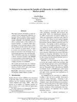
A CT scan demonstrated free air and fluid within the per-
itoneum, air within the intra-hepatic bile ducts and gall-
bladder, and a right-sided pleural effusion [Fig 1]. CT scan
did not show any obvious evidence of matastatic disease.
It was concluded that the gallbladder had perforated and
patient was prepared for emergency laparotomy.
She underwent laparotomy, and was found to have a gan-
grenous, perforated gallbladder containing multiple small
gallstones. Cholecystectomy was performed following an
attempt of intra-operative cholangiogram which was
unsuccessful due to difficulty in cannulating the cystic
duct.
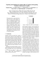
Histologically, the lesion appeared to be a metastatic ade-
nocarcinoma [Fig 2]. The gallbladder showed haemor-
rhagic infarction of the wall, probably caused by an
obstructing metastatic carcinoma near the cystic duct. The
tumour cells were pleomorphic and were forming glandu-
lar structures. Immuno-histochemistry indicated a pri-
mary breast tumour as the cells were strongly positive for
ER, positive for CK19 and EMA and negative for TTF1,
CK20, WT1, CK7, Ca19.9 and Ca125.
Published: 1 June 2009
World Journal of Surgical Oncology 2009, 7:50 doi:10.1186/1477-7819-7-50
Received: 9 March 2009
Accepted: 1 June 2009
This article is available from: />© 2009 Jones et al; licensee BioMed Central Ltd.
This is an Open Access article distributed under the terms of the Creative Commons Attribution License ( />),
which permits unrestricted use, distribution, and reproduction in any medium, provided the original work is properly cited.
World Journal of Surgical Oncology 2009, 7:50 />Page 2 of 3
(page number not for citation purposes)
A 3.2 × 3.0 cm irregular lump suspicious of cancer was
subsequently discovered in the right breast and a 2.9 cm
diameter lymph node was palpable in the ipsilateral
axilla. The patient had been unaware of these lumps.
Post-operative period was uneventful and she made full
recovery. The multidisciplinary team elected to treat her
with endocrine therapy and she was therefore started on
Anastrozole. She remains asymptomatic and her right
sided tumour with axillary metastasis remains stable with
Anastrozole even after 34 months of follow-up.
Discussion
Gall bladder is an uncommon site for metastasis, and in a
large series of autopsies with known cancer, gall bladder
metastasis was identified in 5.8% of cases [5].
Tumours which commonly metastasise to the gall bladder
are malignant melanoma and it occurs in 15% of cases
[6,7]. Other less common primary sites leading to second-
ary metastasis to gall bladder include renal cell cancer, cer-
vical cancer, lung cancer, and breast cancers [8].
Lobular cancers of the breast are well known to metasta-
sise to the gastrointestinal tract compared to ductal can-
cers, and metastasis to the gallbladder has previously been
reported [2-4]. Mechanism behind the affinity for lobular
cancers to metastasise to gastrointestinal tract is not well
understood. A difference in cell size or shape which
favours certain areas of microanatomy that is more contu-
sive to accommodate these cells has been suggested as a
possible explanation [1]. It has also been demonstrated
that loss of expression of cell to cell adhesion molecule E-
cadherin in invasive lobular cancer decreases adhesive-
ness of cells and could contribute to these differences
[9,10].
Bile peritonitis subsequent to metastasis to the gall blad-
der is extremely rare. The only reported case is an elderly
lady with previous history of breast cancer who under-
went mastectomy, radiation and chemotherapy many
years back, presenting acutely as ruptured gall bladder
with associated disseminated metastasis [8].
Conclusion
Here we report the first case of breast cancer initially pre-
senting as a gallbladder perforation. We postulate that the
rupture may be the result of increased pressure in the gall-
bladder due to obstruction of the cystic duct by metastatic
breast carcinoma, which may also explain the difficulty in
performing the intra-operative cholangiogram.
Consent
Written consent was obtained from the patient.
Competing interests
The authors declare that they have no competing interests.
Authors' contributions
MJ wrote the report. JM revised and submitted the report
for publication. KLC conceived the idea and edited the
report. KEA and TMC also helped in editing the report. All
authors read and approved the final manuscript.
CT abdomen showing air in the biliary tree and free air in the peritoneumFigure 1
CT abdomen showing air in the biliary tree and free
air in the peritoneum.
Metastatic lobular breast carcinoma (bottom left) infiltrating the neck of the gallbladder (top right)Figure 2
Metastatic lobular breast carcinoma (bottom left)
infiltrating the neck of the gallbladder (top right).
Publish with BioMed Central and every
scientist can read your work free of charge
"BioMed Central will be the most significant development for
disseminating the results of biomedical research in our lifetime."
Sir Paul Nurse, Cancer Research UK
Your research papers will be:
available free of charge to the entire biomedical community
peer reviewed and published immediately upon acceptance
cited in PubMed and archived on PubMed Central
yours — you keep the copyright
Submit your manuscript here:
/>BioMedcentral
World Journal of Surgical Oncology 2009, 7:50 />Page 3 of 3
(page number not for citation purposes)
References
1. Arpino G, Bardou VJ, Clark GM, Elledge RM: Infiltrating lobular
carcinoma of the breast: tumor characteristics and clinical
outcome. Breast Cancer Res 2004, 6(3):R149-56.
2. Beaver BL, Denning DA, Minton JP: Metastatic breast carcinoma
of the gallbladder. J Surg Oncol 1986, 31(4):240-242.
3. Pappo I, Feigin E, Uziely B, Amir G: Biliary and pancreatic metas-
tases of breast carcinoma: is surgical palliation indicated? J
Surg Oncol 1991, 46(3):211-4.
4. Calafat P, de Diller AB, Sanchez C: [Breast carcinoma metastasis
in ileum-colon and gallbladder simulating inflammatory dis-
eases] [Article in Spanish]. Rev Fac Cien Med Univ Nac Cordoba
1999, 56(2):123-7.
5. Abrams HL, Spiro R, Goldstein N: Metastases in carcinoma; anal-
ysis of 1000 autopsied cases. Cancer 1950, 3(1):74-85.
6. Langley RG, Bailey EM, Sober AJ: Acute cholecystitis from meta-
static melanoma to the gall-bladder in a patient with a low-
risk melanoma. Br J Dermatol 1997, 136(2):279-82.
7. Lee YT: Breast carcinoma: pattern of metastasis at autopsy.
J Surg Oncol 1983, 23(3):175-80.
8. Shah RJ, Koehler A, Long JD: Bile peritonitis secondary to breast
cancer metastatic to the gallbladder. Am J Gastroenterol 2000,
95:1379-1381.
9. Lehr HA, Folpe A, Yaziji H, Kommoss F, Gown AM: Cytokeratin 8
immunostaining pattern and E-cadherin expression distin-
guish lobular from ductal breast carcinoma. Am J Clin Pathol
2000, 114(2):190-6.
10. Sastre-Garau X, Jouve M, Asselain B, Vincent-Salomon A, Beuzeboc
P, Dorval T, Durand JC, Fourquet A, Pouillart P: Infiltrating lobular
carcinoma of the breast. Clinicopathologic analysis of 975
cases with reference to data on conservative therapy and
metastatic patterns. Cancer 1996, 77(1):113-20.