Báo cáo khoa học: "Urachal tumour: case report of a poorly understood carcinoma" pot
Bạn đang xem bản rút gọn của tài liệu. Xem và tải ngay bản đầy đủ của tài liệu tại đây (463.12 KB, 3 trang )
BioMed Central
Page 1 of 3
(page number not for citation purposes)
World Journal of Surgical Oncology
Open Access
Case report
Urachal tumour: case report of a poorly understood carcinoma
Stefano Scabini*
1
, Edoardo Rimini
1
, Emanuele Romairone
1
,
Renato Scordamaglia
1
, Luigi Vallarino
2
, Veronica Giasotto
3
, Carlo Ferro
3
and
Valter Ferrando
1
Address:
1
Department of Emato-Oncology, AOU San Martino Hospital, Genoa, Italy,
2
ASL 3 Genoa, Italy and
3
Department of Radiology, AOU
San Martino Hospital, Genoa, Italy
Email: Stefano Scabini* - ; Edoardo Rimini - ;
Emanuele Romairone - ; Renato Scordamaglia - ;
Luigi Vallarino - ; Veronica Giasotto - ; Carlo Ferro - ;
Valter Ferrando -
* Corresponding author
Abstract
Background: Urachal carcinoma is an uncommon neoplasm associated with poor prognosis.
Case presentation: A 45-year-old man was admitted with complaints of abdominal pain and
pollakisuria. A soft mass was palpable under his navel. TC-scan revealed a 11 × 6 cm tumor, which
was composed of a cystic lesion arising from the urachus and a solid mass component at the urinary
bladder dome. The tumor was removed surgically. Histological examination detected poor-
differentiated adenocarcinoma, which had invaded the urinary bladder. The patient has been
followed up without recurrence for 6 months.
Conclusion: The urachus is the embryological remnant of urogenital sinus and allantois. Involution
usually happens before birth and urachus is present as a median umbilical ligament. The
pathogenesis of urachal tumours is not fully understood. Surgery is the treatment of choice and
role of adjuvant treatment is not clearly understood.
Background
Urachal carcinoma is an uncommon neoplasm associated
with poor prognosis. The estimated annual incidence of
urachal carcinoma in the general population is one in 5
million, or 0.01% of all cancers in adults. Urachal carci-
noma has been estimated to comprise 0.17-0.34% of all
bladder cancers [1]. Adenocarcinoma is common among
urachal carcinomas, whereas squamous cell carcinoma
(SCC) is very rare. We report a case of primary adenocar-
cinoma of the urachus.
Case presentation
A 45-year-old man was referred to our hospital with com-
plaints of micturition pain of 5 months' duration, and
lower abdominal pain and pollakisuria of 1 month's
duration. A physical examination revealed a soft tender
mass under his navel. Periumbilical discharge was not rec-
ognized. Laboratory data: hemoglobin, 15.9 g/dL; eryth-
rocyte count, 4865000/mL; leukocyte count, 11600/mL;
platelet count, 259000/mL. CEA: 5.1 ng/ml. Urinalysis:
hemoglobin (++). Urine cytology: negative. Cystoscopy
detected mucosal edema and erosion at the bladder
Published: 7 November 2009
World Journal of Surgical Oncology 2009, 7:82 doi:10.1186/1477-7819-7-82
Received: 9 September 2009
Accepted: 7 November 2009
This article is available from: />© 2009 Scabini et al; licensee BioMed Central Ltd.
This is an Open Access article distributed under the terms of the Creative Commons Attribution License ( />),
which permits unrestricted use, distribution, and reproduction in any medium, provided the original work is properly cited.
World Journal of Surgical Oncology 2009, 7:82 />Page 2 of 3
(page number not for citation purposes)
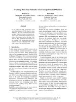
dome. TC-scan showed a soft-tissue mass along the
umbilicus, which extended from the bladder dome to the
posterior wall of the bladder and the peritoneum. The size
of the tumor was 11 × 6 cm (Fig. 1).
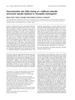
It was noted that the tumor extended from the bladder
dome to the umbilicus. Open laparotomy revealed blad-
der edema and intraoperative cytological examination
was negative. The tumor was removed en bloc together
with the umbilicus, lateral umbilical ligaments, adjacent
peritoneum and bladder dome (Fig. 2).
On gross inspection, the suprapubic mass consisted of slid
and cystic lesions. Microscopically, poor-differentiated
adenocarcinoma of the solid lesion was observed (Fig. 3).
According to CT scan and pathologist report the tumor
can be staged as Sheldon II, confined only to urachus. The
grading of this tumor was impossible.
Tumor cells not invaded the bladder. Follow-up abdomi-
nal computerized tomography at 6 months showed no
tumor recurrence.
Discussion
The urachus is the embryological remnant of urogenital
sinus and allantois. Involution usually happens before
birth and urachus is present as a median umbilical liga-
ment. The pathogenesis of urachal tumours is not fully
understood. It is believed that urachal carcinomas arise
from malignant transformation of columnar or glandular
metaplastic epithelium. They lie in the space of Retzius,
between transversalis fascia anteriorly and peritoneum
posteriorly, extending from the dome of the bladder to the
umbilicus.
Urachal cancer was first described in 1863 by Hue and Jac-
quin in a report translated and summarized by Sheldon
[1]. Although this tumour has now become a recognisable
'neoplastic entity', its origin and pathophysiology remain
unknown [2]. The estimated annual incidence of urachal
carcinoma is 0.01% of all cancers in adults. The incidence
of the disease ranges from 0.55 to 1.20% of bladder
tumors in Japan and 0.07-0.70% of bladder tumors in
TC-scan: a soft-tissue mass along the umbilicus, which extended from the bladder dome to the posterior wall of the bladder and the peritoneumFigure 1
TC-scan: a soft-tissue mass along the umbilicus,
which extended from the bladder dome to the poste-
rior wall of the bladder and the peritoneum. The size
of the tumor was 11 × 6 cm
Tumor removed en bloc together with the umbilicus, lateral umbilical ligaments, adjacent peritoneum and bladder domeFigure 2
Tumor removed en bloc together with the umbilicus,
lateral umbilical ligaments, adjacent peritoneum and
bladder dome
Macroscopic aspect of tumor: the suprapubic mass consisted of slid and cystic lesionsFigure 3
Macroscopic aspect of tumor: the suprapubic mass
consisted of slid and cystic lesions
Publish with BioMed Central and every
scientist can read your work free of charge
"BioMed Central will be the most significant development for
disseminating the results of biomedical research in our lifetime."
Sir Paul Nurse, Cancer Research UK
Your research papers will be:
available free of charge to the entire biomedical community
peer reviewed and published immediately upon acceptance
cited in PubMed and archived on PubMed Central
yours — you keep the copyright
Submit your manuscript here:
/>BioMedcentral
World Journal of Surgical Oncology 2009, 7:82 />Page 3 of 3
(page number not for citation purposes)
Western countries. Histopathologically, adenocarcinoma
accounts for 80-90% of the tumors. The 5-year cancer-spe-
cific survival rate, depending of pathologic stage [3], is 6%
in Japan [4] but Ashley [5] in a 50 years of experience at
Mayo Clinic reported a 5-year cancer-specific survival rate
of 49%. The poor prognosis of this cancer is due to: (a)
late presentation of symptoms leading to advanced stage
at diagnosis; (b) a propensity for early local invasion; and
(c) distal metastasis.
The new Mayo staging system was less complicated than
the Sheldon system, although both systems predicted can-
cer-specific mortality equally well. Positive surgical mar-
gins (hazard ratio [HR], 4.7), high tumor grade (HR, 3.6),
positive local lymph nodes (HR, 5.1), metastases at diag-
nosis (HR, 3.3), advanced tumor stage (HR, 4.8), failure
to perform umbilectomy (HR, 3.0), and primary radiation
therapy (HR, 2.9) were all univariately associated with
death (P < .05). Only grade and margins were significant
in the multivariate analysis. Modern therapeutic regimens
have offered minimal benefit, especially when unresecta-
ble. No survival benefit was noted for lymphadenectomy
or adjuvant therapy. Salvage surgery resulted in a long-
term cure for 50% of patients who had local recurrences.
No effective treatment was identified for patients with
metastatic UrC. The question as to whether partial or rad-
ical cystectomy is suitable for localised disease is difficult
to answer since urachal tumours are rare. Furthermore,
within this group, urachal adenocarcinoma is uncommon
in those under 40 years. However, in order to evaluate
which surgical approach is correct or whether new chem-
otherapeutic agents will induce objective responses and
improve long-term survival requires co-operation
between physicians and centres internationally so that
larger studies can be conducted. This approach can only
benefit patients [6-8].
Conclusion
The urachus is the embryological remnant of urogenital
sinus and allantois. Involution usually happens before
birth and urachus is present as a median umbilical liga-
ment. The pathogenesis of urachal tumours is not fully
understood. Surgery is the treatment of choice and role of
adjuvant treatment is not clearly understood.
Consent
Written informed consent was obtained from the patient
for publication of this case report. A copy of the consent is
available with editorial office
Competing interests
The authors declare that they have no competing interests.
Authors' contributions
SS, ER, ER, RS and VF are surgeons of the Unit of Surgical
Oncology (Chief: VF) and have performed the operation.
LV is General Psysician of the patient. VG and CF are radi-
ologists of the Department of Radiology. All authors read
and approved the final manuscript.
References
1. Sheldon CA, Clayman RV, Gonzalez R, Williams RD, Fraley EE:
Malignant urachal lesions. J Urol 1984, 131:1-8.
2. Mostfi FK, Thomson RV, Dean AL: Mucinous Adenocarcinoma of
the urinary bladder. Cancer 1955, 8:741-58.
3. Gopalan A, Sharp DS, Fine SW, Tickoo SK, Herr HW, Reuter VE,
Olgac S: Urachal carcinoma: a clinicopathologic analysis of 24
cases with outcome correlation. Am J Surg Pathol 2009,
33(5):659-68.
4. Ghazizadeh M, Yamamoto S, Kurokawa K: Clinical features of
urachal carcinoma in Japan: review of 157 patients. Urol Res
1983, 11:235-8.
5. Ashley RA, Inman BA, Sebo TJ, Leibovich BC, Blute ML, Kwon ED,
Zincke H: Urachal carcinoma: clinicopathologic features and
long-term outcomes of an aggressive malignancy. Cancer
2006, 107:712-20.
6. Tian J, Ma JH, Li CL, Xiao ZD: Urachal mass in adults: clinical
analysis of 33 cases. Zhonghua Yi Xue Za Zhi 2008, 88(12):820-2.
7. Paras FA Jr, Maclennan GT: Urachal adenocarcinoma. J Urol 2008,
180(2):720.
8. Sugarbaker PH, Verghese M, Yan TD, Brun E: Managment of muci-
nous urachal neoplasm presenting as pseudomyxoma peri-
tonei. Tumori 2008, 94(5):732-6.