Báo cáo khoa học: "Comparison of immunohistochemistry (IHC) and fluorescence in situ hybridization (FISH) assessment for Her-2 status in breast cancer" ppsx
Bạn đang xem bản rút gọn của tài liệu. Xem và tải ngay bản đầy đủ của tài liệu tại đây (1.36 MB, 6 trang )
BioMed Central
Page 1 of 6
(page number not for citation purposes)
World Journal of Surgical Oncology
Open Access
Research
Comparison of immunohistochemistry (IHC) and fluorescence in
situ hybridization (FISH) assessment for Her-2 status in breast
cancer
Weiguo Sui
†1
, Minglin Ou
†1,2
, Jiejing Chen
†1
, Youhua Wan
†3
,
Hongbo Peng
†1
, Minfang Qi
†3
, He Huang
†1
and Yong Dai*
1
Address:
1
Laboratory Center of Guangzhou Military Area Command, 181st Hospital of People's Liberation Army, Guilin, Guangxi, PR China,
2
College of Life Science, Guangxi Normal University, Guilin, Guangxi, PR China and
3
Pathology Department of Guangzhou Military Area
Command, 181st Hospital of People's Liberation Army, Guilin, Guangxi, PR China
Email: Weiguo Sui - ; Minglin Ou - ; Jiejing Chen - ;
Youhua Wan - ; Hongbo Peng - ; Minfang Qi - ;
He Huang - ; Yong Dai* -
* Corresponding author †Equal contributors
Abstract
Background: The concordance rate between IHC and FISH according to clinical performance is
still controversial. We report a prospective study to reflect the concordance between IHC and
FISH in Guilin city, People's Republic of China.
Methods: Fifty cases of invasive ductal carcinoma of breast tested by IHC and scored as 0, 1+, 2+
and 3+ by pathologists were further analyzed by FISH using a commercially available double-color
probe, and the FISH findings were compared with IHC test results.
Results: A total concordance of 82.0% was observed with a Kappa coefficient of 0.640 (P < 0.001).
A high discordance was observed in 30.0% of the patients with IHC 2+, 7.1% in IHC 3+, 19.2%
overall in IHC 0 and 1+.
Conclusion: The IHC can be used firstly to screen the HER-2 status, and FISH can be used as a
supplementary role to IHC and 2+ and some negative cases. And only those cases with Her-2 status
of IHC 3+ or FISH positive should be treated with Herceptin.
Background
Breast cancer is one of the most common malignancy in
the world. According to the global cancer statistics, Europe
and America has the high incidence and mortality of
breast cancer [1]. The incidence of breast cancer in China
is 20 per 100,000 population, and the incidence is grow-
ing [2]. Researches have shown that about 20%-30% of
the breast cancer patients have Her-2 amplification or
over expression, that is associating with a more aggressive
phenotype and decreased survival [3-7]. The benefit of
humanized anti-Her-2 monoclonal antibody trastuzu-
mab (Herceptin) in Her-2-positive breast cancers has been
well documented as noted by prolonged survival [8]. But
this therapy is effective only if the detection of Her-2 sta-
tus is accurate.
There are several methods available to detect the Her-2
status like polymerase chain reaction (PCR), immunohis-
Published: 9 November 2009
World Journal of Surgical Oncology 2009, 7:83 doi:10.1186/1477-7819-7-83
Received: 30 August 2009
Accepted: 9 November 2009
This article is available from: />© 2009 Sui et al; licensee BioMed Central Ltd.
This is an Open Access article distributed under the terms of the Creative Commons Attribution License ( />),
which permits unrestricted use, distribution, and reproduction in any medium, provided the original work is properly cited.
World Journal of Surgical Oncology 2009, 7:83 />Page 2 of 6
(page number not for citation purposes)
tochemistry (IHC), fluorescence in situ hybridization
(FISH), chromogenic in situ hybridisation (CISH) [9,10].
Protein over-expression detected by IHC or amplification
of Her-2 gene analyzed by FISH are the two main methods
used to detect Her-2 status in clinical practice. FISH is con-
sidered as a gold standard because of its sensitivity and
specificity. But FISH has disadvantages as it requires a
modern and expensive fluorescence microscope equipped
with multi-band-pass fluorescence filters, and the fluores-
cence fades so quickly that it could not provide a perma-
nent record [10]. Compared with FISH, IHC is widely
used in china as it is cheaper and convenient to operate
and conserve; the morphology is clear. Comparative stud-
ies of IHC and FISH have generally shown a high concord-
ance rate by some researches [11]. But protein
overexpression may be found without gene amplification
or gene amplification can be found in negative IHC [12].
Research has documented that the discordance rate
between Her-2 by FISH and IHC is high in all four IHC
scores (0, 1+, 2+, 3+), and a FISH-alone screening strategy
has been alternatively suggested [13]. Our objective was to
perform a prospective study in our own local setting and
record the concordance between IHC and FISH in 50 cases
of invasive ductal carcinoma of breast.
Methods
Study Design
The study population consists of 50 cases of invasive duc-
tal carcinoma of breast treated between July, 2008 and
March, 2009 at The 181 Hospital and Traditional Chinese
Medicine Hospital which are the two main hospitals for
the treatment of breast cancer in Guilin city of China. The
specimens were fixed in 10% neutral-buffered formalin
(pH7.4) for 24 hours. Only cases with sufficient invasive
carcinoma for multiple assays were included in the study.
For each case, 2-4 μm thick tissue sections were cut from a
representative paraffin block and applied to positively
charged slides. Her-2 protein expression was measured
using a commercial available S-P kit. FISH for Her-2 gene
amplification was performed in the key laboratory of 181
Hospital using a commercial available double-color
probe. The interpretation of IHC and FISH were each per-
formed by investigators blinded to the results of the other
assay
IHC analysis
IHC study was performed on paraffinem-bedded, forma-
lin-fixed tissue sections using a commercial available
Ultra Sensitive™ S-P kit (Maixin-Bio Co., Fuzhou, China),
following the manufacturer's instructions and American
Society of Clinical Oncology/College of American Pathol-
ogists guideline recommendations for human Epidermal
Growth Factor Receptor 2 testing in breast cancer [14].
Briefly, this procedure included the deparaffinization and
rehydration steps, followed by an epitope retrieval step in
which the tissue sample was incubated in a citrate buffer
solution at 90-95°C for 20 minutes. The slides were then
subjected to a series of alternating washes in tris
(hydroxymethyl) aminomethane hydrochloride buffer
and incubation steps with, first, a peroxidase-blocking
reagent for 5 minutes and then with Her-2 primary anti-
body, followed by a visualization reagent for 30 minutes
each, and finally with a 3,3'-diaminobenzidine chro-
mogen solution. After a finally wash, the slides were coun-
terstained with haematoxylin [15].
Tumor cells with circumferential membranous positivity
were considered as Her-2 protein over expression, and
scoring was performed according to the manufacturer's
recommendations by pathologists in a number of differ-
ent practice groups (Figure 1), each with at least 10 years
of experience in clinical practice; in order to truly reflect
the concordance or discordances between IHC and FISH
in our daily practices, the results of IHC were all from the
data bank of the two hospitals and without revision again.
FISH for Her-2 gene amplification
FISH analysis was modified in cooperation with the man-
ufacturer of China Medical Technologies, Inc. (Beijing,
China). The commercially available double-color FISH
probe consisted of two probes: 17q11.2-q12 (labeled with
Spectrum Orange) covering the whole Her-2 gene and the
control, centromeric chromosome 17p11.1-q11.1
(labeled with Spectrum Green). The FISH fixed glass
microscope slides with tissue sections were baked over-
night at 65°C, deparaffinized in two 10-minute changes
of xylene, transferred through two 3-minute changes of
100% ethanol, one 3-minute changes of 85% ethanol,
one 3-minute changes of 70% ethanol and immersed for
15 minutes in pure water at 90°C. The slides were then
incubated for 7-15 minutes in protease solution at 37°C.
Then the slides were briefly washed in 2× sodium saline
citrate (2× SSC; pH 7.2) at room temperature, dehydrated
through 70%, 85%, 100% ethanol and acetone, then
allowed to air dry. To denature DNA, the slides were
placed in 78.5°C preheated 70% formamide/2× SSC for 8
min and then dehydration in a graded series of concentra-
tions of ethanol which were precooling in -20°C. After
drying in the open-air, 10 μl of probe which was destruc-
tured at 75.5°C for 7 min was applied onto each slide,
cover slip was placed and sealed with rubber cement, then
hybridized overnight at 42.8°C. After 16-18 h of hybridi-
zation, the slides were washed in 46°C preheated post-
hybridization buffer (2× SSC/0.1% sodium dodecyl sul-
fate) for 5 min and rinsed in 70% ethanol. After air-drying
(out of direct light), the slides were counterstained with
15 μL DAPI/anti-fade solution and cover slip applied.
FISH analysis was performed by two cytotechnologistes
who were blinded to the clinical diagnoses at the time of
World Journal of Surgical Oncology 2009, 7:83 />Page 3 of 6
(page number not for citation purposes)
evaluation. The slides were scanned using a OLYMPUS
BX51 fluorescent microscope (OLYMPUS BX51, Japan)
equipped with a 100-watt mercury lamp and single band
pass filter set to detect DAPI, Rhodamine (17q11.2-q12),
and FITC (chromosome 17) at 1000×. Thirty randomly
selected invasive tumor nuclei in each of two separate, dis-
tinct microscopic areas were evaluated. Cases were scored
as negative by FISH when the Her-2 with a Her-2 to CEP
17 ratio < 1.8 by counting at least 30 interphase nuclei,
and those cases with a Her-2 to CEP 17 ratio > 2.2 were
scored as positive (Figure 2). In addition, more randomly
selected invasive tumor nuclei (for example, a total of 100
nuclei) would be evaluated if the Her-2 with a Her-2 to
CEP 17 ratio was between 1.8 and 2.2.
Results
Of the 50 specimens in our study (invasive ductal carci-
noma with varying tumor grades and clinical stages), 9
were classified as IHC 0, 17 were classified as IHC 1+, 10
were classified as IHC 2+, and 14 were classified as IHC
3+. Five of the IHC 0 and 1+ cases, seven of the 10 IHC 2+
cases and 13 of the 14 IHC 3+ cases were found to be Her-
2 FISH positive. They had a total concordance of 82.0%
and a Kappa coefficient of 0.640 (P < 0.001), which was
defined as IHC 2+/3+ and Her-2 FISH positive, or IHC 0/
1+ and Her-2 FISH negative.
Discordance was defined as a discrepancy between the
IHC and Her-2 FISH, including the following two condi-
tions: (1) IHC 2+ or 3+ but Her-2 FISH negative; (2) IHC
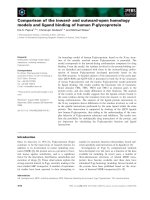
IHC for Her-2 protein expressionFigure 1
IHC for Her-2 protein expression. Figure 1 (A) Completely negative, IHC0; (B) Faint membranous positivity, ICH1+; (C)
Moderate membranous positivity, IHC2+; (D)Strong, more than 30% tumor cells with circumferential membranous positivity,
IHC3+.
World Journal of Surgical Oncology 2009, 7:83 />Page 4 of 6
(page number not for citation purposes)
0 or 1+ but Her-2 FISH positive. For example, the discord-
ance rate according to IHC 0 and 1+ was defined as the
number of discrepant IHC 0 and 1+ cases divided by the
total number of IHC 0 and 1+ cases and was 19.2% (5/
26). Following the same way of counting, the discordance
rate according to IHC 2+ was 30.0% (3/10), IHC 3+ was
7.1% (1/14). The overall discordance rate by IHC was
therefore 18.0% (9/50) (Table 1).
Discussion
Reliable laboratory data in evaluating Her-2 status is
essential, because the treatment is beneficial for advanced
breast cancer and can avoid potential cardiotoxic effects in
women not showing amplification or overexpression
[16]. Her-2 status studied at the levels of DNA using FISH
and protein using IHC are the two most accessible and
feasible methods used in clinical diagnosis, and certain
kits or antibodies are approved by the FDA (U.S. Food and
Drug Administration). IHC is easy to perform and rela-
tively cheap, and is predominantly used to evaluate Her-2
status. However, a wide range of sensitivity and specificity
has been observed among various commercially available
antibodies [17]. As an alternative, FISH is also recognized
as a modality in cases with an equivocal IHC status with
higher sensitivity and specificity.
Considering FISH as a gold standard, research reports that
positive FISH results could be found in 91.7%, 23.2%,
7.4% and 4.1% in cases with IHC respectively diagnosed
as 3+, 2+, 1+ and 0 [18]. In addition, some clinical trials
FISH for Her-2 gene amplificationFigure 2
FISH for Her-2 gene amplification. Figure 2 (A) Negative amplification of human epidermal growth factor receptor-2/neu
(Her-2) gene case with the ratio of Her-2 (red signals) to CEP 17 (green signals) is obviously smaller than 1.8; (B) Positive
amplification of human epidermal growth factor receptor-2/neu (Her-2) gene case with the ratio of Her-2 (red signals) to CEP
17 (green signals) is obviously larger than 2.2.
Table 1: Comparison of the results of IHC and FISH
IHC scoring Her-2 FISH amplified Her-2 FISH non-amplified Concordance by IHC Discordance by IHC
0 and 1+ (n = 26) 5 21 (21/26)80.8% (5/26)19.2%
2+ (n = 10) 7 3 (7/10)70.0% (3/10)30.0%
3+ (n = 14) 13 1 (13/14)92.9% (1/14)7.1%
IHC = immunohistochemistry; FISH = fluorescence in situ hybridization
World Journal of Surgical Oncology 2009, 7:83 />Page 5 of 6
(page number not for citation purposes)
have shown that amplification by FISH is more predictive
of response to trastuzumab than IHC [12,19,20].
Our objective in this study was not to try and compare the
sensitivity and specificity of these two tests (IHC and
FISH). This study aimed to investigate the concordance
and discordance rates between IHC and Her-2 FISH. Some
similar studies adopting a similar strategy have been
reported. Dolan and Snover found that the concordance
between the IHC and FISH scores (defined as cases that
were IHC negative/FISH nonamplified or IHC positive/
FISH amplified) was found in 35 cases (27.1%) and dis-
cordance in 94 cases (72.9%) [15]. Lan et al. used FISH to
ascertain the prevalence of erb-b2 gene amplification in
221 cases of breast cancer specimens read as 2+ in IHC
analysis, and found 96 (44.4%) cases were detected to be
erb-b2 amplified [21]. Kuo et al. compared FISH and IHC
in breast cancer patients and found that the discordance
rates by IHC were high (46.7% in IHC 2+, 16.7% in IHC
3+, 30.3% overall in IHC 2+ or 3+) [22]. All these
researches indicated that the concordance between IHC
and FISH is still controversial.
There are some factors leading to false IHC test results,
including variability in tissue fixation and processing, var-
iable sensitivity and specificity of commercially available
antibodies, and differences in scoring criteria with consid-
erable interobserver variability in interpretation of results
[23]. Our study is trying to answer a simpler question
about the concordance between IHC and FISH in our
local setting in Guilin city of China, using some commer-
cially available antibodies, which have not been approved
by the FDA. Compared with some studies [15,22,23], our
concordance in the cases of IHC 3+ is similar with theirs,
but there are some differences in the cases of IHC 0, 1+,
2+. The concordance in this study of IHC 0 and 1+ is
lower, and IHC 2+ is higher than theirs. This may due to
the different sensitivity and specificity of the antibodies
and probe used in this study. The different sensitivity and
specificity of the antibodies and probe need to be further
researched.
Conclusion
In order to improve the therapeutic effect of herceptin in
Her-2-positive breast cancers, the current algorithm using
FISH as a supplementary role to IHC 2+ need to be mod-
ified according to this study in our setting. The IHC can be
used firstly to screen the HER-2 status, and FISH can be
used as a supplementary role to detect IHC and 2+ and
some negative cases, especially those with a high tumour
grades. And only those cases with Her-2 status of IHC 3+
or FISH positive are proposed to be treated with herceptin.
Competing interests
This work is supported by the funding of Ministry of
Health, P. R. China (Funding NO. WKJ 2007-3-001).
Authors' contributions
WS carried out the studies design, helped to draft the man-
uscript. MO drafted the manuscript and participated in
FISH analysis. JC and HP carried out FISH analysis. YW,
MQ and HH carried out IHC analysis. YD conceived of the
study, and participated in its design and coordination and
helped to draft the manuscript. All authors read and
approved the final manuscript.
Acknowledgements
We thank Xuefeng Shi of Guilin Traditional Chinese Medicine Hospital and
Runqiang Wu of Guilin 181 hospital for their assistance in this work.
References
1. Parkin DM, Bray F, Ferlay J: Global cancer statistics, 2002. CA
Cancer J Clin 2005, 55(2):74-108.
2. Zheng Y, Li DL, Xiang YM, Li XJ: The status and trend of breast
cancer incidence in Shanghai. J Surg Concepts Pract 2001,
6:219-221.
3. Paterson MC, Dietrich KD, Danyluk J, Paterson AH, Lees AW, Jamil
N, Hanson J, Jenkins H, Krause BE, McBlain WA: Correlation
between c-erbB-2 amplification and risk of recurrent disease
in node-negative breast cancer. Cancer Res 1991, 51(2):556-67.
4. Seshadri R, Firgaira FA, Horsfall DJ, McCaul K, Setlur V, Kitchen P:
Clinical significance of HER-2/neu oncogene amplification in
primary breast cancer. The South Australian Breast Cancer
Study Group. J Clin Oncol 1993, 11(10):1936-42.
5. Press MF, Pike MC, Chazin VR, Hung G, Udove JA, Markowicz M,
Danyluk J, Godolphin W, Sliwkowski M, Akita R: Her-2/neu expres-
sion in node-negative breast cancer: direct tissue quantita-
tion by computerized image analysis and association of
overexpression with increased risk of recurrent disease. Can-
cer Res 1993, 53(20):4960-70.
6. Press MF, Bernstein L, Thomas PA, Meisner LF, Zhou JY, Ma Y, Hung
G, Robinson RA, Harris C, El-Naggar A, Slamon DJ, Phillips RN, Ross
JS, Wolman SR, Flom KJ: HER-2/neu gene amplification charac-
terized by fluorescence in situ hybridization: poor prognosis
in node-negative breast carcinomas. J Clin Oncol 1997,
15(8):2894-904.
7. Ross JS, Fletcher JA: The HER-2/neu oncogene: prognostic fac-
tor, predictive factor and target for therapy. Semin Cancer Biol
1999, 9(2):125-38.
8. Dean-Colomb W, Esteva FJ: Her2-positive breast cancer: her-
ceptin and beyond. Eur J Cancer 2008, 44(18):2806-12.
9. Tanner M, Gancberg D, Di Leo A, Larsimont D, Rouas G, Piccart MJ,
Isola J: Chromogenic in situ hybridization: a practical alterna-
tive for fluorescence in situ hybridization to detect HER-2/
neu oncogene amplification in archival breast cancer sam-
ples. Am J Pathol 2000, 157(5):1467-72.
10. Sáez A, Andreu FJ, Seguí MA, Baré ML, Fernández S, Dinarés C, Rey
M:
HER-2 gene amplification by chromogenic in situ hybridi-
sation (CISH) compared with fluorescence in situ hybridisa-
tion (FISH) in breast cancer-A study of two hundred cases.
Breast 2006, 15(4):519-27.
11. Reed W, Hannisdal E, Boehler PJ, Gundersen S, Host H, Nesland JM:
The prognostic value of p53 and c-erb B-2 immunostaining is
overrated for patients with lymph node negative breast car-
cinoma: a multivariate analysis of prognostic factors in 613
patients with a follow-up of 14-30 years. Cancer 2000,
88(4):804-13.
12. Pauletti G, Dandekar S, Rong H, Ramos L, Peng H, Seshadri R, Slamon
DJ: Assessment of methods for tissue-based detection of the
HER-2/neu alteration in human breast cancer: a direct com-
parison of fluorescence in situ hybridization and immunohis-
tochemistry. J Clin Oncol 2000, 18(21):3651-64.
Publish with BioMed Central and every
scientist can read your work free of charge
"BioMed Central will be the most significant development for
disseminating the results of biomedical research in our lifetime."
Sir Paul Nurse, Cancer Research UK
Your research papers will be:
available free of charge to the entire biomedical community
peer reviewed and published immediately upon acceptance
cited in PubMed and archived on PubMed Central
yours — you keep the copyright
Submit your manuscript here:
/>BioMedcentral
World Journal of Surgical Oncology 2009, 7:83 />Page 6 of 6
(page number not for citation purposes)
13. Tubbs RR, Pettay JD, Roche PC, Stoler MH, Jenkins RB, Grogan TM:
Discrepancies in clinical laboratory testing of eligibility for
trastuzumab therapy: apparent immunohistochemical false-
positives do not get the message. J Clin Oncol 2001,
19(10):2714-21.
14. Wolff AC, Hammond EH, Schwartz JN, Hagerty KL, Allred DC, Cote
RJ, Dowsett M, Fitzgibbons PL, Hanna WM, Langer A, McShane LM,
Paik S, Pegram MD, Perez EA, Press MF, Rhodes A, Sturgeon C, Taube
SE, Tubbs R, Vance GH, Vijver M, Wheeler TM, Hayes DF: Ameri-
can Society of Clinical Oncology/College of American
Pathologists guideline recommendations for human epider-
mal growth factor receptor 2 testing in breast cancer. J Clin
Oncol 2007, 25(1):118-45.
15. Dolan M, Snover D: Comparison of immunohistochemical and
fluorescence in situ hybridization assessment of HER-2 sta-
tus in routine practice. Am J Clin Pathol 2005, 123(5):766-70.
16. Keefe DL: Trastuzumab-associated cardiotoxicity. Cancer
2002, 95(7):1592-600.
17. Bempt I Vanden, Vanhentenrijk V, Drijkoningen M, Vandenberghe P,
De WC: Real-time reverse transcription-PCR and fluores-
cence in-situ hybridization are complementary to under-
stand the mechanisms involved in HER-2/neu
overexpression in human breast carcinomas. Histopathology
2005, 46(4):431-41.
18. Owens MA, Horten BC, Da Silva MM: HER2 amplification ratios
by fluorescence in situ hybridization and correlation with
immunohistochemistry in a cohort of 6556 breast cancer tis-
sues. Clin Breast Cancer 2004, 5(1):63-9.
19. Dybdal N, Leiberman G, Anderson S: Determination of HER2
gene amplification by fluorescence in situ hybridization and
concordance with the clinical trials immunohistochemical
assay in women with metastatic breast cancer evaluated for
treatment with trastuzumab. Breast Cancer Res Treat 2005,
93(1):3-11.
20. Mass RD, Press MF, Anderson S, Cobleigh MA, Vogel CL, Dybdal N,
Leiberman G, Slamon DJ: Evaluation of clinical outcomes
according to HER2 detection by fluorescence in situ hybridi-
zation in women with metastatic breast cancer treated with
trastuzumab. Clin Breast Cancer 2005, 6(3):240-6.
21. Lan C, Liu JM, Liu TW, Hsu D, Liang S, Chen JR, Peng JW: Erb-b2
amplification by fluorescence in situ hybridization in breast
cancer specimens read as 2+ in immunohistochemical analy-
sis. Am J Clin Pathol 2005,
124(1):97-102.
22. Kuo SJ, Wang BB, Chang CS, Chen TH, Yeh KT, Lee DJ, Yin PL, Chen
M: Comparison of immunohistochemical and fluorescence in
situ hybridization assessment for HER-2/neu status in Tai-
wanese breast cancer patients. Taiwan J Obstet Gynecol 2007,
46(2):146-51.
23. Jacobs TW, Gown AM, Yaziji H, Barnes MJ, Schnitt SJ: HER-2/neu
protein expression in breast cancer evaluated by immuno-
histochemistry. A study of interlaboratory agreement. Am J
Clin Pathol 2000, 113(2):251-8.