Báo cáo khoa học: "Late cutaneous metastases to the face from malignant pleural mesothelioma: A case report and review of the literature" docx
Bạn đang xem bản rút gọn của tài liệu. Xem và tải ngay bản đầy đủ của tài liệu tại đây (541.65 KB, 5 trang )
BioMed Central
Page 1 of 5
(page number not for citation purposes)
World Journal of Surgical Oncology
Open Access
Case report
Late cutaneous metastases to the face from malignant pleural
mesothelioma: A case report and review of the literature
Alaaeldeen M Elbahaie*
1
, Dia E Kamel
2
, Julia Lawrence
3
and
Neville G Davidson
1
Address:
1
Clinical Oncology Department, Mid Essex Hospital Services NHS Trust, Broomfield Hospital, Court Road, Chelmsford, CM1 7ET, UK,
2
Histopathology Department, Mid Essex Hospital Services NHS Trust, Broomfield Hospital, Court Road, Chelmsford, CM1 7ET, UK and
3
Radiotherapy Department, Colchester Hospital University NHS Foundation Trust, Essex County Hospital, Lexden Road, Colchester, CO3 3NB,
UK
Email: Alaaeldeen M Elbahaie* - ; Dia E Kamel - ;
Julia Lawrence - ; Neville G Davidson -
* Corresponding author
Abstract
Background: Malignant Mesothelioma is a rare primary neoplasm affecting the serosal
membranes. During its relative short course, this malignant neoplasm can give local and, rarely,
distant haematogenous metastases in different organs. The reported metastatic sites include liver,
lung, heart, brain, thyroid, adrenals, kidneys, pancreas, bone, soft tissue, skin and lymph nodes.
Case Presentation: We report a sixty one year-old man with a history of malignant pleural
epithelioid mesothelioma treated with six cycles of Pemetrexed and Carboplatin completed 03/11/
04 followed by radiotherapy to the drain site 250 Kv/TD20Gy/5F completed 13/12/2004. Then he
developed multiple facial skin lesions 4 years later. These lesions were proved to be metastatic
malignant sarcomatoid mesothelioma.
Conclusion: Mesothelioma metastases should be suspected in any known Mesothelioma patient
with newly developed skin lesion.
Background
Malignant Mesothelioma is a rare primary neoplasm
affecting the serosal membranes. During its relative short
course, this malignant neoplasm can give local and,
rarely, distant haematogenous metastases in different
organs. The reported metastatic sites include liver, lung,
heart, brain, thyroid, adrenals, kidneys, pancreas, bone,
soft tissue, skin and lymph nodes. The increased incidence
of malignant mesothelioma and the improvement of sur-
vival rates due to the newly introduced chemotherapeutic
agents bring to light the importance of studying its
amended natural history.
Case Presentation
A 61 year-old white man with known history of pleural
mesothelioma on regular follow up was found to develop
multiple facial skin lesions with no clinical evidence of
local recurrence 4 years after the primary diagnosis.
On March 2004, this non-smoker, semi-retired boat
builder with significant asbestos exposure history, pre-
sented with 4 months history of progressive shortness of
breath. The inhalers had not helped and this seemed to be
clearly a different problem to his original asthma. He also
complained of some right chest pain, easy fatigue, dry
Published: 9 November 2009
World Journal of Surgical Oncology 2009, 7:84 doi:10.1186/1477-7819-7-84
Received: 27 August 2009
Accepted: 9 November 2009
This article is available from: />© 2009 Elbahaie et al; licensee BioMed Central Ltd.
This is an Open Access article distributed under the terms of the Creative Commons Attribution License ( />),
which permits unrestricted use, distribution, and reproduction in any medium, provided the original work is properly cited.
World Journal of Surgical Oncology 2009, 7:84 />Page 2 of 5
(page number not for citation purposes)
cough and weight loss. On examination, there were only
signs of pleural effusion. The chest X-rays showed increas-
ing right pleural effusion and CT chest showed a large
right simple pleural effusion with no pleural thickening or
masses. He was admitted and the effusion drained. The
cytology of the effusion was highly suggestive of mesothe-
lioma, but pleural biopsy was insufficient. Few weeks
later, the pleural effusion recurred and an US guided
biopsy on 08/06/04 showed features consistent with a
malignant Mesothelioma, epithelioid type (Fig. 1). The
biopsy contains some skeletal muscle and some pleura
with a thick layer of malignant epithelioid cells which are
positive for mesothelial markers CK5/6 and Calretinin
and negative for lung cancer markers TTF-1 and CEA.
The patient took part on the ALIMTA trial, he received 6
cycles of Pemetrexed 500 mg/m
2
+ Carboplatin AUC 5 day
1 every 3 weeks; the last cycle date was 03/11/04. This was
followed by radiotherapy to the drain site "250 Kv/TD20
Gy/5F" completed 13/12/2004.
Then, the patient underwent close follow up and he
remained well and asymptomatic with no clinical or radi-
ological evidence of disease recurrence until the end of
December 2007, when he noticed small subcutaneous
lesion on his right check and some nasal symptoms. Few
weeks later, he developed fever 38°C with dry cough and
the cheek lesion increased in size. CT scan on 17/03/2008
recorded several sites of disease; notably in the right
hemithorax and right para-renal space consistent with
recurrence of the Mesothelioma. Clinical Examination of
the face revealed 3 skin lesions: right cheek 24 × 24 mm
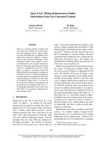
Original pleural biopsy on June 2004, showing infiltration by epithelioid tumour cells (a), which are positive with immuno-histochemical staining for Calretinin (b), consistent with epi-thelioid mesotheliomaFigure 1
Original pleural biopsy on June 2004, showing infiltra-
tion by epithelioid tumour cells (a), which are posi-
tive with immunohistochemical staining for
Calretinin (b), consistent with epithelioid mesotheli-
oma.
(a)
(b)
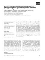
Photos of skin lesion in the right cheekFigure 2
Photos of skin lesion in the right cheek.
World Journal of Surgical Oncology 2009, 7:84 />Page 3 of 5
(page number not for citation purposes)
annular raised red area with eroded central area (Fig. 2), a
small erythematous plaque posterior to the right ear and
a 1 cm very firm subcutaneous lesion on the frontal area.
CT guided core biopsy from the right posterior basal pleu-
ral mass showed a thick layer of malignant epithelioid
cells. A panel of immunohistochemical markers were per-
formed and these malignant cells are positive for mes-
othelial markers CK5/6 and Calretinin and negative for
lung cancer markers TTF-1 and CEA. Overall, these fea-
tures are consistent with a malignant Mesothelioma, Epi-
thelioid type.
The three skin lesions were biopsied. The microscopic
examination showed sarcomatous atypical spindle cell
proliferation within the dermis extending into the subcu-
taneous adipose tissue (Fig. 3). Mitotic figures including
atypical forms are noted. The architecture of the tumour is
partly storiform. Immunohistochemistry showed positiv-
ity with Vimentin, CAM 5.2 and CD10 and focally with
SMA and Calretinin. The histopathological diagnosis was
in keeping with Metastatic Mesothelioma of sarcomatous
type.
Discussion
Malignant Mesothelioma is a rare primary neoplasm
affecting the serosal membranes with a relative increase of
its incidence rate during the last decades [1]. Most cases of
mesotheliomas are related to asbestos exposure [2]. Its
incidence has been rising steadily over the past few dec-
ades [1]. Approximately 1000 people die of mesotheli-
oma each year in the UK, and it is predicted to rise to 3000
by the year 2020 [3].
Histologically, Mesothelioma is divided into epithelial,
sarcomatous and mixed or biphasic subtypes. In several
series epithelial type has a significantly improved progno-
sis compared to sarcomatous variant [4]. The primary
diagnosis of our patient was epithelioid type; then the
local recurrence was also epithelioid, while the skin
metastases are of sarcomatous type; which may be
explained by the heterogeneous nature of the disease or
the known fact that malignant cells may loose some of
there differentiation when metastasis.
Systemic therapy is the only treatment option for the
majority of mesothelioma patients. For many years,
chemotherapy had a minimal impact on the natural his-
tory of this cancer. Countless drugs were evaluated, most
of which achieved response rates below 20% and median
survival of <1 year [5]. In recent years, there has been a
surge of optimism regarding systemic treatment of this
disease. Several cytotoxic agents have been shown to gen-
erate reproducible responses, improve quality of life, or
prolong survival in mesothelioma. Drugs with single-
agent activity include pemetrexed, raltitrexed, vinorel-
bine, and vinflunine [5]. The combination of pemetrexed
plus cisplatin is considered the benchmark front-line reg-
imen for this disease, based on a phase III trial in 456
patients that yielded a response rate of 41% and a median
survival of 12.1 months compared to 9.3 months for sin-
gle agent cisplatin [6]. A recent large International
Expanded Access Program confirmed the activity of peme-
trexed plus cisplatin and pemetrexed plus carboplatin in
chemonaive patients with Malignant Pleural Mesotheli-
oma, demonstrating clinically similar time to progressive
disease and 1-year survival rates [7]. Our patient received
6 cycles of Pemetrexed 500 mg/m
2
+Carboplatin AUC 5
day 1 every 21 days, completed 03/11/04 with >37
months disease free survival. This long disease free sur-
vival in mesothelioma patients is rare; however this may
be explained by the small disease burden on primary pres-
entation and/or treatment received.
During its relative short course, this malignant neoplasm,
independently of the therapy, can give local or distant
haematogenous metastases in different organs. The
reported metastatic sites include liver, lung, heart, brain,
thyroid, adrenals, kidneys, pancreas, bone, soft tissue,
skin and lymph nodes [8,9].
Only small number of cases of subcutaneous metastases
of malignant Mesothelioma has been reported. However,
the majority of the reported cases were considered as local
invasion of the disease. To our knowledge, in English lan-
guage published articles, there are only 10 reported cases
of pleural Mesothelioma with distant subcutaneous
metastases [8,10-17]; seven of them had metastases to the
face and/or scalp. So, our case is the 11
th
reported pleural
Mesothelioma case with a distant subcutaneous metasta-
sis and it is the 8
th
case with face and/or scalp subcutane-
ous metastases [8,10-14].
Conclusion
With the increased incidence of malignant Mesothelioma
and the improvement of survival rates due to the newly
introduced chemotherapeutic agents, the number of
recorded distant skin metastases is likely to increase. Met-
astatic disease from mesothelioma should be suspected in
any known mesothelioma patient who develops a new
malignant skin lesion.
World Journal of Surgical Oncology 2009, 7:84 />Page 4 of 5
(page number not for citation purposes)
Skin biopsy on May 2008 showing dermal infiltration by a spindle cell tumour (a), which was positive with cytokeratin CAM5.2 immunohistochemical staining (b), and focally positive with Calretinin (c) and mesothelin (d) consistent with sarcomatoid mes-otheliomaFigure 3
Skin biopsy on May 2008 showing dermal infiltration by a spindle cell tumour (a), which was positive with
cytokeratin CAM5.2 immunohistochemical staining (b), and focally positive with Calretinin (c) and mesothelin
(d) consistent with sarcomatoid mesothelioma.
(a) (b)
(c) (d)
Publish with BioMed Central and every
scientist can read your work free of charge
"BioMed Central will be the most significant development for
disseminating the results of biomedical research in our lifetime."
Sir Paul Nurse, Cancer Research UK
Your research papers will be:
available free of charge to the entire biomedical community
peer reviewed and published immediately upon acceptance
cited in PubMed and archived on PubMed Central
yours — you keep the copyright
Submit your manuscript here:
/>BioMedcentral
World Journal of Surgical Oncology 2009, 7:84 />Page 5 of 5
(page number not for citation purposes)
Competing interests
The authors declare that they have no competing interests.
Authors' contributions
AE is the main author; he did a major part in the clinical
work, all the literature review, all the editing work and
publication submission. ND is the head of the depart-
ment who supervised all the steps of the work and his
invaluable advices were essential in finalizing the article.
DK did the histopathological work. JL shared in the clini-
cal work.
Consent statement
Written informed consent was obtained from the patient's
next of kin (his widow; as the patient is deceased) for pub-
lication of this case report and accompanying images. A
copy of the written consent is available for review by the
Editor-in-Chief of this journal.
References
1. Antman K: Natural history and epidemiology of malignant
mesothelioma. Chest 1993, 103(Suppl):373-376.
2. Raptopoulos V: Peritoneal mesothelioma. Crit Rev Diagn Imaging
1985, 24:293.
3. Attanoos RL, Gibbs AR: Pathology of malignant mesothelioma.
Histopathology 1997, 30:403-418.
4. Fusco V, Ardizzoni A, Merlo F, Cinquegrana A, Faravelli B, De Palma
M, Chessa L, Nicolò G, Serra M, Capaccio A, et al.: Malignant pleu-
ral mesothelioma. Multivariate analysis of prognostic factors
on 113 patients. Anticancer Research 1993, 13:683-689.
5. Kindler HL: Systemic Treatments for Mesothelioma: Stand-
ard and Novel. Curr Treat Options Oncol 2008.
6. Vogelzang NJ, Rusthoven JJ, Symanowski J, Denham C, Kaukel E,
Ruffie P, Gatzemeier U, Boyer M, Emri S, Manegold C, Niyikiza C,
Paoletti P: Phase III study of pemetrexed in combination with
cisplatin versus cisplatin alone in patients with malignant
pleural mesothelioma. J Clin Oncol 2003, 21(14):2636-2644.
7. Santoro A, O'Brien ME, Stahel RA, Nackaerts K, Baas P, Karthaus M,
Eberhardt W, Paz-Ares L, Sundstrom S, Liu Y, Ripoche V, Blatter J,
Visseren-Grul CM, Manegold C: Pemetrexed plus cisplatin or
pemetrexed plus carboplatin for chemonaïve patients with
malignant pleural mesothelioma: results of the International
Expanded Access Program. J Thorac Oncol 2008, 3(7):756-63.
8. Edstrom L, Dawson P, Kohler J, DeMester T: Malignant mesothe-
lioma: A metastasis to the face. J Surg Oncol 1980, 14:251-254.
9. Walters K, Martinez A: Malignant fibrous Mesothelioma meta-
static to brain and liver. Acta Neuropathol 1975, 33:173-177.
10. Beer TW, Heenan PJ: Malignant mesothelioma presenting as a
lip tumor: report of two cases with one unrecognized by 166
pathologists. Am J Dermatopathol 2007, 29(4):388-91.
11. Kanbay A, Oguzulgen KI, Ozturk C, Memis L, Demircan S, Kurkcuoglu
C, Akyurek N, Kurul C: Malignant pleural mesothelioma with
scalp, cerebellar, and finger metastases: a rare case. South
Med J 2007, 100(1):63-5.
12. Cassarino DS, Xue W, Shannon KJ: Widespread cutaneous and
perioral metastases of mesothelioma. J Cutan Pathol 2003,
30:582-585.
13. Müller C, Reichrath J, Tilgen W: Disseminated cutaneous metas-
tasis of a biphasic pleural mesothelioma. J Eur Acad Dermatol
Venereol 2008.
14. Dutt PL, Baxter JW, O'Malley FP, Glick AD, Page DLl: Distant cuta-
neous metastasis of pleural malignant mesothelioma. J Cutan
Pathol 1992, 19:490-495.
15. Prieto VG, Kenet BJ, Varghese M: Malignant mesothelioma met-
astatic to the skin, presenting as inflammatory carcinoma.
Am J Dermatopathol 1997, 19:261-265.
16. Patel T, Bansal R, Trivedi P, Modi L, Shah MJ: Subcutaneous metas-
tases of sarcomatoid mesothelioma with its differential diag-
nosis on fine needle aspiration a case report. Indian J Pathol
Microbiol 2005, 48(4):482-4.
17. Maiorana A, Giusti F, Cesinaro AM, Conti A, Rossi G: Cutaneous
metastases as the first manifestation of pleural malignant
mesothelioma. J Am Acad Dermatol 2006, 54:363-365.