Báo cáo khoa học: "Prediction of post-operative necrosis after mastectomy: A pilot study utilizing optical diffusion imaging spectroscopy" pot
Bạn đang xem bản rút gọn của tài liệu. Xem và tải ngay bản đầy đủ của tài liệu tại đây (310.93 KB, 6 trang )
BioMed Central
Page 1 of 6
(page number not for citation purposes)
World Journal of Surgical Oncology
Open Access
Research
Prediction of post-operative necrosis after mastectomy: A pilot
study utilizing optical diffusion imaging spectroscopy
Roshni Rao*
1
, Michel Saint-Cyr
2
, Aye Moe Thu Ma
1
, Monet Bowling
1
,
Daniel A Hatef
2
, Valerie Andrews
1
, Xian-Jin Xie
3
, Theresa Zogakis
1
and
Rod Rohrich
2
Address:
1
Department of Surgery, Division of Surgical Oncology, University of Texas, Southwestern Medical Center, 5323 Harry Hines Blvd, Dallas,
TX 75390-9155, USA,
2
Department of Plastic Surgery, University of Texas Southwestern Medical Center, 5323 Harry Hines Blvd., Dallas, TX 75390-
9155, USA and
3
Department of Clinical Sciences-Division of Biostatistics, University of Texas Southwestern Medical Center, 5323 Harry Hines
Blvd, Dallas, TX 75390-9155, USA
Email: Roshni Rao* - ; Michel Saint-Cyr - ; Aye Moe
Thu Ma - ; Monet Bowling - ; Daniel A Hatef - ;
Valerie Andrews - ; Xian-Jin Xie - ;
Theresa Zogakis - ; Rod Rohrich -
* Corresponding author
Abstract
Introduction: Flap necrosis and epidermolysis occurs in 18-30% of all mastectomies.
Complications may be prevented by intra-operative detection of ischemia. Currently, no technique
enables quantitative valuation of mastectomy skin perfusion. Optical Diffusion Imaging
Spectroscopy (ViOptix T.Ox Tissue Oximeter) measures the ratio of oxyhemoglobin to
deoxyhemoglobin over a 1 × 1 cm area to obtain a non-invasive measurement of perfusion (StO
2
).
Methods: This study evaluates the ability of ViOptix T.Ox Tissue Oximeter to predict
mastectomy flap necrosis. StO
2
measurements were taken at five points before and at completion
of dissection in 10 patients. Data collected included: demographics, tumor size, flap length/
thickness, co-morbidities, procedure length, and wound complications.
Results: One patient experienced mastectomy skin flap necrosis. Five patients underwent
immediate reconstruction, including the patient with necrosis. Statistically significant factors
contributing to necrosis included reduction in medial flap StO
2
(p = 0.0189), reduction in inferior
flap StO
2
(p = 0.003), and flap length (p = 0.009).
Conclusion: StO
2
reductions may be utilized to identify impaired perfusion in mastectomy skin
flaps.
Synopsis
In this pilot study of ten patients, increased mastectomy
flap length, a significant drop in medial and inferior StO
2
measurements by Optical Diffusion Imaging Spectros-
copy (ViOptix T.Ox Tissue Oximeter) intra-operatively
predicted post-operative mastectomy skin flap necrosis.
Published: 25 November 2009
World Journal of Surgical Oncology 2009, 7:91 doi:10.1186/1477-7819-7-91
Received: 23 September 2009
Accepted: 25 November 2009
This article is available from: />© 2009 Rao et al; licensee BioMed Central Ltd.
This is an Open Access article distributed under the terms of the Creative Commons Attribution License ( />),
which permits unrestricted use, distribution, and reproduction in any medium, provided the original work is properly cited.
World Journal of Surgical Oncology 2009, 7:91 />Page 2 of 6
(page number not for citation purposes)
Introduction
Breast cancer is diagnosed in approximately 200,000
women in the United States every year. Surgical treatment
for breast cancer involves either breast conserving surgery
(BCT) or total mastectomy. Although recent studies [1]
indicate that the majority of patients diagnosed with
breast cancer receive BCT, 33% of patients continue to
undergo mastectomy [1]. There also appears to be a signif-
icant improvement in the utilization of post-mastectomy
reconstruction across the country [2]. Although the bene-
fits of immediate reconstruction after mastectomy are
well-documented [3], it has also been demonstrated that
immediate reconstruction does increase the rate of post-
operative wound complications [4]. Wound complica-
tions following mastectomy are estimated to be between
18-30% [5,6]. Common complications include partial
flap necrosis, epidermolysis and eschar formation.
Overall cosmetic outcome is highly dependent on the via-
bility of mastectomy skin flaps. There is currently no
accepted standard for evaluating skin flaps in the intra-
operative setting. Techniques which are utilized include
the injection of fluorescein, evaluation of "bleeding
edges", and subjective assessment of capillary refill. Near
Infrared Spectroscopy is a non-invasive method used to
monitor blood perfusion to skin flaps. The unit of meas-
urement is StO
2
. This is a measurement of the ratio of oxy-
hemoglobin (HgbO
2
) and deoxyhemoglobin (Hgb) in
order to obtain noninvasive, real-time measurement of
tissue pO
2
. This technique has previously been validated
and is commonly used by plastic and reconstructive sur-
geons to assess the perfusion and viability of donor digital
implants and microsurgical free tissue transfers [7-9]. The
current pilot study evaluates the ability of near infrared
spectroscopy to predict post-mastectomy skin flap necro-
sis in 10 patients.
Methods
Approval for the protocol was obtained from the Institu-
tional Review Board at the University of Texas Southwest-
ern Medical Center. Ten patients undergoing mastectomy
at a single institution were selected for the study. Data
recorded included patient age, height/weight, co-morbid-
ities, smoking history, medical history, tumor size,
pathology and stage.
Tissue Oximeter
The ViOptix T.Ox Tissue Oximeter Tissue Oximeter
®
made
by ViOptix, Inc. (Fremont, CA) was used to obtain tissue
oxygen saturation (StO
2
) measurements. Near-infrared
lights of 690-nm and 830-nm wavelengths are emitted at
a scan rate of up to 40 Hz and are transmitted to the tissue
through a special quartz fiberglass cable. The light is
absorbed, scattered, and reflected in the layers of the tissue
up to 10 mm deep, including the capillary loops and der-
mal plexus. The light is absorbed by biological com-
pounds known as chromophores, whose absorption
properties are oxygen-dependent. Common chromo-
phores include hemoglobin, myoglobin, and cytochrome
c oxidase. The volume of tissue under investigation is
determined by the depth of near infrared light penetration
(10 mm). The amount of light recovered from tissues is
dependent on the intensity of incident light, separation of
the optodes, degree of light scattering in tissues, and
amount of absorption by chromophores. Since the inten-
sity, distance between the optodes and light scattering are
controlled, the changes in recovered light can be attrib-
uted to the variation in the concentration of chromo-
phores. The recovered light is then processed by an
integrated computer performing a fingerprint analysis of
the spectral data. The data is then displayed in real-time,
numerically, on a monitor.
Patients
A cohort of patients was selected who were undergoing
mastectomy both skin-sparing and traditional mastec-
tomy patients were chosen to more accurately reflect the
heterogeneity encountered by the practicing surgeon.
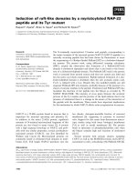
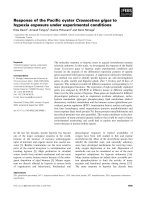
Measurements were made preoperatively, and immedi-
ately after dissection at the following locations: superior
mastectomy skin flap; lateral mastectomy skin flap;
medial mastectomy skin flap; inferior mastectomy skin
flap; and 2 cm inferior to the clavicle (Figure 1). Method
of reconstruction, mastectomy operative time, measure-
ments of the thickness of each skin flap, and length from
clavicle to superior edge of the mastectomy skin flap were
all recorded.
Measurements
Flap thickness was measured by allowing the skin to lie in
a neutral position against the chest wall and then utilizing
an intra-operative ruler to measure the skin flap at its most
distal aspect. Flap length was defined as the superior flap
length, this area was measured since this is typically the
longest flap in a mastectomy. It was measured by allowing
all skin to lie in a neutral position and measuring the dis-
tance, in cm, from the edge of the superior portion of the
incision at the 12 o'clock position to the clavicle, care was
taken to ensure that a straight line was maintained during
this measurement. All complications were noted; pres-
ence and total area of epidermolysis was noted and
recorded. Patients were followed for four weeks post-
operatively to estimate the area of necrosis, evaluate for
wound infection, and seroma formation. De-identified
data was entered into a Microsoft Excel
®
database. Statisti-
cal analysis was performed using Wilcoxon Rank Sum test
and Student's t-test.
World Journal of Surgical Oncology 2009, 7:91 />Page 3 of 6
(page number not for citation purposes)
Results
Of the 10 patients in this study, 1 (10%) developed signif-
icant mastectomy skin flap necrosis. Measurements were
obtained during the operation, the first one just prior to
dissection, and the 2
nd
at the completion of the mastec-
tomy, comparisons were then performed between these
numbers. Statistically significant factors predicting post-
op necrosis included reductions in medial (p = 0.0189)
and inferior (p = 0.003) StO
2
levels, and flap length (p =
0.009) (Table 1). In the patient who experienced necrosis,
medial StO
2
reduction was 61% (p = 0.049), correspond-
ing with an absolute medial StO
2
reduction of 42 points.
Patients who did not have necrosis actually had an
increase in their medial StO
2
of 14.6%, corresponding
with an absolute medial StO
2
increase of 6.7 points. The
patient with necrosis had a 69% decrease in inferior StO
2
levels, corresponding with a 65.5 point drop (p = 0.003).
Patients without necrosis demonstrated a 20% increase in
inferior StO
2
levels, corresponding with a 9.8 point
increase in absolute StO
2
levels. The patient with necrosis
had a 15 cm flap length, as opposed to a 11.9 cm average
flap length in the other 9 patients (p = 0.009).
Patient demographics are displayed in Table 2. Fifty per-
cent of patients were African-American, 40% were His-
panic, 10% were White. The average age was 49, average
body mass index (BMI) was 27.9. There were two patients
with diabetes and five with hypertension. None of the
patients had chronic obstructive pulmonary disease
(COPD) or admitted to smoking. Only one patient had
evidence of tumor skin involvement. The stage of the pri-
mary tumor ranged from DCIS to T4D. Three patients had
DCIS, and five had invasive ductal cancer. Five patients
had undergone neoadjuvant chemotherapy, and one had
previously received radiation to the chest wall. Average
operative time was 109 minutes (60-180 min), a factor
which was not significantly different between the two
groups. The one patient with necrosis did have an
expander in place, four of the patients without necrosis
also had expanders, all of these patients underwent skin-
sparing mastectomy. There were no nipple-sparing mas-
tectomies in this cohort. The remaining five patients did
not undergo immediate reconstruction and underwent
mastectomy with a standard elliptical incision. Operative
time, BMI, tumor pathology, tumor size, patient age and
operating surgeon were not significant factors in predict-
ing necrosis.
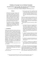
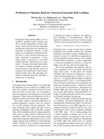
The patient with 108 cm
2
of necrosis (Figure 2) underwent
skin-sparing mastectomy, sentinel node biopsy and
immediate reconstruction with expander placement. The
expander was not filled intra-operatively. This patient had
uniquely significant drops in StO
2
measurements post-
operatively (Figure 2). This patient had full thickness
necrosis in several areas of the mastectomy skin flap. She
did have a personal history of Hepatitis C, sarcoidosis,
and hypertension. Intraoperative fluorescein dye injection
was also used to assess mastectomy skin flap viability and
did indicate a possible perfusion deficit at the 2 o' clock
position. Due to the overlying skin necrosis and conse-
quent exposed expander, she required expander removal
and skin graft two months after her mastectomy.
Discussion
One commonly used tool to evaluate mastectomy flap
viability intra-operatively is the intravenous sodium fluo-
rescein test (Wood's lamp method). This involves intrave-
nous injection of fluorescein followed by intra-operative
evaluation with a Wood's lamp. Although it has been
available since 1931, its application is prone to subjective
errors, and is limited to over/under reading by as much as
30% [10]. It is also a test of vascularity - not viability, and
subject to changes in vascularity such as vasospasm, intra-
vascular clotting, or alterations in the distribution of the
microcirculation. Alternatively the use of infrared spec-
troscopy takes into account metabolic changes of the dis-
sected tissue, and potentially allows trends to be followed
for flap evaluation post-operatively.
The arterial supply of the breast is generally defined as an
anastomotic plexus of vessels originating from the axillary
artery, the internal mammary artery, the intercostal arter-
ies, and lateral thoracic artery. The contribution of each
individual artery and the consequences of vascular inter-
ruption are poorly understood, but the course of the
nerves and vessels may be related to the ligamentous
apparatus [11]. One such horizontal ligamentous suspen-
sion originates from the pectoral fascia along the 5
th
rib
[12]. Our finding that the decrease in perfusion from the
Cardinal points of measurement pre-operativelyFigure 1
Cardinal points of measurement pre-operatively.
World Journal of Surgical Oncology 2009, 7:91 />Page 4 of 6
(page number not for citation purposes)
inferior portion of the breast most accurately predicted
post-operative epidermolysis may be supportive of this
finding.
In addition, there currently does not exist any standard-
ized method for measuring mastectomy skin flap thick-
ness during an operation, further refinements in this
technique-i.e. the use of calipers, may be helpful for future
trials.
Traditionally, surgeons are careful to avoid transection of
medial perforators. Consistent with this, our data demon-
strate an increased likelihood of necrosis in the patient
who had a significant decrease in medial StO
2
measure-
ments. This may be particularly important in those
patients who undergo disruption of the medial perfora-
tors secondary to internal mammary node dissection.
There are significant limitations to this study. Most nota-
ble is the small sample size. Contributions from underly-
ing co-morbidities (coronary artery disease, diabetes) may
be more readily apparent with a larger sample size. In
addition, this study population was predominately a
minority population; there is an under-representation of
Caucasian patients. Although the ViOptix T.Ox Tissue
Oximeter system has been validated in several racial
groups, there may be variability in StO
2
measurements
between races which can only be further elucidated with a
large sample size. For further studies, assuming a 10%
necrosis rate, a sample of 40 patients will provide more
than 90% power to detect a two standard deviation differ-
ence of the mean StO
2
measures (significance level is held
at 0.05, two sided). Clearly a group of patients undergoing
skin-sparing mastectomy with immediate reconstruction
Table 1: Analysis of patients with and without necrosis
Necrosis
Yes (1) No (9) p-value
Age 57 48 0.278
Seroma 01
Infection 01
Diabetes 02
Radiation 01
Hypertension 14
Flap Length (cm) 15 11.9 0.009
Thickness of flap (mm)
Superior 3.0 4.2 0.201
Inferior 3.0 4.4 0.100
Lateral 4.0 40 1.000
Medial 4.0 4.0 1.000
Pre-operative Tissue Oxygenation (StO2)
Superior 59.0 60.9 0.910
Inferior 94.0 49.1 0.0017
Lateral 73.5 58.2 0.371
Medial 68.5 58.5 0.540
Post-operative Tissue Oxygenation (StO2)
Superior 28 54.4 0.083
Inferior 29 59.0 0.199
Lateral 50 62.2 .0586
Medial 27 65.2 0.058
Changes in Tissue Oxygenation
StO2 percent change (absolute StO2 change)
Superior -53% (-31.5) -5.9% (-6.5) 0.280
Inferior -69% (-65.5) +20% (+9.8) 0.003
Lateral -32% (-23.5) +7.17% (+4.1) 0.145
Medial -61% (-42) +14.6% (+6.7) 0.018
Clavicular +6% (+2) +14.6% (+6.8) 0.850
Variables analyzed, statistically significant variables are bold and
italicized
Table 2: Patient Demographics
Factor % (n)
Average Age 49
Race
White 10% (1)
African American 50% (5)
Hispanic 40% (4)
Body Mass Index (BMI)
Average 27.9
Smoking 0% (0)
Co-morbidities
Diabetes 20% (2)
Hypertension 50% (5)
COPD 0% (0)
Skin Involvement
None 80% (8)
Skin retraction 10% (1)
Clinical T Size
Tis 30% (3)
T0 10% (1)
T1 30% (3)
T3 10% (1)
T4A 10% (1)
T4D 10% (1)
Histology
DCIS 30% (3)
Invasive Ductal 50% (5)
Invasive Lobular 10% (1)
Other 10% (1)
Neoadjuvant Chemo 50% (5)
Radiation to Chest Wall 10%(10)
World Journal of Surgical Oncology 2009, 7:91 />Page 5 of 6
(page number not for citation purposes)
would provide the most useful clinical information as
these patients are more likely to have difficulties with
wound healing and face the greatest consequences
(implant extrusion, flap failure) from poor wound heal-
ing.
It is known that the perfusion to the subdermal plexus of
the skin is controlled by the autonomic nervous system in
response to variations in metabolic demands and envi-
ronment. All patients in this study were stable intra-oper-
atively. However, the actual oxygen saturation and blood
pressure measurements at the time of StO
2
measurement
were not evaluated, the influence of these factors will be
examined in future studies. The patient with necrosis had
drops in StO
2
measurement, which also may be an indica-
tor of failure to compensate for injury, whereas the
patients who did not have necrosis, for the most part, had
increased StO
2
levels after dissection, potentially indicat-
ing an ability to increase perfusion appropriately to the
area of injury.
Similarly, wound healing is a complicated process. Factors
contributing to or complicating the wound healing proc-
ess include body habitus, age, co-morbidities, prolonged
operative time, collagen disorders, infection, history of
radiation exposure, immune status, and steroid use [13-
15].
Lastly, a review of the patient response to the ViOptix
T.Ox Tissue Oximeter system indicates that the patient
having necrosis also had a longer flap length. This would
appear to be consistent with the concept that the blood
supply of longer flaps is more tenuous, likely due to the
greater area of vascular disruption required when a mas-
tectomy is performed.
Conclusion
Commonly used intraoperative methods to determine
flap viability include detection of skin discoloration,
wound edge bleeding and intra-operative assessment with
fluorescein and a Wood's Lamp. The use of near-infrared
reflection spectroscopy to monitor myocutaneous flaps
has been previously validated in humans [9]. Our study
indicates that ViOptix T.Ox Tissue Oximeter is a non-inva-
sive method which may be utilized to identify impaired
perfusion in mastectomy skin flaps. It could potentially
add valuable information to clinical observation, and
may be able to detect early vascular complications. Areas
which demonstrate sub-optimal perfusion can therefore
be excised intra-operatively to potentially decrease wound
complications and improve cosmetic outcome, alterna-
tively, reconstruction may also be postponed until a later
date or potentially an autologous reconstruction may be
considered. Further studies are planned with a larger sam-
ple size for validation, and to establish standards.
Consent
Written informed consent was obtained from the patient
for publication of this case report and accompanying
images. A copy of the written consent is available for
review by the Editor-In-Chief of this journal.
Competing interests
The authors declare that they have no competing interests.
Authors' contributions
RR initiated this research & enrolled patients, & wrote the
initial manuscript, MS-C designed the study, assisted with
writing the manuscript & enrolled patients, AMTM col-
lected data and wrote portions of the manuscript, MB col-
lected data, DH collected data and assisted with study
design, VA enrolled patients and performed measure-
ments, X-JX performed all statistical analysis, TZ enrolled
patients and performed measurements, RR enrolled
patients and assisted with manuscript writing. All authors
have read and approved the final manuscript.
Acknowledgements
The authors are grateful to the invaluable assistance of our colleagues: Wil-
liam Brooks MD, Fiemu Nwariaku MD, Lisa Lilley NP, William Lodrigues
NP, Victoria Warren RN, and Fatemah Youssefi PhD.
A patient with significant intraoperative decrease in StO
2
Figure 2
A patient with significant intraoperative decrease in
StO
2
. Decreases were: 53%, 69%, 61%, and 32% at superior,
inferior, medial, and lateral, respectively.
53%
69%
61% 32%
Publish with Bio Med Central and every
scientist can read your work free of charge
"BioMed Central will be the most significant development for
disseminating the results of biomedical research in our lifetime."
Sir Paul Nurse, Cancer Research UK
Your research papers will be:
available free of charge to the entire biomedical community
peer reviewed and published immediately upon acceptance
cited in PubMed and archived on PubMed Central
yours — you keep the copyright
Submit your manuscript here:
/>BioMedcentral
World Journal of Surgical Oncology 2009, 7:91 />Page 6 of 6
(page number not for citation purposes)
Presented at the 24
th
Annual Miami Breast Cancer Conference, February
21
st
-24
th
, 2007. Miami, FL.
Presented at the 9
th
Annual University of Texas Southwestern Department
of Surgery Surgical Research Forum, June 6, 2007. Dallas, TX.
References
1. Malin JL, Schneider EC, Epstein AM, Adams J, Emanuel EJ, Kahn KL:
Results of the National Initiative for Cancer Care Quality:
how can we improve the quality of cancer care in the United
States? J Clin Oncol 2006, 24:626-634.
2. Christian CK, Niland J, Edge SB, Ottesen RA, Hughes ME, Theriault
R, Wilson J, Hergrueter CA, Weeks JC: A multi-institutional anal-
ysis of the socioeconomic determinants of breast recon-
struction: a study of the National Comprehensive Cancer
Network. Ann Surg 2006, 243:241-249.
3. Rowland JH, Desmond KA, Meyerowitz BE, Belin TR, Wyatt GE,
Ganz PA: Role of breast reconstructive surgery in physical and
emotional outcomes among breast cancer survivors. J Natl
Cancer Inst 2000, 92:1422-1429.
4. Mortenson MM, Schneider PD, Khatri VP, Stevenson TR, Whetzel TP,
Sommerhaug EJ, Goodnight JE Jr, Bold RJ: Immediate breast
reconstruction after mastectomy increases wound compli-
cations: however, initiation of adjuvant chemotherapy is not
delayed. Arch Surg 2004, 139:988-991.
5. Margulies AG, Hochberg J, Kepple J, Henry-Tillman RS, Westbrook K,
Klimberg VS: Total skin-sparing mastectomy without preser-
vation of the nipple-areola complex. Am J Surg 2005,
190:907-912.
6. Garwood ER, Moore D, Ewing C, Hwang ES, Alvarado M, Foster RD,
Esserman LJ: Total skin-sparing mastectomy: complications
and local recurrence rates in 2 cohorts of patients. Ann Surg
2009, 249:26-32.
7. Scheufler O, Exner K, Andresen R: Investigation of TRAM flap
oxygenation and perfusion by near-infrared reflection spec-
troscopy and color-coded duplex sonography. Plast Reconstr
Surg 2004, 113:141-152. discussion 153-145
8. Stranc MF, Sowa MG, Abdulrauf B, Mantsch HH: Assessment of tis-
sue viability using near-infrared spectroscopy. Br J Plast Surg
1998, 51:210-217.
9. Mancini DM, Bolinger L, Li H, Kendrick K, Chance B, Wilson JR: Val-
idation of near-infrared spectroscopy in humans. J Appl Physiol
1994, 77:2740-2747.
10. Myers B, Donovan W: An evaluation of eight methods of using
fluorescein to predict the viability of skin flaps in the pig.
Plast
Reconstr Surg 1985, 75:245-250.
11. Wuringer E, Mader N, Posch E, Holle J: Nerve and vessel supply-
ing ligamentous suspension of the mammary gland. Plast
Reconstr Surg 1998, 101:1486-1493.
12. Wueringer E, Tschabitscher M: New aspects of the topographi-
cal anatomy of the mammary gland regarding its neurovas-
cular supply along a regular ligamentous suspension. Eur J
Morphol 2002, 40:181-189.
13. Cruse PJ, Foord R: A five-year prospective study of 23,649 sur-
gical wounds. Arch Surg 1973, 107:206-210.
14. Cruse PJ, Foord R: The epidemiology of wound infection. A 10-
year prospective study of 62,939 wounds. Surg Clin North Am
1980, 60:27-40.
15. Nandi PL, Soundara Rajan S, Mak KC, Chan SC, So YP: Surgical
wound infection. Hong Kong Med J 1999, 5:82-86.