Báo cáo y học: "γ Paradoxical roles of IFN-γ in models of Th1-mediated autoimmunity" potx
Bạn đang xem bản rút gọn của tài liệu. Xem và tải ngay bản đầy đủ của tài liệu tại đây (47.48 KB, 4 trang )
333
CIA = collagen-induced arthritis; IL = interleukin; IFN-γ = interferon γ; IFN-γR = interferon γ receptor; MHC = major histocompatibility complex; PG
= proteoglycan; PGIA = proteoglycan-induced arthritis; R = receptor; RA = rheumatoid arthritis; Th = T helper cell.
Available online />Introduction
Cytokines play critical roles in regulating the outcome of
antigen-specific T-cell responses, and thus have been a
major focus in the study of the pathogenesis of auto-
immunity. On the basis of the original description by
Mosmann et al. [1], we know that the cytokine profile of a
T-cell response to an antigen is indicative of which T
helper (Th) cell pathway is stimulated by the antigen-pre-
senting cell. Th1 responses, generally characterized as
cell-mediated immune responses, are identified primarily
by the presence of IL-12, IL-2 and IFN-γ, whereas Th2
responses, generally characterized as humoral responses,
are defined primarily by the production of IL-4 and IL-10.
In addition to these characterizations, there is convincing
evidence that these two pathways are antagonistic, in
other words Th1 cytokines repress Th2 responses, and
Th2 cytokines repress Th1 responses. Learning how to
regulate these responses therapeutically, therefore, has
become an important focus in autoimmunity research.
Most autoimmune diseases and models of autoimmunity in
which susceptibility is associated with the expression of
specific MHC class II allotypes appear to be of the Th1
type, based on these cytokine definitions of Th function.
Thus considerable emphasis has been placed on develop-
ing means of altering the course of the autoimmune Th1
response to become that of a Th2 response, with the goal
of downregulating the autoimmune pathogenesis. These
cytokine networks not only influence the function of T cells
involved in the pathogenesis of the autoimmune disease,
however, but also affect qualitative differences in the anti-
body responses that are often associated with autoim-
mune disease, or, in some cases, represent the actual
pathogenic mechanism. For example, IFN-γ, a cytokine
strongly associated with a Th1 response, is an important
regulator of the production of IgG2a antibody, a subclass
frequently associated with a pathogenic autoantibody
response, while IgG1 production (promoted by IL-4) pre-
dominates in a Th2 response. IFN-γ, therefore, has been
T-cell responses to antigens are classified on the basis of the cytokines they produce as either Th1
(IFN-γ, IL-2) or Th2 (IL-4, IL-10), with these Th types being indicative of either cell-mediated or
antibody-mediated responses, respectively. Using this classification, T-cell responses in MHC-class-II-
restricted autoimmune diseases appear to be predominantly of the Th1 type, based on the presence of
high levels of IFN-γ. This simplistic classification has recently been challenged, however, as disease
incidence and severity are frequently elevated in animals that have a deficient IFN-γ response. The
recent data discussed here indicate that the cytokine circuits involved in the regulation of cell-mediated
and humoral immune responses during the development of autoimmune arthritis are more complex
than originally proposed; perhaps our characterization of autoimmune responses as strictly Th1 or Th2
is overly simplistic, especially as it pertains to the role of IFN-γ.
Keywords: arthritis, autoimmunity, cytokines, IFN-γ
Commentary
Paradoxical roles of IFN-
γγ
in models of Th1-mediated
autoimmunity
Edward F Rosloniec
1,2,3
, Kary Latham
3
and Yajaira B Guedez
2
1
VA Medical Center, Research Service (151), Memphis, Tennessee, USA
2
Department of Medicine, University of Tennessee Health Science Center, Memphis, Tennessee, USA
3
Department of Pathology, University of Tennessee Health Science Center, Memphis, Tennessee, USA
Corresponding author: Edward F Rosloniec (e-mail: )
Received: 13 May 2002 Revisions received: 3 July 2002 Accepted: 3 July 2002 Published: 17 July 2002
Arthritis Res 2002, 4:333-336 (DOI 10.1186.ar432)
© 2002 BioMed Central Ltd (
Print ISSN 1465-9905; Online ISSN 1465-9913)
Abstract
334
Arthritis Research Vol 4 No 6 Rosloniec et al.
considered a prime target for modulating autoimmunity,
with the hypothesis being that if IFN-γ expression can be
downregulated, then both the Th pathway and the produc-
tion of pathogenic autoantibody can be altered. Data gen-
erated using models of autoimmunity have revealed a
much more complex role of the Th1 cytokine IFN-γ in
autoimmune pathogenesis than expected. As we discuss
below for several models of autoimmunity, IFN-γ, despite
being a component of a ‘pathogenic’ Th1 response, can
also play a protective role in the development of an
autoimmune response. Furthermore, the regulation of
IgG2a and IgG1 isotypes appears to be more complex
than previously thought.
IFN-
γγ
and models of autoimmunity
The vast majority of autoimmune diseases studied and their
corresponding animal models have been characterized as
being mediated by the Th1 pathway, based on cytokine
expression patterns in lymphoid and targeted tissues.
Although the original paradigm of Th1 and Th2 cell function
described a distinct separation of cytokines and function in
T-cell-dependent immune responses, through the use of a
number of models of autoimmunity it has become clear that
these stereotyped roles do not accurately reflect the com-
plexities of the biological immune responses. These com-
plexities are perhaps best demonstrated in the analysis of
the role of IFN-γ in models of autoimmunity. Early attempts
to define the role of IFN-γ in autoimmunity by the adminis-
tration of IFN-γ or neutralizing antibodies specific for IFN-γ
yielded conflicting results. In some experimental systems
disease was accelerated, while in others it was prevented,
with these differences being confounded by a number of
experimental variables. The use of models in which either
IFN-γ or the IFN-γ receptor (IFN-γR) had been genetically
deleted revealed that, in the absence of a competent IFN-γ
response, disease incidence and severity were enhanced
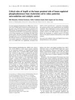
in the majority of autoimmune models tested (Table 1).
These data include both models in which T cells are the
primary mechanism of pathogenesis [2–4] as well as those
in which autoantibody is the primary pathogenic mecha-
nism [5–8]. Yet perhaps even more puzzling is that, for at
least two models with seemingly different pathogenic
mechanisms (experimental myasthenia gravis [9,10] and
proteoglycan-induced arthritis [11]), expression of the
experimental disease was found to be dependent on IFN-γ.
IFN-
γγ
in collagen-induced autoimmune
arthritis
One model of autoimmunity in which the role of IFN-γ has
been studied in detail is collagen-induced arthritis (CIA) in
the mouse. In this model, an autoimmune arthritis resem-
bling rheumatoid arthritis (RA) is induced by immunization
of genetically susceptible strains of mice with type II colla-
gen [12]. Disease susceptibility is restricted by the murine
class II molecule I-A (specifically I-A
r
and I-A
q
haplotypes)
[13], and subsequently, CD4
+
T cells play a central role in
the immunopathogenesis of this experimental autoimmune
disease. The collagen type II (CII)-specific T-cell response
is predominated by Th1 cells producing IL-2 and IFN-γ
that in turn drive the production of complement-fixing CII-
specific IgG2a, a major component in the pathogenesis of
this experimental disease [14].
Early attempts to define the role of IFN-γ in CIA by the
administration of IFN-γ or neutralizing antibodies specific
for IFN-γ yielded conflicting results [15–19], probably
because of variations in timing, sites and means of admin-
istration. Studies of CIA development in genetically sus-
ceptible mice in which IFN-γ (Y Guedez and E Rosloniec,
unpublished observations) or the IFN-γ receptor (IFN-γR)
[6,7] had been deleted revealed that autoimmune arthritis
develops faster and is more severe in the absence of an
IFN-γ response. Despite efforts by numerous investigators,
a clear consensus on how the absence of a potent Th1
cytokine such as IFN-γ renders an animal more suscepti-
ble to a Th1-mediated autoimmune response is still
lacking. Although it might be predicted that, in the
absence of IFN-γ, there would be a compensatory
increase in Th2 cytokines, such as IL-4 and IL-10, no evi-
dence for enhanced expression of any of the Th2
Table 1
Role of IFN-
γγ
in various models of autoimmunity.
Immunopathogenic Genetic Effect on disease
Autoimmune model mechanism deletion incidence or severity References
Experimental autoimmune encephalomyelitis T cells IFN-γ Increased [10]
Experimental autoimmune myasthenia gravis Antibody IFN-γR Decreased [10]
Experimental autoimmune thyroiditis T cells IFN-γR Accelerated, less severe [5]
Collagen-induced arthritis Antibody IFN-γR & IFN-γ Increased [6–8]
Uveitis T cells IFN-γ Increased [4]
Proteoglycan-induced arthritis T cells IFN-γ Decreased [11]
335
cytokines in these models has been demonstrated. Thus,
despite the association of CIA with a strong Th1
response, the absence of an IFN-γ response in genetically
susceptible mice enhances the development of auto-
immune arthritis, and this occurs despite the lack of CII-
specific IgG2a that has been presumed to be a major
factor in the initiation of the pathogenesis.
Analogous to the paradoxical role of IFN-γ in CIA is the
apparent surprising role of its counterpart, IL-4. When the
function of IL-4 was neutralized either by antibody admin-
istration or genetic deletion, the onset and severity of CIA
were greatly reduced [20]. Similar results were obtained
in a complementary approach using DBA/1 mice express-
ing an IL-2Rb/IL-4R chimeric transgene. In this approach,
IL-2 binding of the receptor transmits a signal via the IL-4
pathway [21]. Like the IFN-γ-deficient mice, arthritis devel-
oped in these chimeric transgenic mice at an accelerated
rate and with increased severity. The autoimmune disease
was associated with an increase in type 2 cytokines (IL-4,
IL-5, IL-10), and an increase in CII-specific IgG1 levels, with
IgG2a levels comparable to those in nontransgenic mice.
Despite the elevated levels of Th2 cytokines, however, IFN-γ
production was not significantly affected, again indicating
the complex relationships among these mediators.
A regulatory role of IFN-γ in models of autoimmune arthritis
is also supported by studies using strains genetically non-
susceptible to CIA. Although CIA susceptibility is
restricted to strains expressing H-2
q
and H-2
r
class II
alleles, other strains, such as C57BL/6 (B6, H-2
b
),
develop marginal T-cell and B-cell immune responses to
CII without developing autoimmune arthritis [8,13]. Yet
when IFN-γ is genetically deleted from the B6 genome (B6
IFN-γ
–/–
), these mice become acutely susceptible to the
development of CIA [8,20]. The arthritis in the B6 IFN-γ
–/–
mice is accompanied by an enhanced T-cell response and
high amounts of IgG1 and IgG2b CII-specific antibody.
Like the studies in the CIA susceptible models, cytokine
analysis did not reveal any significant changes in the
remaining Th1 or Th2 cytokines but did reveal elevated
levels of IL-1β in the lymph nodes and synovial cells of
arthritic B6 IFN-γ
–/–
mice. The elevated levels of IL-1β
appear to be important for the development of the
disease, as treatment of B6 IFN-γ
–/–
mice with anti-IL-1β
significantly reduced the incidence and the severity of the
arthritis [8]. In all, these data serve as a clear example of
the complexity of both the dynamics of the cytokine milieu
as well as the complex relationships that exist between the
Th1 and Th2 cytokines regulating the development of an
autoimmune and inflammatory response.
Role of IFN-
γγ
in proteoglycan-induced arthritis
Recently, Kaplan et al. [11] examined the role of IFN-γ in
another model of autoimmune arthritis, proteoglycan-
induced arthritis (PGIA). Like CIA, the induction of PGIA is
based upon Th1-mediated cross-reactive immune
responses between the heterologous immunogen (proteo-
glycan) and the self-antigen located in the articular joints
[22–24]. The arthritis in PGIA is characterized by a pro-
gressive disease course with intermittent exacerbations
and remissions reminiscent of the clinical appearance of
RA. To date, only Balb/c mice have been found to be sus-
ceptible to PGIA [22–24], which is interesting in that this
strain has a genetic predisposition to generating Th2
responses [25]. Although PGIA is considered a Th1-medi-
ated experimental disease, it is clear that the immuno-
pathogenesis involves a complex pattern of Th1 and Th2
cytokines with elevated levels of PG-specific IgG1 domi-
nating in comparison to IgG2a, yet a strong predominance
of IFN-γ over IL-4 in inflamed paws [11,26].
Despite the fact that both CIA and PGIA are considered
to be Th1 models of RA, the role of IFN-γ appears to be
totally different in these two models. Based on the recent
report by Kaplan et al. [11], Balb/c mice genetically defi-
cient in IFN-γ (knockout) are resistant to the development
of PGIA. Arthritis incidence and severity were both found
to be reduced in these mice in comparison to wild-type
Balb/c mice, and, as would be expected, the amount of
PG-specific IgG2a was also significantly decreased in the
IFN-γ-deficient mice. Thus these data indicate that IgG2a
is likely to be a major factor in the pathogenesis of this
model, despite the observation that IgG1 predominates in
the immune response to PG. These data were supported
by studies of PGIA in IL-4-deficient Balb/c mice [11]. In
the absence of IL-4, Balb/c mice developed an acceler-
ated and very severe PGIA that was accompanied by an
increase in IFN-γ and a sixfold increase in PG-specific
IgG2a. Surprisingly, the levels of PG-specific IgG1 were
only minimally decreased in the IL-4-deficient mice, sug-
gesting that, at least in Balb/c mice, IgG1 production may
be heavily influenced by yet other cytokines.
Conclusion
Animal models of autoimmune disease are providing a
valuable means of analyzing the functional roles of
cytokines in the pathogenesis of autoimmunity. The origi-
nal description of Th1 and Th2 responses has provided us
with a valuable framework for advancing our understand-
ing of pathogenic T cell responses, and we now are begin-
ning to understand the complexities that regulate these
responses. Although the data from Kaplan et al. [11]
provide some interesting insight into the development of a
pathogenic autoimmune response, they represent single
time point analyses, making it difficult to decipher the
complex regulation of the humoral responses and the
complex dynamics of the cytokine milieu present during
the development of autoimmunity. Multiple time point
analyses of antibody production and multiplexed cytokine
expression profiling may help to increase our understand-
ing of the complexities of the regulation of autoimmune
Available online />336
humoral responses and the role they play in mediating
susceptibility to autoimmune disease. For example, mice
treated with IL-18 or IL-18 plus IL-12 produced markedly
more collagen-specific IgG1 and IgG2a than did controls,
whereas IL-12 treatment alone enhances only the IgG2a
responses [27]. Regardless, it is clear from these data and
the studies from many others using murine models of
autoimmunity that cytokine circuits involved in the regula-
tion of humoral and cell-mediated immune responses in
the development of autoimmune diseases are more
complex than originally proposed, and perhaps our charac-
terization of autoimmune responses as Th1 or Th2 is overly
simplistic, especially as it pertains to the role of IFN-γ.
Acknowledgements
The work of the authors was supported in part by grants from the
Department of Veterans Affairs, Memphis, TN, USA and by US Public
Health Service Grants AR45201, AR47379, AR39166 from the
National Institute for Arthritis and Musculoskeletal Diseases.
References
1. Mosmann TR, Cherwinski H, Bond MW, Giedlin MA, Coffman RL:
Two types of murine helper T cell clone. I. Definition accord-
ing to profiles of lymphokine activities and secreted proteins.
J Immunol 1986, 136:2348-2357.
2. Ferber IA, Brocke S, Taylor-Edwards C, Ridgway W, Dinisco C,
Steinman L, Dalton D, Fathman CG: Mice with a disrupted IFN-
gamma gene are susceptible to the induction of experimental
autoimmune encephalomyelitis (EAE). J Immunol 1996, 156:5-7.
3. Caspi RR, Chan CC, Grubbs BG, Silver PB, Wiggert B, Parsa
CF, Bahmanyar S, Billiau A, Heremans H: Endogenous systemic
IFN-gamma has a protective role against ocular autoimmunity
in mice. J Immunol 1994, 152:890-899.
4. Jones LS, Rizzo LV, Agarwal RK, Tarrant TK, Chan CC, Wiggert
B, Caspi RR: IFN-gamma-deficient mice develop experimental
autoimmune uveitis in the. J Immunol 1997, 158:5997-6005.
5. Alimi E, Huang S, Brazillet MP, Charreire J: Experimental
autoimmune thyroiditis (EAT) in mice lacking the IFN-gamma
receptor gene. Eur J Immunol 1998, 28:201-208.
6. Vermeire K, Heremans H, Vandeputte M, Huang S, Billiau A,
Matthys P: Accelerated collagen-induced arthritis in IFN-
gamma receptor-deficient mice. J Immunol 1997, 158:5507-
5513.
7. Manoury-Schwartz B, Chiocchia G, Bessis N, Abehsira-Amar O,
Batteux F, Muller S, Huang S, Boissier MC, Fournier C: High sus-
ceptibility to collagen-induced arthritis in mice lacking IFN-
gamma receptors. J Immunol 1997, 158:5501-5506.
8. Guedez YB, Whittington KB, Clayton JL, Joosten LA, van de Loo
FA, van den Berg WB, Rosloniec EF: Genetic ablation of inter-
feron-gamma up-regulates interleukin-1beta expression and
enables the elicitation of collagen-induced arthritis in a non-
susceptible mouse strain. Arthritis Rheum 2001, 44:2413-
2424.
9. Zhang GX, Xiao BG, Bai XF, Orn A, van der Meide PH, Link H:
IFN-gamma is required to induce experimental autoimmune
myasthenia gravis. Ann NY Acad Sci 1998, 841:576-579.
10. Zhang GX, Xiao BG, Bai XF, van der Meide PH, Orn A, Link H:
Mice with IFN-gamma receptor deficiency are less suscepti-
ble to experimental autoimmune myasthenia gravis. J
Immunol 1999, 162:3775-3781.
11. Kaplan C, Valdez JC, Chandrasekaran R, Eibel H, Mikecz K, Glant
TT, Finnegan A: Th1 and Th2 cytokines regulate proteoglycan-
specific autoantibody isotypes and arthritis. Arthritis Res 2002,
4:54-58.
12. Courtenay JS, Dallman MJ, Dayan AD, Martin A, Mosedale B:
Immunisation against heterologous type II collagen induces
arthritis in mice. Nature 1980, 283:666-668.
13. Wooley PH, Luthra HS, Stuart JM, David CS: Type II collagen
induced arthritis in mice. I. Major histocompatibility complex (I
region) linkage and antibody correlates. J Exp Med 1981, 154:
688-700.
14. Terato K, Hasty KA, Reife RA, Cremer MA, Kang AH, Stuart JM:
Induction of arthritis with monoclonal antibodies to collagen. J
Immunol 1992, 148:2103-2108.
15. Mauritz NJ, Holmdahl R, Jonsson R, Van der Meide PH, Scheynius
A, Klareskog L: Treatment with gamma-interferon triggers the
onset of collagen arthritis in mice. Arthritis Rheum 1988, 31:
1297-1304.
16. Nakajima H, Takamori H, Hiyama Y, Tsukada W: The effect of
treatment with interferon-gamma on type II collagen-induced
arthritis. Clin Exp Immunol 1990, 81:441-445.
17. Boissier MC, Chiocchia G, Bessis N, Hajnal J, Garotta G, Nicoletti
F, Fournier C: Biphasic effect of interferon-gamma in murine
collagen-induced arthritis. Eur J Immunol 1995, 25:1184-1190.
18. Cooper SM, Sriram S, Ranges GE: Suppression of murine col-
lagen-induced arthritis with monoclonal anti-Ia antibodies and
augmentation with IFN- gamma. J Immunol 1988, 141:1958-
1962.
19. Williams RO, Williams DG, Feldmann M, Maini RN: Increased
limb involvement in murine collagen-induced arthritis follow-
ing treatment with anti-interferon-gamma. Clin Exp Immunol
1993, 92:323-327.
20. Ortmann RA, Shevach EM: Susceptibility to collagen-induced
arthritis: cytokine-mediated regulation. Clin Immunol 2001, 98:
109-118.
21. Chen Y, Rosloniec E, Goral MI, Boothby M, Chen J: Redirection
of T cell effector function in vivo and enhanced collagen-
induced arthritis mediated by an IL-2R beta/IL-4R alpha
chimeric cytokine receptor transgene. J Immunol 2001, 166:
4163-4169.
22. Finnegan A, Mikecz K, Tao P, Glant TT: Proteoglycan (aggre-
can)-induced arthritis in BALB/c mice is a Th1-type disease
regulated by Th2 cytokines. J Immunol 1999, 163:5383-5390.
23. Glant TT, Cs-Szabo G, Nagase H, Jacobs JJ, Mikecz K: Progres-
sive polyarthritis induced in BALB/c mice by aggrecan from
normal and osteoarthritic human cartilage. Arthritis Rheum
1998, 41:1007-1018.
24. Glant TT, Mikecz K, Arzoumanian A, Poole AR: Proteoglycan-
induced arthritis in BALB/c mice. Clinical features and
histopathology. Arthritis Rheum 1987, 30:201-212.
25. Reiner SL, Locksley RM: The regulation of immunity to Leish-
mania major. Annu Rev Immunol 1995, 13:151-177.
26. Hollo K, Glant TT, Garzo M, Finnegan A, Mikecz K, Buzas E:
Complex pattern of Th1 and Th2 activation with a preferential
increase of autoreactive Th1 cells in BALB/c mice with pro-
teoglycan (aggrecan)-induced arthritis. Clin Exp Immunol
2000, 120:167-173.
27. Leung BP, McInnes IB, Esfandiari E, Wei XQ, Liew FY: Com-
bined effects of IL-12 and IL-18 on the induction of collagen-
induced arthritis. J Immunol 2000, 164:6495-6502.
Correspondence
Dr Edward F Rosloniec, VA Medical Center, Research Service (151),
1030 Jefferson Avenue, Memphis, TN 38104, USA. Tel: +1 (901) 577-
7281; fax: +1 (901) 577-7273; e-mail:
Arthritis Research Vol 4 No 6 Rosloniec et al.