Báo cáo y học: "Elevated matrix metalloproteinase-9 in patients with systemic sclerosi" doc
Bạn đang xem bản rút gọn của tài liệu. Xem và tải ngay bản đầy đủ của tài liệu tại đây (394.86 KB, 9 trang )
Open Access
Available online />R71
Vol 7 No 1
Research article
Elevated matrix metalloproteinase-9 in patients with systemic
sclerosis
Wan-Uk Kim, So-Youn Min, Mi-La Cho, Kyung-Hee Hong, Yong-Joo Shin, Sung-Hwan Park and
Chul-Soo Cho
Division of Rheumatology, Department of Internal Medicine, Catholic Research Institutes of Medical Science, School of Medicine, The Catholic
University of Korea, Seoul, Republic of Korea
Corresponding author: Chul-Soo Cho,
Received: 19 Mar 2004 Revisions requested: 19 Apr 2004 Revisions received: 23 Sep 2004 Accepted: 1 Oct 2004 Published: 10 Nov 2004
Arthritis Res Ther 2005, 7:R71-R79 (DOI 10.1186/ar1454)
http://arthrit is-research.com /content/7/1/R 71
© 2004 Kim et al.; licensee BioMed Central Ltd.
This is an Open Access article distributed under the terms of the Creative Commons Attribution License ( />2.0), which permits unrestricted use, distribution, and reproduction in any medium, provided the original work is cited.
Abstract
Matrix metalloproteinase-9 (MMP-9) has been implicated in the
pathogenesis of cancer, autoimmune disease, and various
pathologic conditions characterized by excessive fibrosis. In this
study, we investigated the expression of MMP-9 and its clinical
significance in systemic sclerosis (SSc). The patients (n = 42)
with SSc had higher concentrations of MMP-9 and of tissue
inhibitor of metalloproteinase-1 (TIMP-1) and a higher ratio of
MMP-9 to TIMP-1 in sera than healthy controls (n = 32). Serum
MMP-9 concentrations were significantly higher in the diffuse
type (n = 23) than the limited type of SSc (n = 19). Serum
concentrations of MMP-9 correlated well with the degree of skin
involvement, as determined by the Rodnan score and with
serum concentrations of transforming growth factor β.
Moreover, dermal fibroblasts from patients with SSc produced
more MMP-9 than those from healthy controls when they were
stimulated with IL-1β, tumor necrosis factor α, or transforming
growth factor β. Such an increase in MMP-9 production was
partially blocked by treatment with cyclosporin A. In summary,
the serum MMP-9 concentrations were elevated in SSc patients
and correlated well with skin scores. The increased MMP-9
concentrations may be attributable to overproduction by dermal
fibroblasts in SSc. These findings suggest that the enhanced
production of MMP-9 may contribute to fibrogenic remodeling
during the progression of skin sclerosis in SSc.
Keywords: dermal fibroblasts, metalloproteinase-9, skin score, systemic sclerosis
Introduction
Systemic sclerosis (SSc) is a generalized disorder of con-
nective tissue characterized by microvacular damage and
excessive fibrosis in the skin and internal organs, including
the heart, lungs, and gastrointestinal tract. One of the major
hallmarks of the disease is an increased amount of collagen
deposits in the affected tissue. The relative proportion of
two major types of skin procollagen, types I and III, is higher
in SSc lesions than in healthy controls [1,2]. This increase
in collagen deposits may be associated with changes in the
dermal microvasculature in SSc. In particular, alterations in
the structure of the basement membrane, a critical compo-
nent of the vessel, may lead to changes in the surrounding
tissue and to subsequent development of fibrosis in SSc
[3]. The finding that the synthesis of type IV collagen, a
major collagen type in basement membrane, is dispropor-
tionately increased in the dermal fibroblasts and sera of
patients with SSc supports this notion [4,5].
The enhanced expression of matrix collagen is presumably
associated with abnormal immune responses to collagen in
SSc [6-10]. For example, autoantibodies to type IV colla-
gen have been observed in some SSc patients and may be
involved in endothelial injury [7,8]. Immunization of mice
with autologous type IV collagen leads to the activation of
fibroblasts and to fibrosis [9]. Furthermore, type IV collagen
activates T cells from patients with SSc [10], suggesting
that the selective immunity to type IV collagen may influ-
ence the clinical expression of SSc. The excessive produc-
tion of type IV collagen and subsequent autoimmune T-cell
responses to type IV collagen may set off a self-perpetuat-
CsA = cyclosporin A; DMEM = Dulbecco's modified Eagle's medium; ELISA = enzyme-linked immunosorbent assay; IL-1β = interleukin-1β; MMP =
matrix metalloproteinase; PBS = phosphate-buffered saline; SSc = systemic sclerosis; TGFβ = transforming growth factor β; TIMP = tissue inhibitor
of metalloproteinase; TNF-α = tumor necrosis factor α.
Arthritis Research & Therapy Vol 7 No 1 Kim et al.
R72
ing cycle in SSc through the interaction between lym-
phocytes and fibroblasts.
The matrix metalloproteinases (MMPs) are a family of extra-
cellular endopeptidases that selectively degrade the com-
ponents of various extracellular matrixes. Of these, MMP-9
(92–96 kD gelatinase B), whose substrates include type IV
collagen in basement membrane [11], has been thought to
be involved in the cellular invasion of the basement mem-
brane by cells involved in arthritis and cancer (e.g. T cells,
mononuclear phagocytes, synovial fibroblasts, and meta-
static tumor cells) [12-15]. MMP-9 has been associated
with chronic inflammatory autoimmune diseases, including
rheumatoid arthritis, Sjögren's syndrome, idiopathic uveitis,
and systemic lupus erythematosus [16-19]. Moreover, the
overexpression of MMP-9 has been reported in various
pathologic conditions characterized by excessive fibrosis,
including idiopathic pulmonary fibrosis, bronchial asthma,
experimental biliary cirrhosis, and chronic pancreatitis [20-
23], suggesting that elevated MMP-9 is closely linked to
fibrogenic remodeling in target organs. In the present
study, we measured the expression of MMP-9 and tissue
inhibitor of metalloproteinase-1 (TIMP-1), an inhibitor of
MMP-9, in the sera and culture supernatants of dermal
fibroblasts from SSc patients and compared them with
serum concentrations of transforming growth factor β
(TGFβ) and with clinical and laboratory parameters of SSc.
Materials and methods
Patients
This study was conducted in accordance with the princi-
ples embodied in the Declaration of Helsinki and was
approved by the Ethical Committees in the Catholic
Research Institutes of Medical Sciences. Before the study,
informed consent was obtained from all patients and
healthy controls. Forty-two patients (1 man and 41 women),
all of whom fulfilled the criteria of the American Rheuma-
tism Association for SSc [24], were studied; their mean
age was 43.7 years (range 24–69 years). The mean dura-
tion of disease was 80.8 months (range 5–276 months).
The comparisons were made with 32 healthy controls (all
women) who had no rheumatic disease; their mean age
was 44.2 years (range 21–62 years). The ages and sexes
of the patient and control groups did not differ significantly.
Clinical and laboratory evaluation
Clinical and laboratory assessments were done at the time
of sampling. The clinical variables were age, sex, disease
duration, type of SSc [25], modified Rodan score [26],
presence or absence of esophageal involvement on endos-
copy and esophageal manometry, interstitial lung disease
on chest radioagrapy and/or high-resolution computerized
tomography, diffusion capacity (DLCO; diffusion of carbon
monoxide in the lung) on the pulmonary function test, arthri-
tis, sicca syndrome, and antibodies to Scl-70 or centro-
mere using ELISA kits (MBL, Nagoya, Japan). Interstitial
lung disease was defined as bibasilar interstitial fibrosis on
chest radiographs, or, in patients with no abnormalities on
chest radiographs, as the presence of alveolitis on high-
resolution computerized tomography.
ELISA for serum MMP-9, TIMP-1, and TGFβ
The total MMP-9 and TIMP-1 concentrations were deter-
mined in the serum and the culture supernatant using a
commercial ELISA kit (R&D Systems Inc, Minneapolis, MN,
USA). In accordance with the manufacturer's recommen-
dations, the aliquots of serum were diluted to a ratio of
1:100 in the assay buffer. The detection limits of the MMP-
9 and TIMP-1 kits were 0.15 ng/ml and 0.08 ng/ml, respec-
tively. The MMP-9 assay kit detected pro-MMP-9 and com-
plexes of pro-MMP-9 with TIMP-1 and had no significant
cross-reactivity with MMP-1, MMP-2, MMP-3, TIMP-1, or
TIMP-2. Again, the TIMP-1 detection kit detected TIMP-1
either free or in complex with some MMPs and showed no
cross-reactivity or interference with TIMP-2.
Circulating TGFβ was measured in the same samples
using ELISA, as described previously [27]. Briefly, 2 µg/ml
of monoclonal antibodies to TGFβ1, β2, and β3 (R&D Sys-
tems) were added to 96-well plates (Nunc Inc, Roskilde,
Denmark). They were incubated overnight at 4°C and
blocked with PBS containing 1% bovine serum albumin
and 0.05% Tween 20 for 2 hours at room temperature. A
sample (50 µl) of each patient's serum was diluted 1:2 with
PBS, acidified with 50 µl of 2.5 M acetic acid and 10 M
urea for 10 minutes at room temperature and then was neu-
tralized with 50 µl of 2.7 M NaOH and 1 M HEPES. The
patient's sera and the standard recombinant TGFβ (R&D
Systems) were then put into 96-well plates and incubated
at room temperature for 2 hours. Biotinylated polyclonal
antibodies (50 ng/ml) to human TGFβ (R&D Systems) were
added and the reaction was allowed to proceed for 2 hours
at room temperature. Streptavidin–alkaline phosphatase
(Sigma Bioscience, St Louis, MO, USA) diluted 1:2000
with PBS was added, and the reaction was again allowed
to proceed for 2 hours. p-Nitrophenylphosphate (1 mg/ml)
(Sigma Bioscience) dissolved in diethanolamine (Sigma
Bioscience) was added to induce a color reaction, and 1 N
NaOH (Fisher Scientific, Pittsburgh, PA, USA) was used to
stop the reaction. An automated microplate reader (Vmax,
Molecular Devices, Palo Alto, CA, USA) was used to meas-
ure the optical density at 405 nm. Between each of these
steps, the plates were washed four times with PBS con-
taining 0.05% Tween 20. A standard curve was drawn by
plotting the optical density versus the log of the recom-
binant TGFβ concentration. The detection limit for TGFβ
was 30 pg/ml.
Available online />R73
Detection of MMP-9 activities by gel zymography
MMP-9 and MMP-2 activities were also tested by gelatin
zymography. A 0.5-µl sample of serum diluted in 30 µl of
SDS buffer was separated in 10% SDS–PAGE gel polym-
erized with 1 mg/ml gelatin (Invitrogen Life Technologies,
Carlsbad, CA, USA). Culture supernatants of HT1080 cell
lines (malignant human fibroblasts) stimulated with 10 µg/
ml of concanavalin A were used as a positive control. Gels
were washed once for 3 hours in 2.5% Triton X-100 to
remove the SDS and once for 30 minutes in the reaction
buffer containing 50 mM Tris/HCl, 200 mM NaCl, 10 mM
CaCl
2
, and 0.02% (w/v) Brij 35 (pH 7.5). The reaction
buffer was changed to a fresh one, and the gels were incu-
bated at 37°C for 24 hours. Gelatinolytic activity was visu-
alized by staining the gels with 0.5% Coomassie brilliant
blue and was quantified by densitometry.
Isolation and culture of dermal fibroblasts
Dermal fibroblasts were obtained from affected skin of two
SSc patients and from two healthy controls, as described
previously [28]. Fibroblasts were grown from explants in
Dulbecco's modified Eagle's medium (DMEM) at 37°C in
5% CO
2
. The cells were then centrifuged at 500 g, resus-
pended in DMEM supplemented with 10% fetal calf serum
(Gibco-BRL, Grand Island, NY, USA), 2 mM glutamine,
penicillin (100 U/ml), and streptomycin (100 µg/ml), and
plated in 25-cm
2
flasks. The cultures were kept at 37°C in
5% CO
2
and the culture medium was replaced every 3
days. When cells approached confluence, they were
detached with trypsin, passed after dilution 1:3 with fresh
medium, and recultured until use. Cells were housed in a
37°C humidified incubator with 5% CO
2
. Second- or third-
passage cells were used for all experiments. Fibroblasts
were seeded in 24-well plates at 5 × 10
4
cells per well in
serum-free DMEM supplemented with insulin–transferrin–
selenium A (ITSA; Gibco BRL). After the cells had been
grown in selected medium alone for 12 hours, we added
cytokines – IL-1β (10 ng/ml), tumor necrosis factor α (TNF-
α) (10 ng/ml), and TGFβ (10 ng/ml) – to stimulate the
fibroblasts. After 24 hours of incubation, cell-free media
were collected and stored at -20°C until assay. All cultures
were set up in triplicate or quadruplicate.
Statistics
Data are expressed as means ± standard error of the mean
(SEM). Numerical data for groups were compared using
the Mann–Whitney rank sum test, and data for categories
were compared using a chi-square test. Correlation
between two variables was tested using Spearman's rank
correlation coefficient. P values less than 0.05 were con-
sidered statistically significant.
Results
Elevated serum MMP-9 and TIMP-1 concentrations in
SSc patients
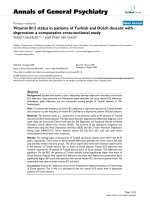
The serum concentrations of MMP-9 were significantly
higher in patients with SSc (n = 42) than in healthy controls
(n = 32) (317.6 ± 33.5 ng/ml versus 81.2 ± 6.8 ng/ml, P
< 0.001) (Fig. 1). The serum concentration of TIMP-1, an
inhibitor of MMP-9, was also higher in SSc patients than in
healthy controls (157.1 ± 13.2 ng/ml versus 77.7 ± 12.5
ng/ml, P < 0.001), but SSc patients had higher MMP-9/
TIMP-1 ratios than healthy controls (233.0 ± 27.1 versus
69.5 ± 24.3, P < 0.001). There was no correlation between
MMP-9 and TIMP-1 concentrations in SSc patients or in
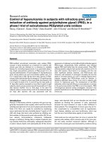
healthy controls. SSc patients with the diffuse type (n = 23)
and had higher concentrations of MMP-9 than those with
the limited type (n = 19) (364.6 ± 32.4 ng/ml versus 260.0
± 34.6 ng/ml, P = 0.034) (Fig. 2). No significant differ-
ences were found between the two groups of patients with
regard to age, sex, disease duration, and prednisolone
usage or the kinds of immunosuppressive agents being
used (e.g. D-penicillamine and cyclosphosphamide) (Table
1).
Table 1
Demographics of patients with systemic sclerosis (SSc), and medications being taken
With diffuse SSc
a
(n = 23) With limited SSc
a
(n = 19)
Age (y) [mean ± SEM] 42.5 ± 2.8 46.5 ± 2.9
% female 95.7 100
Disease duration (mo.) [mean ± SEM] 72.7 ± 15.7 95.4 ± 17.1
Medications (% of sample)
Prednisone 82.6 68.4
D-penicillamine 69.6 57.9
Cyclophosphamide 17.4 5.3
a
P ≥ 0.05 in a comparison of the two groups (Mann–Whitney rank sum test). SEM, standard error of the mean.
Arthritis Research & Therapy Vol 7 No 1 Kim et al.
R74
MMP-9 activities measured by gel zymography
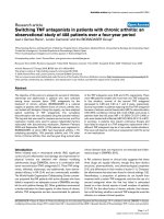
We used gel zymography to study sera of 20 SSc patients
and 10 healthy controls, all selected unsystematically, to
ascertain the serum gelatinase activity of MMP-9. As can
be seen in Fig. 3, the 92 kDa band, consistent with the
latent form of MMP-9, was detected in the sera of all
subjects. The bands in Fig. 3 represent the latent form of
MMP-9 (92 kDa, upper band) and the latent form of MMP-
2 (72 kDa, lower band). The serum MMP-9 activities of SSc
patients were higher than those of healthy controls. Densi-
tometric analysis in sera of 20 SSc patients and 10 healthy
controls indicated that the mean MMP-9 activity for SSc
patients was 137.2 ± 21.7 densitometry units and for
healthy controls, 38.5 ± 4.2 densitometry units (P <
0.001). Furthermore, a good linear correlation was found
between the densitometry units measured by zymogram
and the respective concentrations of MMP-9 measured by
immunoassay in the sera of SSc patients (r = 0.875 and P
< 0.001; data not shown). However, the intensity of the 86
kDa band (active MMP-9) was generally weak and was
often not measurable.
Correlation of serum MMP-9 concentrations with skin
scores
To determine the association of MMP-9 concentrations
with a definite clinical manifestation of SSc, we compared
the serum MMP-9 concentrations with clinical and labora-
tory characteristics in patients (n = 35) with SSc. The
patients with severe skin involvement (n = 18), defined by
a modified Rodnan score ≥20, had significantly higher con-
centrations of circulating MMP-9 than those with mild to
moderate skin involvement (n = 17) (modified Rodnan
score <20) (Table 2). Moreover, the serum MMP-9 concen-
trations correlated well with the Rodnan scores (n = 35, r
= 0.425, P = 0.011) and with the serum TGFβ concentra-
tions (n = 41, r = 0.736, P < 0.001) (Fig. 4a,4b). However,
a correlation between MMP-9 and TGFβ was not found in
the sera from healthy controls (data not shown). There were
no differences in the MMP-9 concentrations with respect to
age, the presence of esophageal involvement, interstitial
lung disease, decrease of diffusion capacity (DLCO <
70%), digital ulcer, arthritis, sicca syndrome, and antibod-
ies to Scl-70 or centromere-B (Table 2).
MMP-9 production by dermal fibroblasts
The finding that MMP-9 concentrations correlated with skin
scores prompted us to investigate the in vitro MMP-9 pro-
duction by dermal fibroblasts from SSc patients. The spon-
taneous MMP-9 concentrations in the culture supernatants
of dermal fibroblasts were not greatly different between
SSc patients and healthy controls (Fig. 5). However, stim-
ulation of SSc fibroblasts with IL-1β, TNF-α, or TGFβ
strongly increased MMP-9 production relative to the
unstimulated concentration, by factors of 3.5, 3.2, and 2.3,
Figure 1
Comparison of serum concentrations of matrix metalloproteinase-9 (MMP-9) and tissue inhibitor of metalloproteinase-1 (TIMP-1) in patients with systemic sclerosis versus healthy controlsComparison of serum concentrations of matrix metalloproteinase-9
(MMP-9) and tissue inhibitor of metalloproteinase-1 (TIMP-1) in
patients with systemic sclerosis versus healthy controls. Data are pre-
sented as means ± standard error of the mean (Mann–Whitney rank
sum test).
Figure 2
Concentrations of circulating matrix metalloproteinase-9 (MMP-9) in patients with diffuse (n = 23) or limited (n = 19) systemic sclerosis and in healthy controls (n = 32)Concentrations of circulating matrix metalloproteinase-9 (MMP-9) in
patients with diffuse (n = 23) or limited (n = 19) systemic sclerosis and
in healthy controls (n = 32). Data are presented as means ± standard
error of the mean (Mann–Whitney rank sum test).
Figure 3
Gelatinase activity of matrix metalloproteinase (MMP)-2 (72 kDa) and MMP-9 (92 kDa) in sera of patients with systemic sclerosis (SSc) and healthy controlsGelatinase activity of matrix metalloproteinase (MMP)-2 (72 kDa) and
MMP-9 (92 kDa) in sera of patients with systemic sclerosis (SSc) and
healthy controls. Sera (0.5 µl) from 20 patients with SSc and 10
healthy controls were analyzed for their MMP-2 and MMP-9 activities by
gel zymography. As a positive control, supernatants from cultured
HT1080 cell lines (HT) stimulated with 10 µg/ml of concanavalin A
were used. Numbers in parentheses are MMP-9 concentrations (ng/ml)
determined by ELISA. The figure shows representative results for
serum samples from the two groups.
Available online />R75
respectively, whereas fibroblasts of healthy controls
responded weakly to these cytokines (by factors of 1.6,
1.5, and 1.2, respectively). The increase in MMP-9 produc-
tion by IL-1β and TNF-α appears to be triggered at least in
part by a cyclosporin A (CsA)-sensitive pathway, since 500
ng/ml CsA limited MMP-9 production in SSc fibroblasts
stimulated with IL-1β or TNF-α to 63% and 57% of original
responses, respectively.
Discussion
We have shown that circulating MMP-9 is higher in patients
with SSc than in healthy controls, particularly in the diffuse
type of SSc, and correlates well with the extent of skin fibro-
sis. This finding supports earlier reports that overexpres-
sion of MMP-9 is closely linked with various diseases
characterized by excessive fibrosis [20-23]. Recent studies
support the evidence for a crucial role of MMP-9 in fibrotic
diseases. For example, MMP-9-deficient mice exhibit signif-
icantly less pulmonary fibrosis in response to bleomycin
than their with MMP-9
+/+
littermates [29]. In the hepatic
fibrosis model infected by Schistosoma mansoni, the
severity of fibrosis was most closely associated with the
increased MMP-9 activity [30]. Similarly, in response to ble-
omycin, mice deficient in γ-glutamyl transpeptidase showed
a reduction in pulmonary fibrosis, in part associated with
lower MMP-9 activity in lung tissues [31].
What, then, are the plausible mechanisms by which MMP-
9 participates in fibrotic response? One possible explana-
tion comes from the role of MMP-9 in chronic inflammation,
resulting in fibrosis. MMP-9 can trigger inflammation
directly, by tissue destruction, or indirectly, by generation of
an inflammatory signal or recruitment of inflammatory cells
[32]. Infiltration of inflammatory cells is closely associated
with an abnormal fibrotic response [33]. Moreover, in mice,
targeted deletion of MMP-9 attenuated collagen accumula-
tion, which was correlated with decreased infiltration of
neutrophils and macrophages in resolving experimental
myocardial infarction [34]. In SSc, several proinflammatory
cytokines activate fibroblasts to increase MMP-9
production, as depicted in Fig. 5. The overproduced MMP-
9 may induce microvascular damage and leakage of sub-
stances that further augment endothelial cell damage or
fibroblast activation in SSc. This damage may facilitate the
movement of inflammatory cells across the basement mem-
brane [11,35], ultimately leading to excessive fibrosis. In
this context, type IV collagen autoimmunity, as mentioned in
the Introduction, would play an additional role in fibroblast
activation through the interaction between T lymphocytes
and fibroblasts [9,10]. Such a hypothesis is supported by
the findings in SSc patients that microvascular injury pre-
cedes fibrosis and that the degree of hypoxia is correlated
with skin fibrosis [36,37].
Although the role of TGFβ in SSc remains elusive, several
reports have suggested that it may be an ideal candidate as
a mediator of skin fibrosis in SSc [38,39]. In the present
study, the circulating TGFβ strongly correlated with the
MMP-9 concentrations, a finding consistent with the obser-
vation that MMP-9 concentrations correlated best with skin
scores of SSc. It is known that TGFβ increases the produc-
tion of MMP-9 in several cell types, possibly through a
process requiring protein synthesis that leads to increased
statility of MMP-9 mRNA [40,41]. On the other hand, the
Table 2
Association of circulating matrix metalloproteinase (MMP)-9 concentrations with laboratory and clinical variables in patients (n =
35) with systemic sclerosis
MMP-9 (ng/ml)
a
Variable Variable present (%)
b
Variable absent
Modified Rodnan score >20 388 ± 29 (51)* 248 ± 38*
Esophageal involvement
c
342 ± 33 (63) 304 ± 41
Interstitial lung disease
d
321 ± 32 (60) 335 ± 41
Decrease of DLCO (<70%)
e
350 ± 42 (43) 314 ± 34
Digital ulcer 364 ± 32 (43) 260 ± 35
Arthritis 333 ± 58 (14) 333 ± 28
Sicca syndrome 358 ± 40 (31) 320 ± 31
Antibodies to Scl-70
f
348 ± 36 (37) 323 ± 45
Antibodies to centromere-B
f
288 ± 34 (37) 370 ± 39
a
Concentrations are presented as mean ± standard error of the mean.
b
Percentage of patients in whom the variable was clearly present; all other
patients are included in the 'Variable absent' group.
c
Determined by endoscopy and esophageal manometry.
d
Evaluated by chest x-ray and/or
high-resolution computerized tomography, if necessary.
e
Abnormal diffusion capacity (DLCO, diffusion of carbon monoxide in the lung) on
pulmonary function test was defined as below 70% of that in healthy controls.
f
Measured by ELISA. *P = 0.006 in a comparison of the two groups
(Mann–Whitney rank sum test). All other differences were not significant.
Arthritis Research & Therapy Vol 7 No 1 Kim et al.
R76
increased MMP, in turn, is able to cleave latent TGFβ,
leading to activation of TGFβ [42], in a process that may
constitute a self-perpetuating cycle. If this is the case in
SSc patients, MMP-9 may indirectly participate in the
fibrotic reaction through the activation of TGFβ, a potent
fibrogenic growth factor.
The expression of MMP-9 has been reported to be elevated
in the culture medium of alveolar macrophages from
patients with idiopathic pulmonary fibrosis or bronchial
asthma [20,21,43]. Serum MMP-9 and the MMP-9/TIMP-1
ratio also correlate with the severity of the airway
inflammation [44]. In the present study, we did not find any
association between serum MMP-9 and the presence or
severity of interstitial lung disease, even in a subgroup of
SSc patients with diffuse or limited disease (data not
shown). The contribution of interstitial lung disease to
MMP-9 elevation may be obscured by the stronger effect of
skin fibrosis.
The sources of MMP-9 are keratinocytes, monocytes, leu-
kocytes, macrophages, and fibroblasts [12-15]. Fibroblasts
from patients with early SSc exhibited higher concentra-
tions of other MMPs (MMP-1 and MMP-3) than fibroblasts
from normal individuals [45]. In addition, the finding that
MMP-9 correlated best with skin scores prompted us to
explore the production of MMP-9 by dermal fibroblasts in
SSc patients. This study has shown that SSc fibroblasts
produced more MMP-9 after stimulation with IL-1β, TNF-α,
and TGFβ than fibroblasts of healthy controls. These find-
ings show that one of the sources for MMP-9 production in
SSc is dermal fibroblasts. Moreover, CsA, a calcineurin
Figure 4
Correlation of circulating matrix metalloproteinase-9 (MMP-9) concen-trations with skin fibrosis and with concentrations of transforming growth factor β (TGFβ)Correlation of circulating matrix metalloproteinase-9 (MMP-9) concen-
trations with skin fibrosis and with concentrations of transforming
growth factor β (TGFβ). (a) Correlation of MMP-9 concentrations with
skin scores. The extent of skin involvement of systemic sclerosis was
determined by modified Rodnan scores. Broken line indicates cutoff
value for patients with severe skin involvement (Rodnan score ≥20).
Bars represent the mean ± standard error of the mean of MMP-9 in
patients with severe versus mild-to-moderate skin involvement (Rodnan
score <20). (b) Correlation of circulatory MMP-9 concentrations with
TGFβ concentrations.
Figure 5
The production of matrix metalloproteinase-9 (MMP-9) and tissue inhib-itor of metalloproteinase-1 (TIMP-1) from cultured dermal fibroblastsThe production of matrix metalloproteinase-9 (MMP-9) and tissue inhib-
itor of metalloproteinase-1 (TIMP-1) from cultured dermal fibroblasts.
Dermal fibroblasts were obtained from affected skin of two patients
with systemic sclerosis (SSc) and two healthy controls. Second- or
third-passage fibroblasts (5 × 10
4
cells) were cultured for 24 hours in
Dulbecco's modified Eagle's medium alone and in the presence of IL-
1β (10 ng/ml), tumor necrosis factor (TNF)-α (10 ng/ml), transforming
growth factor β (TGFβ) (10 ng/ml), IL-1β (10 ng/ml) plus cyclosporin A
(CsA) (500 ng/ml), or TNF-α (10 ng/ml) plus CsA (500 ng/ml). The
concentrations of MMP-9 in the supernatants were determined by
ELISA. Data are expressed as means ± standard error of the mean
(SEM) of two independent experiments performed in triplicate using dif-
ferent cell lines. *P < 0.05, **P < 0.01, ***P < 0.001 versus medium
alone (Mann–Whitney rank sum test).
Available online />R77
inhibitor, partially blocked IL-1β-induced or TNF-α-induced
MMP-9 production by SSc fibroblasts. This finding sug-
gests that activation of calcineurin and further downstream
dephosphorylation of nuclear factor of activated T cells
plays a role in the induction of MMP-9 [46] and that CsA
may exert its therapeutic effect against SSc [47] by modu-
lating MMP-9 activity.
The findings we report here are in sharp contrast to those
in a recently published paper by Kikuchi and colleagues
[48], who found decreased concentrations of the active
form of MMP-9 in the sera of patients with diffuse SSc. It
seems unlikely that this discrepancy is attributable to a dif-
ference in the ELISA method (e.g. assay for total MMP-9 in
this study versus active MMP-9 in the earlier report),
because our patients showed a strong correlation between
total MMP-9 and active MMP-9 in the additional test using
the ELISA kit (R&D Systems; r = 0.745, P < 0.001; data
not shown). In our study, 33 patients (79%) required corti-
costeroid plus penicillamine or cyclosphosphamide to con-
trol the disease, whereas in the study by Kikuchi and
colleagues, only 13 (21%) of 62 patients had been treated
with these drugs, suggesting that our patients were in a
more active and inflammatory stage of the disease. Given
that MMP-9 is abundant in highly inflammatory lesions [32],
differences in the stage of disease and clinical features of
the patients assessed could account for the opposite
results.
Accumulating evidence indicates the importance of TIMP
activities in the progression of fibrosis in various pathologic
conditions, including asthmatic bronchitis, cirrhosis of the
liver, and SSc [49-51]. Moreover, both TIMP1- and TIMP-2
can promote the proliferation of fibroblasts in vitro [52,53].
Therefore, it remains to be defined whether the elevated
expression of MMP-9 relative to that of TIMP-1 in SSc is
directly involved in skin fibrosis or merely reflects biological
compensation for excessive fibrosis. Studies of the effect
of active MMP-9 or its inhibitor on fibrogenic remodeling in
animal models of SSc are needed to clarify this issue.
Conclusion
Circulating MMP-9 concentrations were elevated in the
patients with SSc and correlated best with the skin scores
and serum TGFβ concentrations. The production of MMP-
9 by dermal fibroblasts of SSc patients was strongly upreg-
ulated by stimulation with IL-1β, TNF-α, and TGFβ and
such an increase was suppressed by a CsA-sensitive
mechanism. Our findings suggest that MMP-9 may play a
role in the progression of skin fibrosis in SSc.
Competing interests
This work was supported by grants from the Korea
Research Foundation Grant (KRF-2002-041-E00107) and
the Catholic Research Institutes of Medical Science,
Republic of Korea.
Authors' contributions
W-UK collected the clinical data and analyzed it. S-YM and
Y-JS cultured dermal fibroblasts and measured the MMP-9
concentration in the culture supernatant. M-LC performed
the gel zymography. K-HH determined the concentrations
of MMP-9 and TIMP-1 in the sera. M-LC drafted the manu-
script. C-SC designed the study. All authors read and
approved the final manuscript.
Acknowledgements
This work was supported by grants from Korea Research Foundation
(KRF-2002-041-E00107) and the Catholic Research Institutes of Med-
ical Science.
References
1. Uitto J, Bauer EA, Eisen AZ: Scleroderma: increased biosynthe-
sis of triple-helical type I and type III procollagens associated
with unaltered expression of collagenase by skin fibroblasts in
culture. J Clin Invest 1979, 64:921-930.
2. Jimenez SA, Feldman G, Bashey RI, Bienkowski R, Rosenbloom J:
Co-ordinate increase in the expression of type I and type III
collagen genes in progressive systemic sclerosis fibroblasts.
Biochem J 1986, 237:837-843.
3. Hoyland JA, Newson L, Jayson MI, Freemont AJ: The vascular
basement membrane in systemic sclerosis skin: heterogene-
ity of type IV collagen. Br J Dermatol 1993, 129:384-388.
4. Gay RE, Buckingham RB, Prince RK, Gay S, Rodnan GP, Miller EJ:
Collagen types synthesized in dermal fibroblast cultures from
patients with early progressive systemic sclerosis. Arthritis
Rheum 1980, 23:190-196.
5. Gerstmeier H, Gabrielli A, Meurer M, Brocks D, Braun-Falco O,
Krieg T: Levels of type IV collagen and laminin fragments in
serum from patients with progressive systemic sclerosis. J
Rheumatol 1988, 15:969-972.
6. Kondo H, Rabin BS, Rodnan GP: Cutaneous antigen-stimulat-
ing lymphokine production by lymphocytes of patients with
progressive systemic sclerosis (scleroderma). J Clin Invest
1976, 58:1388-1394.
7. Riente L, Marchini B, Dolcher MP, Puccetti A, Bombardieri S, Migl-
iorini P: Anti-collagen antibodies in systemic sclerosis and in
primary Raynaud's phenomenon. Clin Exp Immunol 1995,
102:354-359.
8. Mackel AM, DeLustro F, Harper FE, LeRoy EC: Antibodies to col-
lagen in scleroderma. Arthritis Rheum 1982, 25:522-531.
9. Mackel AM, DeLustro F, DeLustro B, Fudenberg HH, LeRoy EC:
Immune response to connective tissue components of the
basement membrane. Connect Tissue Res 1982, 10:333-343.
10. Huffstutter JE, DeLustro FA, LeRoy EC: Cellular immunity to col-
lagen and laminin in scleroderma. Arthritis Rheum 1985,
28:775-780.
11. Wilhelm SM, Collier IE, Marmer BL, Eisen AZ, Grant GA, Goldberg
GI: SV40-transformed human lung fibroblasts secrete a 92-
kDa type IV collagenase which is identical to that secreted by
normal human macrophages. J Biol Chem 1989,
264:17213-17221.
12. Weeks BS, Schnaper HW, Handy M, Holloway E, Kleinman HK:
Human T lymphocytes synthesize the 92 kDa type IV colla-
genase (gelatinase B). J Cell Physiol 1993, 157:644-649.
13. Welgus HG, Campbell EJ, Cury JD, Eisen AZ, Senior RM, Wilhelm
SM, Goldberg GI: Neutral metalloproteinases produced by
human mononuclear phagocytes. Enzyme profile, regulation,
and expression during cellular development. J Clin Invest
1990, 86:1496-1502.
14. Unemori EN, Hibbs MS, Amento EP: Constitutive expression of
a 92-kD gelatinase (type V collagenase) by rheumatoid syno-
vial fibroblasts and its induction in normal human fibroblasts
by inflammatory cytokines. J Clin Invest 1991, 88:1656-1662.
Arthritis Research & Therapy Vol 7 No 1 Kim et al.
R78
15. Bernhard EJ, Muschel RJ, Hughes EN: Mr 92,000 gelatinase
release correlates with the metastatic phenotype in trans-
formed rat embryo cells. Cancer Res 1990, 50:3872-3877.
16. Ahrens D, Koch AE, Pope RM, Stein-Picarella M, Niedbala MJ:
Expression of matrix metalloproteinase 9 (96-kd gelatinase B)
in human rheumatoid arthritis. Arthritis Rheum 1996,
39:1576-1587.
17. Konttinen YT, Halinen S, Hanemaaijer R, Sorsa T, Hietanen J,
Ceponis A, Xu JW, Manthorpe R, Whittington J, Larsson A, et al.:
Matrix metalloproteinase (MMP)-9 type IV collagenase/gelati-
nase implicated in the pathogenesis of Sjogren's syndrome.
Matrix Biol 1998, 17:335-347.
18. El-Shabrawi YG, Christen WG, Foster SC: Correlation of metal-
loproteinase-2 and -9 with proinflammatory cytokines inter-
leukin-1β, interleukin-12 and the interleukin-1 receptor
antagonist in patients with chronic uveitis. Curr Eye Res 2000,
20:211-214.
19. Faber-Elmann A, Sthoeger Z, Tcherniack A, Dayan M, Mozes E:
Activity of matrix metalloproteinase-9 is elevated in sera of
patients with systemic lupus erythematosus. Clin Exp Immunol
2002, 127:393-398.
20. Fukuda Y, Ishizaki M, Kudoh S, Kitaichi M, Yamanaka N: Localiza-
tion of matrix metalloproteinases-1, -2, and -9 and tissue
inhibitor of metalloproteinase-2 in interstitial lung diseases.
Lab Invest 1998, 78:687-698.
21. Hoshino M, Takahashi M, Takai Y, Sim J: Inhaled corticosteroids
decrease subepithelial collagen deposition by modulation of
the balance between matrix metalloproteinase-9 and tissue
inhibitor of metalloproteinase-1 expression in asthma. J
Allergy Clin Immunol 1999, 104:356-363.
22. Kossakowska AE, Edwards DR, Lee SS, Urbanski LS, Stabbler AL,
Zhang CL, Phillips BW, Zhang Y, Urbanski SJ: Altered balance
between matrix metalloproteinases and their inhibitors in
experimental biliary fibrosis. Am J Pathol 1998,
153:1895-1902.
23. Ishihara T, Hayasaka A, Yamaguchi T, Kondo F, Saisho H: Immu-
nohistochemical study of transforming growth factor-β1,
matrix metalloproteinase-2,9, tissue inhibitors of metallopro-
teinase-1,2, and basement membrane components at pancre-
atic ducts in chronic pancreatitis. Pancreas 1998, 17:412-418.
24. Subcommittee for scleroderma criteria of the American Rheuma-
tism Association Diagnostic and Therapeutic Criteria Committee:
Preliminary criteria for the classification of systemic sclerosis
(scleroderma). Arthritis Rheum 1980, 23:581-590.
25. LeRoy EC, Black C, Fleischmajer R, Jablonska S, Krieg T, Medsger
TA Jr, Rowell N, Wollheim F: Scleroderma (systemic sclerosis):
classification, subsets and pathogenesis. J Rheumatol 1988,
15:202-205.
26. Clements P, Lachenbruch P, Siebold J, White B, Weiner S, Martin
R, Weinstein A, Weisman M, Mayes M, Collier D, et al.: Inter and
intraobserver variability of total skin thickness score (modified
Rodnan TSS) in systemic sclerosis. J Rheumatol 1995,
22:1281-1285.
27. Marth T, Strober W, Kelsall BL: High dose oral tolerance in oval-
bumin TCR-transgenic mice: systemic neutralization of IL-12
augments TGF-β secretion and T cell apoptosis. J Immunol
1996, 157:2348-2357.
28. Choi SW, Park HY, Rubeiz NG, Sachs D, Gilchrest BA: Protein
kinase C-α levels are inversely associated with growth rate in
cultured human dermal fibroblasts. J Dermatol Sci 1998,
18:54-63.
29. Betsuyaku T, Fukuda Y, Parks WC, Shipley JM, Senior RM: Gela-
tinase B is required for alveolar bronchiolization after intratra-
cheal bleomycin. Am J Pathol 2000, 157:525-535.
30. Vaillant B, Chiaramonte MG, Cheever AW, Soloway PD, Wynn TA:
Regulation of hepatic fibrosis and extracellular matrix genes
by the Th response: new insight into the role of tissue inhibi-
tors of matrix metalloproteinases. J Immunol 2001,
167:7017-7026.
31. Pardo A, Ruiz V, Arreola JL, Ramirez R, Cisneros-Lira J, Gaxiola M,
Barrios R, Kala SV, Lieberman MW, Selman M: Bleomycin-
induced pulmonary fibrosis is attenuated in γ-glutamyl
transpeptidase-deficient mice. Am J Respir Crit Care Med
2003, 167:925-932.
32. Van den Steen PE, Dubois B, Nelissen I, Rudd PM, Dwek RA,
Opdenakker G: Biochemistry and molecular biology of gelati-
nase B or matrix metalloproteinase-9 (MMP-9). Crit Rev Bio-
chem Mol Biol 2002, 37:375-536.
33. Jones HA, Schofield JB, Krausz T, Boobis AR, Haslett C: Pulmo-
nary fibrosis correlates with duration of tissue neutrophil
activation. Am J Respir Crit Care Med 1998, 158:620-628.
34. Ducharme A, Frantz S, Aikawa M, Rabkin E, Lindsey M, Rohde LE,
Schoen FJ, Kelly RA, Werb Z, Libby P, Lee RT: Targeted deletion
of matrix metalloproteinase-9 attenuates left ventricular
enlargement and collagen accumulation after experimental
myocardial infarction. J Clin Invest 2000, 106:55-62.
35. Leppert D, Waubant E, Galardy R, Bunnett NW, Hauser SL: T cell
gelatinases mediate basement membrane transmigration in
vitro. J Immunol 1995, 154:4379-4389.
36. Prescott RJ, Freemont AJ, Jones CJ, Hoyland J, Fielding P:
Sequential dermal microvascular and perivascular changes in
the development of scleroderma. J Pathol 1992, 166:255-263.
37. Silverstein JL, Steen VD, Medsger TA Jr, Falanga V: Cutaneous
hypoxia in patients with systemic sclerosis (scleroderma).
Arch Dermatol 1988, 124:1379-1382.
38. Sfikakis PP, McCune BK, Tsokos M, Aroni K, Vayiopoulos G,
Tsokos GC: Immunohistological demonstration of transform-
ing growth factor-β isoforms in the skin of patients with sys-
temic sclerosis. Clin Immunol Immunopathol 1993, 69:199-204.
39. Ichiki Y, Smith EA, LeRoy EC, Trojanowska M: Basic fibroblast
growth factor inhibits basal and transforming growth factor-β
induced collagen alpha 2(I) gene expression in scleroderma
and normal fibroblasts. J Rheumatol 1997, 24:90-95.
40. Duivenvoorden WC, Hirte HW, Singh G: Transforming growth
factor-β1 acts as an inducer of matrix metalloproteinase
expression and activity in human bone-metastasizing cancer
cells. Clin Exp Metastasis 1999, 17:27-34.
41. Sehgal I, Thompson TC: Novel regulation of type IV collagenase
(matrix metalloproteinase-9 and -2) activities by transforming
growth factor-β1 in human prostate cancer cell lines. Mol Biol
Cell 1999, 10:407-416.
42. Yu Q, Stamenkovic I: Cell surface-localized matrix metallopro-
teinase-9 proteolytically activates TGF-beta and promotes
tumor invasion and angiogenesis. Genes Dev 2000,
14:163-176.
43. Lemjabbar H, Gosset P, Lechapt-Zalcman E, Franco-Montoya ML,
Wallaert B, Harf A, Lafuma C: Overexpression of alveolar mac-
rophage gelatinase B (MMP-9) in patients with idiopathic pul-
monary fibrosis: effects of steroid and immunosuppressive
treatment. Am J Respir Cell Mol Biol 1999, 20:903-913.
44. Bosse M, Chakir J, Rouabhia M, Boulet LP, Audette M, Laviolette
M: Serum matrix metalloproteinase-9: Tissue inhibitor of met-
alloproteinase-1 ratio correlates with steroid responsiveness
in moderate to severe asthma. Am J Respir Crit Care Med
1999, 159:596-602.
45. Kuroda K, Shinkai H: Gene expression of types I and III colla-
gen, decorin, matrix metalloproteinases and tissue inhibitors
of metalloproteinases in skin fibroblasts from patients with
systemic sclerosis. Arch Dermatol Res 1997, 289:567-572.
46. Timmerman LA, Clipstone NA, Ho SN, Northrop JP, Crabtree GR:
Rapid shuttling of NF-AT in discrimination of Ca
2+
signals and
immunosuppression. Nature 1996, 383:837-840.
47. Clements PJ, Lachenbruch PA, Sterz M, Danovitch G, Hawkins R,
Ippoliti A, Paulus HE: Cyclosporine in systemic sclerosis.
Results of a forty-eight-week open safety study in ten patients.
Arthritis Rheum 1993, 36:75-83.
48. Kikuchi K, Kubo M, Hoashi T, Tamaki K: Decreased MMP-9 activ-
ity in the serum of patients with diffuse cutaneous systemic
sclerosis. Clin Exp Dermatol 2002, 27:301-305.
49. Mautino G, Henriquet C, Jaffuel D, Bousquet J, Capony F: Tissue
inhibitor of metalloproteinase-1 levels in bronchoalveolar lav-
age fluid from asthmatic subjects. Am J Respir Crit Care Med
1999, 160:324-330.
50. Iredale JP, Benyon RC, Arthur MJ, Ferris WF, Alcolado R, Win-
wood PJ, Clark N, Murphy G: Tissue inhibitor of metalloprotein-
ase-1 messenger RNA expression is enhanced relative to
interstitial collagenase messenger RNA in experimental liver
injury and fibrosis. Hepatology 1996, 24:176-184.
51. Young-Min SA, Beeton C, Laughton R, Plumpton T, Bartram S,
Murphy G, Black C, Cawston TE: Serum TIMP-1, TIMP-2, and
MMP-1 in patients with systemic sclerosis, primary Raynaud's
phenomenon, and in normal controls. Ann Rheum Dis 2001,
60:846-851.
Available online />R79
52. Kikuchi K, Kadono T, Furue M, Tamaki K: Tissue inhibitor of met-
alloproteinase 1 (TIMP-1) may be an autocrine growth factor in
scleroderma fibroblasts. J Invest Dermatol 1997, 108:281-284.
53. Corcoran ML, Stetler-Stevenson WG: Tissue inhibitor of metal-
loproteinase-2 stimulates fibroblast proliferation via a cAMP-
dependent mechanism. J Biol Chem 1995, 270:13453-13459.