Báo cáo y học: "Intra-articular injections of high-molecular-weight hyaluronic acid have biphasic effects on joint inflammation and destruction in rat antigen-induced arthritis" ppt
Bạn đang xem bản rút gọn của tài liệu. Xem và tải ngay bản đầy đủ của tài liệu tại đây (1.58 MB, 10 trang )
Open Access
Available online />R677
Vol 7 No 3
Research article
Intra-articular injections of high-molecular-weight hyaluronic acid
have biphasic effects on joint inflammation and destruction in rat
antigen-induced arthritis
Andreas Roth
1
, Jürgen Mollenhauer
1,2
, Andreas Wagner
1
, Reneè Fuhrmann
1
, Albrecht Straub
1
,
RudolfAVenbrocks
1
, Peter Petrow
3
, Rolf Bräuer
3
, Harald Schubert
4
, Jörg Ozegowski
5
,
Gundela Peschel
6
, Peter J Müller
6
and Raimund W Kinne
7
1
Department of Orthopaedics, 'Rudolf-Elle' Hospital, Friedrich Schiller University Jena, Eisenberg, Germany
2
Department of Biochemistry, Rush Medical College Head, Chicago, Illinois, USA
3
Institute of Pathology, Friedrich Schiller University Jena, Germany
4
Institute of Animal Studies, Friedrich Schiller University Jena, Germany
5
Institute of Biochemistry 2, Friedrich Schiller University Jena, Germany
6
Hans Knoell Institute for Natural Products Research, Jena, Germany
7
Experimental Rheumatology Unit, Friedrich Schiller University Jena, Germany
Corresponding author: Andreas Roth,
Received: 6 Dec 2004 Revisions requested: 23 Feb 2005 Revisions received: 23 Feb 2005 Accepted: 1 Mar 2005 Published: 31 Mar 2005
Arthritis Research & Therapy 2005, 7:R677-R686 (DOI 10.1186/ar1725)
This article is online at: />© 2005 Roth et al, licensee BioMed Central Ltd.
This is an Open Access article distributed under the terms of the Creative Commons Attribution License ( />2.0), which permits unrestricted use, distribution, and reproduction in any medium, provided the original work is cited.
Abstract
To assess the potential use of hyaluronic acid (HA) as adjuvant
therapy in rheumatoid arthritis, the anti-inflammatory and
chondroprotective effects of HA were analysed in experimental
rat antigen-induced arthritis (AIA). Lewis rats with AIA were
subjected to short-term (days 1 and 8, n = 10) or long-term
(days 1, 8, 15 and 22, n = 10) intra-articular treatment with
microbially manufactured, high-molecular-weight HA (molecular
weight, 1.7 × 10
6
Da; 0.5 mg/dose). In both tests, 10 buffer-
treated AIA rats served as arthritic controls and six healthy
animals served as normal controls. Arthritis was monitored by
weekly assessment of joint swelling and histological evaluation
in the short-term test (day 8) and in the long-term test (day 29).
Safranin O staining was employed to detect proteoglycan loss
from the epiphyseal growth plate and the articular cartilage of
the arthritic knee joint. Serum levels of IL-6, tumour necrosis
factor alpha and glycosaminoglycans were measured by ELISA/
kit systems (days 8 and 29). HA treatment did not significantly
influence AIA in the short-term test (days 1 and 8) but did
suppress early chronic AIA (day 15, P < 0.05); however, HA
treatment tended to aggravate chronic AIA in the long-term test
(day 29). HA completely prevented proteoglycan loss from the
epiphyseal growth plate and articular cartilage on day 8, but
induced proteoglycan loss from the epiphyseal growth plate on
day 29. Similarly, HA inhibited the histological signs of acute
inflammation and cartilage damage in the short-term test, but
augmented acute and chronic inflammation as well as cartilage
damage in the long-term test. Serum levels of IL-6, tumour
necrosis factor alpha, and glycosaminoglycans were not
influenced by HA. Local therapeutic effects of HA in AIA are
clearly biphasic, with inhibition of inflammation and cartilage
damage in the early chronic phase but with promotion of joint
swelling, inflammation and cartilage damage in the late chronic
phase.
Introduction
Rheumatoid arthritis (RA), a chronic systemic disease primarily
affecting the joints, is characterised by progressive destruc-
tion of cartilage and bony structures of the joints [1,2]. Its
social impact results from the personal suffering of patients as
well as from medical and indirect costs [3].
AIA = antigen-induced arthritis; ELISA = enzyme-linked immunosorbent assay; GAG = glycosaminoglycan; HA = hyaluronic acid; HL = HA-treated
AIA rats, long-term test; HS = HA-treated AIA rats, short-term test; IL = interleukin; mBSA = methylated bovine serum albumin; PBS = phosphate-
buffered saline; RA = rheumatoid arthritis; TNF-α = tumour necrosis factor alpha; UL = untreated AIA rats, long-term test; US = untreated AIA rats,
short-term test.
Arthritis Research & Therapy Vol 7 No 3 Roth et al.
R678
Hyaluronic acid (HA) is a large linear glycosaminoglycan com-
posed of repeating disaccharide units of glucuronic acid and
N-acetylglucosamine, linked via the 1–4 position of the sugar
rings [4]. The synovial fluid in the joint consists of ultrafiltrated
plasma and HA, the latter being produced by type-B synovio-
cytes of the lining layer [5]. Inflammatory changes lead to
depolymerisation of HA, resulting in a decrease of its molecu-
lar weight and its concentration [6]. Its lubricant properties
decrease, contributing to the destruction of cartilage and bone
[7].
HA protects cells and anatomical structures against mechani-
cal overloading due to its viscoelastic characteristics [8]. The
viscosity of the synovial fluid is reduced in patients with RA [9],
a deficit that can be balanced by the supply of exogenous HA
[10]. In addition, the production of endogenous synovial HA is
stimulated via the supply of exogenous HA [11].
RA is characterised by a loss of proteoglycans in the affected
joints [12,13]. HA possesses chondroprotective effects
[10,14] and is reported to inhibit the loss of proteoglycans
from the matrix of joint cartilage [15,16]. HA also blocks the
loss of proteoglycans caused by the addition of catalytic
cytokines to cultivated cartilage [17,18] and suppresses the
degradation of cartilage matrix mediated by fibronectin frag-
ments [19,20]. HA is also reported to protect the cartilage
against proteoglycan loss, against chondrocyte cell death
caused by free oxygen radicals, IL-1, or mononuclear-cell-
enriched medium, and against other alterations [14,15,21-24].
Cartilage degradation induced by neutrophil leukocytes is also
reduced by HA in vitro [25]. Injection of exogenous HA
induces a decrease of inflammatory and proliferative proc-
esses within the synovium [26]. Also, HA inhibits the prolifera-
tion [27] and migration of white blood cells [28], and affects
their adherence, chemotaxis, and phagocytosis properties
[11,29,30]. Degradation of HA by reactive oxygen species, on
the other hand, may reduce the protective properties of HA
[14,31].
In spite of the known potential benefits of HA on a number of
pathological features of RA, a general estimate of its validity for
the treatment of RA is still lacking, particularly in terms of
experimental studies in animal models of arthritis. The present
study was therefore designed to examine the effects of HA in
rat antigen-induced arthritis (AIA). This experimental monoar-
ticular arthritis shares some characteristics of RA; for example,
hyperplasia of the synovial membrane, inflammatory infiltration
of the joints, and destruction of cartilage [32]. This model is
also useful to characterise treatment responses; for example,
the reduction of inflammation or changes in the synovial con-
nective tissue [33].
Materials and methods
Animals
Female Lewis rats (10–12 weeks of age) were obtained from
the Institute of Animal Studies, Friedrich Schiller University
Jena, Germany. The rats were housed under standard condi-
tions, in a 12-hour light/dark cycle. The animals were fed with
standard rodent chow and water ad libitum. The rats were
divided into two groups: non-arthritic animals (n = 6) and
arthritic animals (n = 40). The latter were subdivided into the
following groups (each n = 10): untreated AIA rats, short-term
test (US); untreated AIA rats, long-term test (UL); HA-treated
AIA rats, short-term test (HS); and HA-treated AIA rats, long-
term test (HL). All animal studies were approved by the gov-
ernmental committee for animal protection.
Hyaluronic acid
Pyrogen-free, sterile-filtered HA with a molecular weight of 1.7
× 10
6
Da was used, obtained by biotechnological fermentation
from Streptococcus equisimilis ssp. zooepidemicus V 2541.
This bacterial HA, also called non-animal-source hyaluronan, is
completely identical to human HA. The content of pyrogen was
minimised to less than 0.05 IE/ml HA by cleaning steps, there-
fore fulfilling the demands of the European Pharmacopeia
(Supplement 2001, page 1472). The zero-viscosity of the puri-
fied 1.0% high-molecular-weight HA (molecular weight, 1.7 ×
10
6
Da) in 0.9% NaCl solution amounted to h0 = 10.74 Pa s.
The injection units contained 10 mg HA in 1 ml of 0.9% NaCl.
Induction of AIA
All experimental animals were immunised by two subcutane-
ous injections (days -21 and -14) of 0.5 g methylated bovine
serum albumin (mBSA), dissolved in 0.5 ml saline and emulsi-
fied with 0.5 ml complete Freund's adjuvant [32,34]. Knee
monoarticular arthritis was induced 2 weeks after the second
immunisation via a single joint injection of 0.5 mg mBSA (50
µl of 10 mg/ml mBSA dissolved in 0.9% NaCl) into the right
knee joint (day 0 of AIA). The left knee remained without
injection.
Treatment with HA
On day 1 of AIA, all 40 arthritic animals received an intra-artic-
ular injection into the right inflamed knee joint. HA-treated AIA
rats (groups HS and HL) received in each case 0.5 mg HA (50
µl of 10 mg/ml HA in 0.9% NaCl), whereas the untreated AIA
rats (groups US and UL) received 50 µl PBS. The AIA rats of
the long-term test received further injections at the beginning
of each subsequent week (days 8, 15, and 22): the HL group
received 50 µl HA, and the UL group received 50 µl PBS.
The short-term test (groups US and HS) was terminated 1
week after the first injection of HA or PBS (day 8). The long-
term test (groups UL and HL) was terminated 1 week after the
fourth injection (day 29).
Available online />R679
In all cases, the contralateral (left) knee joint remained
untreated. The group of six non-arthritic animals without AIA
(12 weeks of age) served for the collection of normal values.
All injections (including those necessary to induce immunisa-
tion and knee AIA) were performed under ether anaesthesia.
At the end of the experiment, the animals were sacrificed using
an overdose of CO
2
and cervical dislocation.
Collection of samples
Blood samples were collected by heart puncture after opening
the thorax. The blood was centrifuged for 10 min at 3000 × g
and ambient temperature. The serum was divided into three
portions of at least 250 µl and was frozen at -80°C until
analysis.
The knee joints were disconnected from the long bones and
stored in 6% formaldehyde. In order to ensure an optimal
impregnation with formaldehyde, the adhering remainders of
the long bones were kept very short (approximately 1.0 cm
above and below the joint space) and the dorsal joint capsule
was opened.
Evaluation of arthritis
Joint swelling, body weight, and the general state of the ani-
mals were regularly monitored. The measurements of weight
and mediolateral joint diameter took place on days 0, 1, 4, 8,
15, 22, and 29. The mediolateral joint diameter was measured
using a vernier caliper [32,34].
Histological analyses
All preparations were stored in 6% formaldehyde for 24 hours.
Decalcification in ethylenediamine tetraacetic acid subse-
quently took place and the preparations were embedded in
paraffin. After the removal of paraffin, 5-µm thick sections were
cut [35].
For the assessment of the histological arthritis scores, the sec-
tions were stained with haematoxylin and eosin. All slides were
evaluated by an independent observer who was blinded to the
design and details of the study. In all cases, three sections per
knee joint were examined and scored using a semiquantitative
scale.
The extent of acute joint inflammation – as defined by the
degree of infiltration of the synovial membrane by polymorpho-
nuclear leukocytes, and defined by the exudation of granulo-
cytes in the joint space – was evaluated in each case with 0 =
no changes, 1 = mild changes, 2 = moderate changes, and 3
= severe changes. In addition, the presence (score 1) or
absence (score 0) of fibrin exudation in the joint space and
periarticular inflammation was assessed, resulting in a maxi-
mum total score of 8 for acute inflammation.
Chronic joint inflammation – based on the parameters hyper-
plasia of synovial lining cells, infiltration by mononuclear cells,
and fibrosis of synovial membrane or periarticular tissue – was
evaluated with a score of 0–3, resulting in a maximum total
score of 9.
The extent of the damage to articular cartilage and adjacent
bone structures (cell necrosis, structural bone, and cartilage
defects) was evaluated with score 0 = no damage, score 1 =
<5% of the cartilage surface affected, score 2 = 5–10% of the
cartilage surface affected, score 3 = 10–50% of the cartilage
surface affected, and score 4 = >50% of the cartilage surface
affected (maximal total score of 4).
Safranin O staining was performed to estimate the proteogly-
can content in the cartilage [36-38]. In order to obtain compa-
rable histological results, all slides were stained using exactly
the same procedure [39]. The preparations were analysed
under defined conditions using a Zeiss microscope Axiovert
200 M (20 × magnification) (Carl Zeiss, Göttingen, Germany)]
and the results were stored as pixel pictures. The staining
intensity was determined in 175 × 25 mm
2
areas, using Scion
Image software (Scion Corporation, Frederick, MD, USA).
First, the staining intensity (red) at the epiphyseal growth plate
of the femoral condyle of non-arthritic and arthritic animals was
measured (maximum value 255). The arithmetic mean
obtained from these values was used as a reference value
(232 [= 100%]). The measurements of articular cartilage took
place at the most distal point of the curvature of the femoral
condyle. In each case, values were obtained for the superficial
layer, middle layer, and deep layer of the hyaline cartilage, as
well as for the calcified cartilage layer (Fig. 1). Data were
expressed as a percentage of the reference value. Subse-
quently, the values of the contralateral, non-arthritic knee joint
(left) were subtracted from the arthritic knee (right), resulting
in negative values in the case of proteoglycan loss.
Cytokine and serum glycosaminoglycan evaluation
The serum levels of IL-6, tumour necrosis factor alpha (TNF-α)
and glycosaminoglycan (GAG) were determined at the end
point of the short-term test (day 8) and at the endpoint of the
long-term test (day 29).
The serum levels of IL-6 and TNF-α were determined using a
commercial sandwich ELISA kits for rats according to the
manufacturer's instructions (Biosource International, Camal-
liro, CA, USA). The detection limits were 8 pg/ml for IL-6 and
4 pg/ml for TNF-α. According to the manufacturer, there was
no cross-reactivity with other rat cytokines.
The serum levels of total GAG were measured in non-diluted
serum with a commercially available kit. The standard values
for healthy rats were 10.8–17.4 mg/l (Glycane T Labor + Diag-
nostica, Freital, Germany).
Arthritis Research & Therapy Vol 7 No 3 Roth et al.
R680
Statistics
Statistical evaluations were carried out using the programme
SigmaStat 2.0. Since nearly all data were not normally distrib-
uted, the non-parametric Mann–Whitney U test was used.
Data were expressed as means and standard errors of the
means. P ≤ 0.05 was considered statistically significant for α.
In cases in which P values for α were at the limits of signifi-
cance (0.05 ≤ P ≤ 0.1; joint swelling day 29, cartilage damage
day 8), the statistical power of the U test was determined
using the actual difference at a given time point as delta.
Because for the time period from day 0 to day 8 the procedure
and results did not differ between the US and UL groups or
between the HS and HL groups, respectively, the values from
the short-term test and the long-term test were pooled for sta-
tistical evaluation of this period in both cases.
Results
Body weight
At baseline, the body weight was 188 ± 29 g (untreated AIA
rats) and 197 ± 22 g (HA-treated AIA rats). After a plateau
between day 0 and day 8 in both untreated rats and HA-
treated AIA rats, the body weight rose in concomitance with
the decrease of arthritis severity. At the end of the long-term
test (day 29), the animals weighed 213 ± 15 g (untreated AIA
rats) and 230 ± 13 g (HA-treated AIA rats). The differences
between the groups did not reach statistical significance at
any time point.
Joint swelling
On day 1, AIA developed as a significant swelling of the right
knee joint in all animals (Fig. 2). The swelling increased up to
day 4 in untreated AIA rats (P < 0.001, n = 20), significantly
decreasing on day 8 (P < 0.001, n = 20). The swelling then
continued to slowly decrease until day 29 (P < 0.001, n = 10).
At all time points after initiation of AIA, the swelling remained
significantly higher compared with the baseline levels on day
0 (Fig. 2).
Intra-articular treatment with HA did not significantly affect the
degree of joint swelling on days 1, 4, and 8 (Fig. 2). On day 15
(groups UL and HL, n = 10 each) there was a significant
reduction of joint swelling in the HA-treated AIA group com-
pared with the untreated AIA group (P < 0.05). On day 22 the
swelling was no longer significantly different from the
untreated AIA group (P = 0.37); in fact, it was even somewhat
higher. On day 29 (end of the long-term test) the small
increase of joint swelling in the HA-treated AIA group per-
sisted (as compared with the untreated AIA group), although
without reaching statistical significance (power 1 β = 0.851).
In general, therefore, HA seemed to positively affect the early
chronic phase of AIA (day 15), but did not have an influence
on the acute or late chronic phases of AIA, at least in terms of
joint swelling.
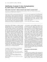
Figure 1
Measurement frames for Safranin O staining of the knee joint cartilageMeasurement frames for Safranin O staining of the knee joint cartilage.
After elimination of green tones and transformation of all red tones into
grey tones, the staining intensity (a measure of the proteoglycan con-
tent) was determined in the following layers: S, superficial layer; M, mid-
dle layer; D, deep layer; and C, calcified cartilage.
Figure 2
Time course of knee joint swellingTime course of knee joint swelling. Joint swelling (difference between
the bilateral diameter of the right knee and the left knee) in untreated
antigen-induced arthritis (AIA) rats and in hyaluronic acid (HA)-treated
AIA rats. V, end of the short-term test (day 8) and end of the long-term
test (day 29). The arrows indicate the days of intra-articular injection of
HA (days 1, 8, 15, and 22). In the short-term test there was no signifi-
cant difference between HA-treated rats and untreated AIA rats. In the
long-term test HA-treated AIA rats showed significantly reduced values
on day 15 (* P < 0.05). On day 29 there were no longer differences
between the two groups; if at all, the swelling in the HA-treated group
was somewhat higher than in untreated AIA group.
Joint swelling
Duration of arthritis (days)
0 5 10 15 20 25 30
Knee swelling (mm)
0
1
2
3
untreated AIA rats
HA-treated AIA rats
nonarthritic animals
*
VV
Available online />R681
Loss of proteoglycans from the epiphyseal growth plate
of the femoral condyle and the articular cartilage
In the short-term test (day 8), the untreated AIA group was
characterised by a significant decrease of the proteoglycan
content in the epiphyseal growth plate of the arthritic right
knee compared with the contralateral knee or with the right
knee joint of non-arthritic animals (in both cases, P < 0.05; Fig.
3). Treatment with HA prevented this loss, maintaining prote-
oglycan levels close to those of non-arthritic animals.
In terms of individual zones of the articular cartilage, the
untreated AIA rats underwent a change of -37% in the super-
ficial layer, -26% in the middle layer, -13% in the deep layer,
and -15% in the calcified cartilage layer (Figs 4b,d and 5). At
this time point, treatment with HA was significantly effective in
preventing the proteoglycan loss in the superficial layer (P <
0.01), the middle layer (P < 0.05), and the calcified cartilage
layer (P < 0.05; Figs 4b,f and 5). In all layers, the proteoglycan
content reached normal levels.
In the long-term test (day 29) there was no significant loss of
proteoglycan content in the epiphyseal growth plate of
untreated AIA rats (see Fig. 3). However, treatment with HA
was characterised by a significant proteoglycan loss in the
growth plate of the arthritic right knee compared with the con-
tralateral knee or with the right knee joint of non-arthritic ani-
mals (P < 0.001; Fig. 3).
In the different layers of the articular cartilage, the untreated
AIA rats no longer showed any significant proteoglycan loss;
that is, there were no significant differences between the right
knee joints and left knee joints of AIA rats, or between the right
knee joint of AIA rats and the right knee joint of non-arthritic
animals (Fig. 5; Safranin O staining data not shown). Treat-
ment with HA did not significantly affect the proteoglycan con-
tent in any layer of the articular cartilage.
Figure 3
Safranin O staining intensity in the epiphyseal growth plate of the femo-ral condyleSafranin O staining intensity in the epiphyseal growth plate of the femo-
ral condyle. The reference value of 232 (100%; continuous line) was
obtained by computing all available values from both non-arthritic rats
and antigen-induced arthritis (AIA) rats. In untreated AIA rats, the right
(arthritic) joint showed a significant reduction of proteoglycan content
of the epiphysis in the short-term test (day 8; *P < 0.05). This loss was
not observed following hyaluronic acid (HA) treatment (day 8). The lat-
ter values were comparable with non-arthritic animals and with the con-
tralateral joint (data not shown). Long-term treatment with HA (day 29)
induced a significant loss of proteoglycans in the epiphyseal growth
plate (*** P < 0.001). In contrast, the arthritic joints of untreated AIA
rats showed values comparable with non-arthritic rats and contralateral
joints (not shown).
Safranin O staining of the epiphysial growth plate
nonarthritic untreated HA-treated untreated HA-treated
Staining intensity (absolute values)
0
50
100
150
200
250
Day 8 Day 29
***
*
Figure 4
Histological findings in synovial tissue and articular cartilage:Histological findings in synovial tissue and articular cartilage: haematox-
ylin and eosin (HE) staining (a, c, e, g, and h) for acute inflammation
(arrowheads), chronic inflammation (*), and cartilage damage (arrows),
as well as Safranin O staining (b, d, and f) for proteoglycan depletion
(arrows). Images are shown for non-arthritic rats (a and b), untreated
antigen-induced arthritis (AIA) rats (day 8, c and d; day 29, g), and
hyaluronic acid hyaluronic acid (HA)-treated AIA rats (day 8, e and f;
day 29, h). The bar indicates the distance in the histological section.
SM, synovial membrane; P, patella; FE, femur. Safranin O staining: S,
superficial layer; M, middle layer; D, deep layer; and C, calcified carti-
lage. N, non-arthritic rats; US, untreated AIA, short-term test; HS, HA-
treated AIA, short-term test; UL, untreated AIA, long-term test; HL, HA-
treated AIA, long-term test.
Arthritis Research & Therapy Vol 7 No 3 Roth et al.
R682
Histological scores of arthritis and cartilage damage
In the short-term test (day 8) there was a strong acute inflam-
mation in untreated AIA rats (Fig. 6a). Treatment with HA sig-
nificantly reduced the acute inflammation compared with the
untreated AIA group (P < 0.05; Figs 4a,c,e and 6a). Notably,
the untreated AIA group underwent a complete, spontaneous
remission of the acute inflammation score from day 8 to day 29
(acute inflammation score nearly 0 in the long-term test; Figs
4c,g and 6a). The HA-treated AIA group in the long-term test
clearly improved compared with the short-term test (P < 0.05),
but it still showed significantly higher, residual acute inflamma-
tion than the untreated AIA group (P < 0.05; Figs 4e,g,h and
6a).
In the short-term test (day 8) a clear score for chronic inflam-
mation was also observed (Figs 4a,c,e and 6b), without signif-
icant differences between untreated and HA-treated AIA
groups. The chronic inflammation significantly decreased from
day 8 to day 29 in untreated and HA-treated AIA (group US
versus group UL, P < 0.001; group HS versus group HL, P <
0.01; Figs 4c,e,g,h and 6b). Unexpectedly, however, on day
29 the chronic inflammation score was more pronounced in
Figure 5
Safranin O staining intensity in different layers of the articular cartilageSafranin O staining intensity in different layers of the articular cartilage.
Comparisons were made for different layers (S, superficial layer; M,
middle layer; D, deep layer; and C, calcified cartilage) between non-
arthritic rats, untreated antigen-induced arthritis (AIA) rats, and
hyaluronic acid (HA)-treated AIA rats in terms of relative differences
between right (arthritic) and left (contralateral) joints. In the short-term
test (day 8) untreated AIA rats showed a reduced proteoglycan content
in the superficial layer (** P = 0.01), middle layer (* P < 0.05), and cal-
cified cartilage (* P < 0.05). HA-treated AIA rats showed proteoglycan
contents comparable with those of non-arthritic rats in all layers. In the
long-term test (day 29), untreated AIA rats also showed reduced prote-
oglycan contents in all layers compared with non-arthritic rats, but no
statistical significance was attained. HA-treated AIA rats showed prote-
oglycan contents comparable with those of non-arthritic rats.
Day 8
Cartilage layer
SMDC SMDC
Difference between right and left (%)
-80
-60
-40
-20
0
20
40
60
80
** * *
Day 29
Safranin O staining intensity of the articular cartilage
untreated AIA rats
HA-treated AIA rats
nonarthritic rats
Figure 6
Histological scoresHistological scores. In the short-term test, hyaluronic acid (HA)-treated
antigen-induced arthritis (AIA) rats showed a significant reduction of (a)
the acute inflammation score compared with untreated AIA rats (* P <
0.05). The score of (b) chronic inflammation and (c) cartilage damage
did not show significant differences between HA-treated rats and
untreated AIA rats. In the long-term test, (a) the acute inflammation was
reduced in both AIA groups compared with that in the short-term test
(HA-treated AIA rats, P < 0.05; untreated AIA rats, P < 0.001; signifi-
cance not indicated); nonetheless, the HA-treated AIA rats showed sig-
nificantly higher scores than untreated AIA rats on day 29 (* P < 0.05).
(b) The scores of chronic inflammation were reduced in both AIA
groups compared with the short-term test (untreated AIA rats, P <
0.001; HA-treated AIA rats, P < 0.01; significance not indicated); none-
theless, the HA-treated rats showed significantly higher scores than
untreated AIA rats on day 29 (* P < 0.05). (c) The cartilage damage
was relatively low in both untreated rats and HA-treated AIA rats, but
HA-treated AIA rats showed a significantly higher damage (* P < 0.05).
Chronic inflammation
nonarthritic untreated HA-treated untreated HA-treated
Sum score
0
1
2
3
4
5
6
7
8
9
Day 8 Day 29
*
Cartilage damage
nonarthritic untreated HA-treated untreated HA-treated
Score
0
1
2
3
4
Day 8
Day 29
*
Acute inflammation
nonarthritic untreated HA-treated untreated HA-treated
Sum score
0
1
2
3
4
5
6
7
8
Day 8 Day 29
*
*
(a)
(b)
(c)
Available online />R683
the animals treated with HA compared with the untreated AIA
group (P < 0.05; Figs 4g,h and 6b).
In terms of cartilage damage, the untreated AIA group was
characterised by a maximum individual score of 3; that is, the
maximal possible score of 4 was not observed (Fig. 6c). On
day 8, the mean cartilage damage was somewhat more pro-
nounced in the untreated AIA group, but without significant dif-
ferences in comparison with the HA-treated AIA group (power
1 - β = 0.821). From day 8 to day 29, the cartilage damage
decreased significantly in untreated rats and HA-treated AIA
rats (group US versus group UL, P < 0.001; group HS versus
group HL, P < 0.01). In the long-term test (day 29), however,
the cartilage damage was significantly higher in the animals
treated with HA than in the untreated AIA group (P < 0.05; Fig.
6c).
Systemic cytokine levels
In non-arthritic animals, the serum IL-6 levels were below the
detection limit of the assay (Fig. 7a). Untreated AIA rats had
significantly elevated IL-6 levels both in the short-term test and
in the long-term test (P < 0.001 in both cases; significance not
indicated in Fig. 7). Treatment with HA did not significantly
influence IL-6 levels at either time point (Fig. 7a).
As for TNF-α, non-arthritic animals had mean serum levels of
5.45 ± 4.56 pg/ml (Fig. 7b). In the short-term test these values
were increased both in the untreated and in the HA-treated
AIA groups, but not to a significant degree. In the long-term
test, the mean TNF-α levels were very similar to those of non-
arthritic animals. Treatment with HA did not significantly influ-
ence TNF-α levels at either time point (Fig. 7b).
Serum GAG levels
In non-arthritic animals, the mean serum levels of GAG were
12.70 ± 3.30 µg/ml (Fig. 7c). Untreated AIA rats had
significantly higher GAG levels than non-arthritic animals in the
short-term test and in the long-term test (P < 0.05 and P <
0.001, respectively; significance not indicated in Fig. 7). Treat-
ment with HA had no influence on this parameter at either time
point (Fig. 7c).
Discussion
Clinical parameters of arthritis
The time course of AIA was similar to that described by other
authors [32,34], confirming that the present results were rep-
resentative of previous studies.
Treatment with HA did not reduce joint swelling in the acute
phase, as significant reduction of joint swelling was found only
on day 15 (i.e. in the early chronic phase of AIA). The tempo-
rary reduction of joint swelling may be a result of the reduced
acute inflammation observed histologically at an earlier time
point (day 8). This anti-inflammatory effect of HA is consistent
Figure 7
Serum levels of IL-6, tumour necrosis factor alpha (TNF-α) and glycosaminoglycanSerum levels of IL-6, tumour necrosis factor alpha (TNF-α) and gly-
cosaminoglycan. During the course of antigen-induced arthritis (AIA),
(a) IL-6 levels in non-arthritic rats showed values below the detection
limit of the assay. Untreated and hyaluronic acid (HA)-treated AIA rats
showed significantly increased values in the short-term test and in the
long-term test compared with non-arthritic rats (all P < 0.001; data not
shown), but HA treatment resulted in IL-6 levels comparable with those
of untreated AIA. (b) Regarding TNF-α levels, the only change was a
short-term, non-significant increase in all AIA rats, whether HA-treated
or untreated. (c) Serum values of glycosaminoglycan increased signifi-
cantly in all AIA rats compared with non-arthritic animals (non-arthritic
animals versus untreated AIA rats [short-term test], P < 0.05, non-
arthritic animals versus HA-treated AIA rats, untreated AIA rats [long-
term test] and HA-treated AIA rats [long-term test], P < 0.001; signifi-
cance not indicated). There were no differences between short-term
tests and long-term tests, or between HA-treated rats and untreated
AIA rats.
TNF-α
nonarthritic untreated HA-treated untreated HA-treated
(pg/ml)
0
2
4
6
8
10
12
14
Day 8 Day 29
IL-6
nonarthritic untreated HA-treated untreated HA-treated
(pg/ml)
0
2
4
6
8
10
12
Day 8 Day 29
Glycosaminoglycans
nonarthritic u ntreated HA-treated untreated HA-treated
(µg/ml)
0
10
20
30
40
Day 8 Day 29
(a)
(b)
(c)
Arthritis Research & Therapy Vol 7 No 3 Roth et al.
R684
with the effects previously reported in collagen-induced arthri-
tis [22,40] and human RA [41,42].
Interestingly, however, while the joint swelling continued to
progressively and spontaneously decrease in untreated AIA, it
persisted in HA-treated animals after day 15, although the
difference remained at the limits of significance (P = 0.06).
The persistence of inflammation after prolonged application of
HA (day 29) is further substantiated by significant proteogly-
can loss in the epiphyseal growth plate, by significant persist-
ence of both acute and chronic inflammation, and by
significantly increased histological signs of articular cartilage
damage. This biphasic course regarding joint inflammation
and destruction after repeated application of high-molecular-
weight HA has not been previously described. HA therefore
probably shows only a limited temporal window of anti-inflam-
matory activity in arthritis.
Adverse reactions to HA have been described in human oste-
oarthritis, either after the first injection and subsequent intra-
articular injections or at the beginning of a new treatment
course. These adverse events consisted of pain and/or tran-
sient swelling of the injected joint, mostly mild or moderate in
intensity [43-45]. The adverse reactions observed upon intra-
articular treatment of human osteoarthritis are not comparable
with the biphasic effects in the present study, since they were
only short-lasting and limited to a period immediately following
injection.
Proteoglycans in the epiphyseal growth plate of the
femoral condyle and the articular cartilage
AIA was accompanied by a significant loss of proteoglycans in
the epiphyseal growth plate of untreated AIA rats in the short-
term test (significantly ameliorated by HA; day 8). This prote-
oglycan loss is probably caused by the inflammatory micromi-
lieu in the arthritic joint [46] and the adjacent periarticular bone
marrow [47], in analogy to the alterations of the primary and
secondary spongiosa in AIA [48]. The significant prevention of
proteoglycan loss from the epiphyseal growth plate by HA at
this time point may be due to the clear anti-arthritic effects of
HA, as also documented by the significant decrease of acute
inflammation on day 8 (Fig. 6a). Whether intra-articular treat-
ment with HA indirectly influences the inflammatory changes in
the bone marrow, thereby preventing proteoglycan loss in the
epiphyseal growth plate, remains to be investigated.
Unexpectedly, repeated intra-articular HA treatment induced
proteoglycan loss in the epiphyseal growth plate in the long-
term test (day 29). This late loss of proteoglycans in the epiph-
ysis under prolonged HA treatment suggests late
proinflammatory effects of HA, as also indicated by signifi-
cantly elevated levels of acute and chronic inflammation, as
well as cartilage damage (Fig. 6).
Regarding the proteoglycan content within the articular carti-
lage, HA completely prevented the proteoglycan loss in the
short-term test. This applied to the severe loss of proteogly-
cans in the superficial layer — the layer most strongly affected
in the present study (37%) and also that most strongly
affected in the fibronectin-mediated arthritis model [19]. This
prevention also applied to deeper layers of the calcified carti-
lage, however, indicating either deep-reaching effects of intra-
articular HA (up to 100 µm; see Fig. 1) or an indirect effect on
the micromilieu in the epiphyseal bone core. Such profound
chondroprotective effects of HA have not previously been
reported in an in vivo arthritis model. Previous in vitro studies,
in which the proteoglycan release from the cell matrix of bovine
chondrocyte cultures was inhibited by HA, have suggested a
covering of the matrix of as a mechanism for the chondropro-
tective capacity of HA [19].
Acute and chronic inflammation, and cartilage damage
HA reduced the histological signs of acute inflammation in the
short-term test (day 8). At this point, HA also showed a ten-
dency (P = 0.06) to protect the joint again against cartilage
damage (Fig. 6c). The tendency for decreased cartilage dam-
age in the short-term test supports the assumption that HA
forms a temporary protecting barrier over the cartilage, and
thereby protects it against degradation [16,19,49]. The known
reduction of free oxygen radicals [31], as well as the reduction
of cytokines and other mediators of acute inflammation by HA
[15,21], could contribute to both its chondroprotective capac-
ity (Fig. 6c) and its anti-inflammatory effects (Fig. 6a) in early
chronic AIA (day 8).
In the long-term test (day 29) HA treatment was associated
with significantly higher histological signs of acute and chronic
inflammation and, more importantly, with more severe cartilage
damage. The late proinflammatory effects of repeated HA
application, in parallel to the late loss of epiphysis proteogly-
cans and late damage of articular cartilage (see earlier), point
to an interdependence of inflammation and damage in HA-
treated AIA rats at this stage.
Significantly higher histological signs of articular cartilage
damage after repeated HA application (day 29) are in contrast
to a non-altered proteoglycan content of the articular cartilage
at this time (Fig. 5). A seemingly normal proteoglycan content
may therefore not be sufficient to exclude structural damage of
the articular cartilage in arthritis. Consequently, more sensitive
in vivo procedures will have to be established to reliably
assess functional or structural cartilage alterations before irre-
versible damage occurs [50].
Serum levels of cytokines and GAG
In the present study, the systemic levels of IL-6, TNF-α and
GAG were not significantly influenced by intra-articular admin-
istration of high-molecular-weight HA, indicating that both anti-
Available online />R685
inflammatory effects (day 8) and proinflammatory effects (day
29) are locally restricted.
Conclusion
In conclusion, HA appears to have a limited therapeutic win-
dow for local treatment of arthritis, as shown by amelioration
of clinical signs (day 15), by prevention of proteoglycan loss in
the articular cartilage and epiphyseal growth plate, and by
prevention of structural cartilage damage, as well as by the
reduction of acute inflammation in the arthritic joint (day 8).
Late aggravation of clinical signs (not significant, day 29), pro-
teoglycan loss from the epiphyseal growth plate, and acute/
chronic inflammation and structural cartilage damage at this
time point strongly indicate biphasic effects of local HA
treatment.
Whether these biphasic effects are due to accumulation of HA
beyond pathological levels (which may be avoidable by single
injection instead of repeated injection) or whether certain
phases of the clinical course render the animals sensitive to
the proinflammatory effects of HA remains the subject of future
research, both in animal models and in human RA.
Competing interests
The co-authors J-H Ozegowski, P-J Müller, S Möller, G
Peschel, A Roth, and RA Venbrocks published a patent for the
use of sulfated hyaluronic acid for the prevention of inflamma-
tory arthritis in 2000. This substance is not the same as that
used for the experiments in the present study. (Ozegowski J-
H, Müller P-J, Möller S, Peschel G, Roth A, Venbrocks RA:
Pharmazeutische Formulierungen zur Hemmung von entzünd-
lichen Arthritiden [Use of hyaluronic acid derivatives for the
prevention of inflammatory arthritis], HA 00-52 2000).
A Roth has to publish papers in peer-reviewed journals as a
part of the process to finish his thesis (habilitation in Ger-
many). This is a non-financial academic interest.
Authors' contributions
AR carried out all experiments, the measurements and evalua-
tion of the data, as well as the statistics, and drafted, revised,
finalised, and submitted the manuscript. JM developed the
experimental basis for the measurements of proteoglycans in
the serum and supervised the analysis, trained and supervised
AR in the analysis of proteoglycan loss from cartilage, actively
participated in the analysis and evaluation of the data, and
reviewed and contributed to the final version of the manu-
script. AW participated in the preparation of the animals, fixa-
tion of the specimens, and blood collection, and reviewed the
manuscript. RF actively participated in the experimental and
organisational design of the study, gave valuable advice for the
evaluation and interpretation of the experimental results, and
reviewed the manuscript. AS reviewed all experimental data,
gave valuable advice for the evaluation and interpretation of
the experimental results and the subsequent conclusions, and
reviewed the manuscript. RAV participated in the experimental
and organisational design of the study, supervised the evalua-
tion and interpretation of the experimental results, reviewed
the manuscript, and organised financial support and laboratory
space for the experiments. PP performed and summarised all
histological analyses, and reviewed the manuscript. RB
actively participated in the underlying animal experiments and
the evaluation of the experimental results, and reviewed and
contributed to the final version of the manuscript. HS actively
participated in the underlying animal studies, provided and
monitored all experimental animals, and reviewed the manu-
script. JO, GP, and PJM developed the method for the
production of HA, produced, purified, and quality-controlled
the microbially manufactured, high-molecular-weight HA, and
reviewed the manuscript. RWK participated in the experimen-
tal design of the study and the interpretation of the results, and
reviewed and contributed to the initial and final version of the
manuscript.
Acknowledgements
The authors are grateful to Dr Frank Brand, Mrs K Neumann, and Mrs K
Axt (Department of Clinical Chemistry and Laboratory Diagnostics,
Rudolf-Elle Hospital) for determination of laboratory values, and to Cor-
dula Müller and Jana Schömburg (Research Department, Rudolf-Elle
Hospital) for preparation of the histological sections. They are also
grateful to Dr A Notni, Dr K Bergmann, and Dr R Winter (Department of
Orthopaedics, Rudolf-Elle Hospital), as well as to Dr C Wicher and Mrs
P Dobermann (Institute of Animal Studies, Friedrich-Schiller University
Jena) for assistance in the animal experiments.
References
1. Firestein GS, Zvaifler NJ: Rheumatoid arthritis: a disease of dis-
ordered immunity. In Inflammation: Basic Principles and Clincal
Correlates Edited by: Gallin J, Goldstein I, Willis K. New York:
Raven Press; 1992:959-975.
2. Gay S, Gay RE, Koopman WJ: Molecular and cellular mecha-
nisms of joint destruction in rheumatois arthritis: two cellular
mechanisms explain joint destruction? Ann Rheum Dis 1993,
52(Suppl 1):39-47.
3. Mau W, Bornmann M, Weber H, Weidemann HF, Hecker H,
Raspe HH: Prediction of permanent work disability in a follow-
up study of early rheumatoid arthritis: results of a tree struc-
tured analysis using RECPAM. Br J Rheumatol 1996,
35:652-659.
4. Balazs EA, Iozzo RV: Matrix proteoglycans: from molecular
design to cellular function. Annu Rev Biochem 1998,
67:609-652.
5. Laurent TC, Laurent UB, Fraser JR: The structure and function of
hyaluronan: an overview. Immunol Cell Biol 1996, 74:1-7.
6. Miossec P, Dinarello CA, Ziff M: Interleukin-1 lymphocyte chem-
otactic activity in rheumatoid arthritis synovial fluid. Arthritis
Rheum 1986, 29:461-470.
7. Dahl LB, Dahl IM, Engström-Laurent A, Granath K: Concentration
and molecular weight of sodium hyaluronate in synovial fluid
from patients with rheumatoid arthritis and other
arthropathies. Ann Rheum Dis 1985, 44:817-822.
8. Balazs EA, Denlinger JL: Sodium hyaluronate and joint function.
J Equine Vet Sci 1985, 5:217-228.
9. Duffy JM, McNally GM, O'Gorman A, Spedding PL, Grimshaw J,
Grimshaw JT, Walker B, Guthrie DJS, Mollan RAB: Viscometic
studies of pathological human synovial fluids. J Orthop
Rheumatol 1994, 7:99-106.
10. Namiki O, Toyoshima H, Morisaki N: Therapeutic effect of intra-
articular injection of high molecular weight hyaluronic acid on
Arthritis Research & Therapy Vol 7 No 3 Roth et al.
R686
osteoarthritis of the knee. Int J Clin Pharmacol Ther Toxicol
1982, 20:501-507.
11. Watterson JR, Esdaile JM: Viscosupplementation: therapeutic
mechanisms and clinical potential in osteoarthritis of the knee.
J Am Acad Orthop Surg 2000, 8:277-284.
12. Saxne T, Wollheim FA, Pettersson H, Heinegard D: Proteoglycan
concentration in synovial fluid: predictor of future cartilage
destruction in rheumatoid arthritis? Br Med J (Clin Res Ed)
1987, 295:1447-1448.
13. Bensouyad A, Hollander AP, Dularay B, Bedwell AE, Cooper RA,
Hutton CW, Dieppe PA, Elson CJ: Concentrations of gly-
cosaminoglycans in synovial fluids and their relation with
immunological and inflammatory mediators in rheumatoid
arthritis. Ann Rheum Dis 1990, 49:301-307.
14. Sato H, Takahashi T, Ide H, Fukushima T, Tabata M, Sekine F,
Kobayashi K, Negishi M, Niwa Y: Antioxidant activity of synovial
fluid, hyaluronic acid, and two subcomponents of hyaluronic
acid. Synovial fluid scavenging effect is enhanced in rheuma-
toid arthritis patients. Arthritis Rheum 1988, 31:63-71.
15. Larsen NE, Lombard KM, Parent EG, Balazs EA: Effect of hylan
on cartilage and chondrocyte cultures. J Orthop Res 1992,
10:23-32.
16. Shimazu A, Jikko A, Iwamoto M, Koike T, Yan W, Okada Y, Shinmei
M, Nakamura S, Kato Y: Effects of hyaluronic acid on the
release of proteoglycan from the cell matrix in rabbit chondro-
cyte cultures in the presence and absence of cytokines. Arthri-
tis Rheum 1993, 36:247-253.
17. Morris ES, Wilcon S, Treadwell BV: Inhibition of interleukin 1-
mediated proteoglycan degradation in bovine articular carti-
lage explants by addition of sodium hyaluronate. Am J Vet Res
1992, 53:1977-1982.
18. Akatsuka M, Yamamoto Y, Tobetto K, Yasui T, Ando T: In vitro
effects of hyaluronan on prostaglandin E2 induction by inter-
leukin-1 in rabbit articular chondrocytes. Agents Actions 1993,
38:122-125.
19. Homandberg GA, Hui F, Wen C, Kuettner KE, Williams JM:
Hyaluronic acid suppresses fibronectin fragment mediated
cartilage chondrolysis: I. In vitro. Osteoarthritis Cartilage 1997,
5:309-319.
20. Williams JM, Plaza V, Hui F, Wen C, Kuettner KE, Homandberg
GA: Hyaluronic acid suppresses fibronectin fragment medi-
ated cartilage chondrolysis: II. In vivo. Osteoarthritis Cartilage
1997, 5:235-240.
21. Presti D, Scott JE: Hyaluronan-mediated protective effect
against cell damage caused by enzymatically produced
hydroxyl (OH.) radicals is dependent on hyaluronan molecular
mass. Cell Biochem Funct 1994, 12:281-288.
22. Campo GM, Avenoso A, Campo S, Ferlazzo AM, Altavilla D, Cala-
troni : Efficacy of treatment with glycosaminoglycans on exper-
imental collagen-induced arthritis in rats. Arthritis Res Ther
2003, 5:R122-R131.
23. Abatangelo G, Cortivo R, Govoni E, De Galateo A, Brun P: Hyalur-
onate reverses inhibition of proteoglycan synthesis by oxygen
free radicals in cultured chick embryonic cartilage [abstract].
Eur J Cell Biol 1989, 49(Suppl 28):18.
24. Cortivo R, Brun P, Cardarelli L, O'Regan M, Radice M, Abatangelo
G: Antioxidant effects of hyaluronan and its alpha-methyl-
prednisolone derivative in chondrocyte and cartilage cultures.
Semin Arthritis Rheum 1996, 26:492-501.
25. Tobetto K, Nakai K, Akatsuka M, Yasui T, Ando T, Hirano S: Inhib-
itory effects of hyaluronan on neutrophil-mediated cartilage
degradation. Connect Tissue Res 1993, 29:181-190.
26. Goldberg RL, Toole BP: Hyaluronate inhibition of cell
proliferation. Arthritis Rheum 1987, 30:769-778.
27. Anastassiades T, Robertson W: Modulation of mitogen-depend-
ent lymphocyte stimulation by hyaluronic acid. J Rheumatol
1984, 11:729-734.
28. Corrado EM, Peluso GF, Gigliotti S, Durante C, Palmieri D, et al.:
The effects of intraarticular administration of hyaluronan on
osteoarthritis of the knee: a clinical study with immunological
and biochemical evaluations. Eur J Rheum Inflamm 1993,
15:547-576.
29. Forrester JV, Balazs EA: Inhibition of phagocytosis by high
molecular weight hyaluronate. Immunology 1980, 40:435-446.
30. Håkansson L, Hällgren R, Venge P: Regulation of granulocyte
function by hyaluronic acid. In vitro and in vivo effects on
phagocytosis, locomotion, and metabolism. J Clin Invest 1980,
66:298-305.
31. Greenwald RA: Oxygen radicals, inflammation, and arthritis:
pathophysiological considerations and implications for
treatment. Semin Arthritis Rheum 1991, 20:219-240.
32. Bräuer R, Kittlick PD, Thoss K, Henzgen S: Different immunolog-
ical mechanisms contribute to cartilage destruction in antigen-
induced arthritis. Exp Toxicol Pathol 1994, 46:383-388.
33. Griffiths RJ: The use of animals in the search for anti-inflamma-
tory drugs. In Mechanisms and Models in Rheumatoid Arthritis
Edited by: Henderson B, Edwards JCW, Pettpher ER. London:
Academic Press; 1995.
34. Buchner E, Bräuer R, Schmidt C, Emmrich F, Kinne RW: Induction
of flare-up reactions in rat antigen-induced arthritis. J
Autoimmun 1995, 8:61-74.
35. Romeis B: Mikroskopische Technik. In Mikroskopische Technik
17th edition. Edited by: Böck P. Urban-Schwarzenberg, München;
1989.
36. Rosenberg L: Chemical basis for the histological use of
safranin O in the study of articular cartilage. J Bone Joint Surg
Am 1971, 53:69-82.
37. Kiviranta I, Jurvelin J, Tammi M, Saamanen AM, Helminen HJ:
Microspectrophotometric quantitation of glycosaminoglycans
in articular cartilage sections stained with Safranin O. Histo-
chemistry 1985, 82:249-255.
38. Arokoski J, Kiviranta I, Jurvelin J, Tammi M, Helminen HJ: Long-dis-
tance running causes site-dependent decrease of cartilage
glycosaminoglycan content in the knee joints of beagle dogs.
Arthritis Rheum 1993, 36:1451-1459.
39. Meyer P, Burkhardt H, Palombo-Kinne E, Grunder W, Bräuer R,
Stiller KJ, Kalden JR, Becker W, Kinne RW: 123I-antileukoprotei-
nase scintigraphy reveals microscopic cartilage alterations in
the contralateral knee joint of rats with 'monarticular' antigen-
induced arthritis. Arthritis Rheum 2000, 43:298-310.
40. Campo GM, Avenoso A, Campo S, Ferlazzo A, Altavilla D, Micali
C, Calatroni A: Aromatic trap analysis of free radicals produc-
tion in experimental collagen-induced arthritis in the rat: pro-
tective effect of glycosaminoglycans treatment. Free Radic Res
2003, 37:257-268.
41. Tanaka N, Sakahashi H, Sato E, Hirose K, Ishima T, Ishii S: Intra-
articular injection of high molecular weight hyaluronan after
arthrocentesis as treatment for rheumatoid knees with joint
effusion. Rheumatol Int 2002, 22:151-154.
42. Goto M, Hanyu T, Yoshio T, Matsuno H, Shimizu M, Murata N,
Shiozawa S, Matsubara T, Yamana S, Matsuda T: Intra-articular
injection of hyaluronate (SI-6601D) improves joint pain and
synovial fluid prostaglandin E2 levels in rheumatoid arthritis: a
multicenter clinical trial. Clin Exp Rheumatol 2001, 19:377-383.
43. Puttick MP, Wade JP, Chalmers A, Connell DG, Rangno KK:
Acute local reactions after intraarticular hylan for osteoarthri-
tis of the knee. J Rheumatol 1995, 22:1311-1314.
44. Allen E, Krohn K: Adverse reaction to Hylan GF-20 [Letter]. J
Rheumatol 2000, 27:1572.
45. Martens PB: Bilateral symmetric inflammatory reaction to
hylan G-F 20 injection. Arthritis Rheum 2001, 44:978-979.
46. Simon J, Surber R, Kleinstauber G, Petrow PK, Henzgen S, Kinne
RW, Brauer R: Systemic macrophage activation in locally-
induced experimental arthritis. J Autoimmun 2001, 17:127-136.
47. Kinne RW, Brauer R, Stuhlmuller B, Palombo-Kinne E, Burmester
GR: Macrophages in rheumatoid arthritis. Arthritis Res 2000,
2:189-202.
48. Oelzner P, Bräuer R, Henzgen S, Thoss K, Wunsche B, Hersmann
G, Abendroth K, Kinne RW: Periarticular bone alterations in
chronic antigen-induced arthritis: free and liposome-encapsu-
lated clodronate prevent loss of bone mass in the secondary
spongiosa. Clin Immunol 1999, 90:79-88.
49. Bräuer R, Kette H, Henzgen S, Thoss K: Influence of cyclosporin
A on cytokine levels in synovial fluid and serum of rats with
antigen-induced arthritis. Agents Actions 1994, 41:96-98.
50. Gray ML, Eckstein F, Peterfy C, Dahlberg L, Kim YJ, Sorensen AG,
Smith RL: Toward imaging biomarkers for osteoarthritis. Clin
Orthop 2004, 427(Suppl):175-181.