Báo cáo y học: " Clinical evaluation of autoantibodies to a novel PM/Scl peptide antigen" doc
Bạn đang xem bản rút gọn của tài liệu. Xem và tải ngay bản đầy đủ của tài liệu tại đây (478.72 KB, 10 trang )
Open Access
Available online />R704
Vol 7 No 3
Research article
Clinical evaluation of autoantibodies to a novel PM/Scl peptide
antigen
Michael Mahler
1
, Reinout Raijmakers
2
, Cornelia Dähnrich
3
, Martin Blüthner
4
and Marvin J Fritzler
5
1
Dr Fooke Laboratorien GmbH, Neuss, Germany
2
Radboud University Nijmegen, The Netherlands
3
Euroimmun GmbH, Lübeck, Germany
4
Labor Seelig und Kollegen, Karlsruhe, Germany
5
Faculty of Medicine, University of Calgary, Canada
Corresponding author: Michael Mahler,
Received: 8 Jan 2005 Revisions requested: 16 Feb 2005 Revisions received: 22 Feb 2005 Accepted: 4 Mar 2005 Published: 1 Apr 2005
Arthritis Research & Therapy 2005, 7:R704-R713 (DOI 10.1186/ar1729)
This article is online at: />© 2005 Mahler et al, licensee BioMed Central Ltd.
This is an Open Access article distributed under the terms of the Creative Commons Attribution License ( />2.0), which permits unrestricted use, distribution, and reproduction in any medium, provided the original work is cited.
Abstract
Anti-PM/Scl antibodies represent a specific serological marker
for a subset of patients with scleroderma (Scl) and polymyositis
(PM), and especially with the PM/Scl overlap syndrome (PM/
Scl). Anti-PM/Scl reactivity is found in 24% of PM/Scl patients
and is found in 3–10% of Scl and PM patients. The PM/Scl
autoantigen complex comprises 11–16 different polypeptides.
Many of those proteins can serve as targets of the anti-PM/Scl
B-cell response, but most frequently the PM/Scl-100 and PM/
Scl-75 polypeptides are targeted. In the present study we
investigated the clinical relevance of a major alpha helical PM/
Scl-100 epitope (PM1-α) using a newly developed peptide-
based immunoassay and compared the immunological
properties of this peptide with native and recombinant PM/Scl
antigens. In a technical comparison, we showed that an ELISA
based on the PM1-α peptide is more sensitive than common
techniques to detect anti-PM/Scl antibodies such as
immunoblot, indirect immunofluorescence on HEp-2 cells and
ELISA with recombinant PM/Scl polypeptides. We found no
statistical evidence of a positive association between anti-PM1-
α and other antibodies, with the exception of known PM/Scl
components. In our cohort a negative correlation could be found
with anti-Scl-70 (topoisomerase I), anti-Jo-1 (histidyl tRNA
synthetase) and anti-centromere proteins. In a multicenter
evaluation we demonstrated that the PM1-α peptide represents
a sensitive and reliable substrate for the detection of a subclass
of anti-PM/Scl antibodies. In total, 22/40 (55%) PM/Scl
patients, 27/205 (13.2%) Scl patients and 3/40 (7.5%) PM
patients, but only 5/288 (1.7%) unrelated controls, tested
positive for the anti-PM1-α peptide antibodies. These data
indicate that anti-PM1-α antibodies appear to be exclusively
present in sera from PM/Scl patients, from Scl patients and, to
a lesser extent, from PM patients. The anti-PM1-α ELISA thus
offers a new serological marker to diagnose and discriminate
different systemic autoimmune disorders.
Introduction
Systemic autoimmune diseases such as scleroderma (Scl),
polymyositis (PM), rheumatoid arthritis, systemic lupus ery-
thematosus (SLE) and mixed connective tissue disease are
characterized by the occurrence of circulating antibodies to
defined intracellular targets [1]. Some of these autoantibodies
represent useful diagnostic markers for a variety of systemic
autoimmune diseases [1,2].
Antibodies targeting the PM/Scl complex serve as a marker for
the PM/Scl overlap syndrome, where they are found in 24% of
sera, but they are also seen in 8% of PM patients and in 3% of
Scl patients [3-6]. The PM/Scl complex was identified as the
human counterpart of the yeast exosome and consists of 11–
16 polypeptides with molecular masses ranging from 20 to
110 kDa [7-11]. PM/Scl-100, the human equivalent of the
yeast Rrp6p, has been cloned by two independent groups and
its key function during the 5.8 S rRNA end formation has been
described [12-14].
DM = dermatomyositis; ELISA = enzyme-linked immunosorbent assay; HCV = hepatitis C virus; IIF = indirect immunofluorescence; PBS = phosphate-
buffered saline; PM = polymyositis; PM1-α = alpha helical PM/Scl-100 epitope; RU = relative units; Scl = scleroderma; SLE = systemic lupus
erythematosus.
Arthritis Research & Therapy Vol 7 No 3 Mahler et al.
R705
In previous studies, the human immune response targeting the
PM/Scl complex has been reported to be predominantly
directed against two polypeptides with apparent molecular
masses of 100 kDa and 75 kDa [15]. In the past it has been
shown that nearly all PM/Scl-positive sera contain autoanti-
bodies to the 100 kDa protein and that only about 50–60%
react with the 75 kDa protein [7,8,15-17]. A more recent study
has shown that the PM/Scl-75 protein contains a previously
unidentified N-terminal region that is important for the anti-
genicity of the protein [18]. The reactivity of sera with this new
isoform of PM/Scl-75c is similar to the conventional PM/Scl-
100 protein [18]. Several other components of the human exo-
some, including hRrp4p, hRrp40p, hRrp41, hRrp42p,
hRrp46p and hCsl4p, are also recognized by anti-PM/Scl anti-
bodies, but to a lesser extent [10,19].
In several studies during the past decade, we and others have
attempted to identify the epitopes on PM/Scl-100 that are rec-
ognized by the cognate autoantibodies [12,20-23]. The prime
reactivity of anti-PM/Scl-100 sera was localized to a domain of
the protein represented by amino acids 231–245 using mem-
brane-bound peptide arrays [22,23]. The amino acids contrib-
uting to the antibody binding were identified by mutational
analysis [22,23]. Based on these observations and on second-
ary structure predictions, a local alpha-helical structure has
been proposed for this major PM/Scl-100 epitope [22,23].
The aim of this study was to develop an ELISA with a 15-mer
peptide comprising the PM/Scl-100 major epitope as a sub-
strate, and to evaluate its sensitivity and specificity for the
detection of anti-PM/Scl antibodies.
Materials and methods
Serum samples
In the present study three different serum panels were used to
analyze the accuracy of the alpha helical PM/Scl-100 epitope
(PM1-α) peptide in the ELISA. For the technical comparative
study, 33 sera with anti-PM/Scl reactivity were preselected by
indirect immunofluorescence on HEp-2 cells and cryopre-
served monkey liver sections (Euroimmun, Lübeck, Germany)
and by immunoblot with total cell extracts (Panel I). Panel II
consisted of sera from a previous study and included patients
with PM/Scl, patients with PM, patients with Scl, patients with
dermatomyositis (DM) patients with melanoma and normal
donors [18]. For the multicenter evaluation, serum samples
were collected from patients with PM/Scl overlap syndrome (n
= 40), from patients with Scl (n = 50), from patients with PM
(n = 40) and from patients with various control diseases
including rheumatoid arthritis (n = 69), SLE (n = 114), undif-
ferentiated connective tissue disease (n = 10), mixed connec-
tive tissue disease (n = 6), Hashimoto thyroiditis (n = 11),
Grave's disease (n = 12), other autoimmune disorders (n = 8),
and hepatitis C virus infection (HCV) (n = 48) (Panel III).
PM/Scl patients were diagnosed based on the official PM and
Scl criteria and were only considered true overlap patients if
they fulfilled both the criteria for PM and for Scl [24,25]. All
other patients with autoimmune disorders were classified
according to the official criteria for each disease as also
applied in a recent investigation [26]. Sera were stored in aliq-
uots at -80°C until use and were shipped on dry ice. Collection
of patient samples was carried out according to local ethics
committee regulations.
Antigens for ELISA
The identified sequence LDVPPALADFIHQQR of the PM/Scl-
100 (accession number JH0796) major B-cell epitope cover-
ing amino acids 231–245 was used to synthesize the PM1-α
peptide with an additional cysteine residue at the C-terminus
using Fmoc chemistry [22]. Crude peptide obtained from pep-
tide synthesis was purified by high-performance liquid chro-
matography. The quality and purity of the peptide was
assessed by mass spectrometry and analytical high-perform-
ance liquid chromatography. The molecular mass was found at
1824.1167 Da (average; monoisotopic mass = 1822.9274
Da) and a purity of 100% was determined. The isoelectric
point of the peptide was 4.0. Recombinant PM/Scl-100
(Diarect AG, Freiburg, Germany), was expressed in
Escherichia coli and purified via a His-tag, and the quality was
ensured by immunoblot and checkerboard analysis of positive
and negative sera in the ELISA [27].
Indirect immunofluorescence
Indirect immunofluorescence (IIF) was carried out using Bio-
Chip-mosaics with HEp-2 cells and primate liver as substrates
(lot number 10116D; Euroimmun GmbH). Antibody titers
were determined using 10-fold serial dilutions in PBS and the
assay was performed according the manufacturer's
instructions.
Immunoblotting
Total cell extracts from HEp-2 cells that were separated by
SDS-PAGE and transferred onto nitrocellulose were used as
substrate for immunoblotting (lot numbers 01011a-88 and
01011a-89; Euroimmun GmbH). The identity of the PM/Scl
antigens was ensured using PM/Scl index sera, which were
previously characterized by several methods. Sera were
diluted and incubated according to the manufacturer's
instruction.
ELISA
The PM1-α peptide was absorbed onto 96-well polystyrene
plates (maxisorb; Nunc, Rosilke, Denmark) by overnight incu-
bation at 4°C in 0.1 M carbonate buffer (pH 9.5). Different
coating concentrations and different blocking, washing and
incubation conditions were compared to optimize the assay
conditions. Finally, the evaluation of antibody binding to the
PM1-α peptide was performed as follows. Serum samples
diluted 1:100 in dilution buffer at a volume of 100 µl/well were
Available online />R706
incubated for 30 min. After washing three times with washing
buffer, anti-human IgG conjugate was added to the wells (100
µl/well) and incubated for 30 min. Surplus conjugate was
removed by three washing cycles. The substrate was finally
added to each well (100 µl/well) and incubated for 15 min.
After stopping the color reaction with stop solution, the
absorbance was measured at 450 nm. All steps were carried
out at room temperature.
A highly positive index patient serum that was available in
larger quantities was used to generate a calibrator. The sam-
ple was diluted 1:200 to yield an optical density of about 2.0
in the ELISA. The optical density of each patient sample was
divided by the optical density of the calibrator and the result
was multiplied by 10. For the technical comparison, the cut-off
value of the prototype kits was based on the mean ± three
standard deviations of 12 healthy blood donors. During the
multicenter study the cut-off was validated and optimized by
receiver operating characteristic analysis (see later).
All ELISAs using recombinant proteins were performed as
already described, using recombinant proteins expressed in E.
coli and purified using either a His-tag or ion-exchange chro-
matography [17,18].
Addressable laser bead immunoassay
Microspheres embedded with laser reactive dyes (Luminex
Corporation, Austin, TX, USA) that were coupled with autoan-
tigens were part of a commercial kit (QUANTA Plex 8 TM;
INOVA Diagnostics Inc., San Diego, CA, USA). This profile
test allows for the semiquantitative detection of autoantibod-
ies to chromatin, Jo-1, Rib-P, RNP, Scl-70, Sm, SS-A (Ro) and
SS-B (La). The assay was performed according to the manu-
facturer's instructions. Briefly, each test serum was diluted to
1/1000 and 50 µl was added to a well of a microtiter plate,
mixed with the antigen-coated beads that were preserved in
the well, and incubated for 30 min. Then 50 µl phycoerythrin-
conjugated goat anti-human IgG (Jackson ImmunoResearch
Laboratories Inc., West Grove, PA, USA) was added to each
well and incubated for an additional 30 min. The reactivity of
the antigen-coated beads was determined on a Luminex 100™
dual laser flow cytometer (Luminex Corporation). The cut-off
for a positive test result was based on the reactivity of control
samples. The control samples were titrated to provide high,
medium, low and negative values. Further information is avail-
able online />.
Statistical evaluation of the results
The results obtained from the comparative study were evalu-
ated using Analyse-it software (Version 1.62; Analyse-it Soft-
ware, Ltd, Leeds, UK). Receiver operating characteristic
curves, positive predictive values and negative predictive val-
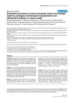
Figure 1
Correlation diagrams of PM1-α, PM/Scl-75a, PM/Scl-75c and PM/Scl-100Correlation diagrams of PM1-α, PM/Scl-75a, PM/Scl-75c and PM/Scl-100. A panel of sera tested previously for reactivity to recombinant polymy-
ositis/scleroderma (PM/Scl) components (PM/Scl-75a, PM/Scl-75c and PM/Scl-100) was assayed for anti-PM1-α peptide reactivity in an ELISA
[18]. Correlation diagrams are shown comparing the peptide ELISA with the recombinant proteins (a)–(c) for all sera (n = 81) and (b)–(f) for only
the sera of PM/Scl patients (n = 36).
Arthritis Research & Therapy Vol 7 No 3 Mahler et al.
R707
Table 1
Results of the technical comparison of indirect immunofluorescence (IIF), immunoblot and ELISA for the detection of anti-
polymyositis/scleroderma (anti-PM/Scl) antibodies
Sample number IIF Immunoblot ELISA
Titer Pattern PM/Scl-75 PM/Scl-100 Other PM/Scl-100 PM1-α
1 1:10000 FG, N ++ +++ 15.3 17.7
2 1:1000 FG, N + + 2.9 7.6
3 1:3200 FG, N ++ ++ 14.4 29.2
1:320 AMA
41:320FG, N-+ 1.14.7
5 1:1000 FG, N + + 2.2 12.1
6 1:3200 FG, N - - 0.6 2.3
7 1:1000 FG, N ++ +++ 5.2 13.3
8 1:1000 FG, N + + 2.3 12.7
9 1:3200 FG, N + +++ P38 15.5 36.1
10 - - + 1.7 4.0
11 1:1000 FG, N - ++ 19.2 2.4
12 1:1000 FG, N ++ + 1.2 3.2
13 1:3200 FG, N +++ +++ Ku86, Jo-1 6.5 10.6
14 1:3200 FG, N - ++ 2.1 5.9
15 1:3200 FG, N ++ +++ 11.4 22.5
16 1:1000 FG, N - + 3.6 8.5
17 1:1000 FG, N + ++ 3.2 15.0
18 1:3200 FG, N ++ ++ 6.1 11.8
19 n.d. n.d. - ++ 7.1 19.4
20 1:320 N - + 2.1 5.6
21 n.d. n.d. +++ ++ 4.1 12.8
22 1:10000 FG - + P38 0.7 1.5
1:10000 SPA
23 1:1000 N n.d. n.d. 0.5 2.7
24 n.d. n.d. ++ +++ 18.2 34.7
25 1:10000 N n.d. n.d. P38 0.5 2.0
1:1000 Rib
26 1:1000 N n.d. n.d. 1.9 8.4
27 n.d. n.d. + ++ 5.9 19.4
Available online />R708
ues, as well as the test efficiency, were calculated. Further-
more, the correlation coefficients between the immunoassays
based on the different antigens were calculated.
Results
Technical comparison of IIF, immunoblot and ELISA for
the detection of anti-PM/Scl antibodies
To compare the different techniques, 33 anti-PM/Scl sera
preselected on the basis of their IIF pattern and/or immunoblot
result were tested in prototype ELISA kits based on the full-
length recombinant PM/Scl-100 polypeptide expressed in E.
coli and on the synthetic PM1-α peptide. In total, 26/33
(78.8%) were positive in the ELISA with the recombinant pro-
tein and 32/33 (97.0%) were positive in the ELISA with the
synthetic peptide. Results are summarized in Table 1. Based
on the high sensitivity of the peptide-based ELISA in this tech-
nical comparison, we evaluated the clinical accuracy of the
assay in an extended multicenter study using clinically defined
sera from various centers.
Correlation of anti-PM1-α with anti-PM/Scl-75a, PM/Scl-
75c and PM/Scl-100 reactivity in ELISA
A panel of sera (n = 81) tested previously for reactivity to
recombinant PM/Scl proteins (Panel II) was assayed for anti-
PM1-α peptide reactivity in the ELISA. The results were com-
pared with the known reactivity of these sera with the recom-
binant proteins [18]. When all assays were adjusted to the
same specificity (91.1%), the clinical sensitivity for the PM/Scl
overlap syndrome was 36.1% for PM1-α, was 27.8% for PM/
Scl-75c and was 25.0% for PM/Scl-100.
There was a clear correlation between the peptide reactivity
and the reactivity of the sera with the recombinant proteins.
Not surprisingly, the strongest correlation was observed with
the anti-PM/Scl-100 reactivity (Fig. 1). Whereas the majority of
the sera showed comparable reactivity in all four assays, some
individual samples showed a higher reactivity to the
recombinant proteins than to the synthetic peptide, and vice
versa. Overall, only one sample (from a patient with DM) was
found that tested positive for the recombinant proteins but
negative for the synthetic peptide. However, 11 sera that
tested positive in the peptide ELISA remained undetected
using the recombinant proteins.
When analyzing only the PM/Scl patients from this panel (36/
81), the correlation between the reactivity of the peptide and
the recombinant proteins was even higher (PM/Scl-100, R
2
=
0.82). Very importantly, no sera were found positive for PM/
Scl-75c and/or PM/Scl-100 but negative for the PM/Scl pep-
tide in the PM/Scl patient group. Two samples were PM/Scl-
75c-positive (new isoform), PM1-α-positive and PM/Scl-100-
negative. One sample reacted with PM/Scl-100 and PM1-α
but not with the PM/Scl-75 proteins. Of the PM/Scl sera,
27.8% (10/36) was positive for the peptide but was negative
for all recombinant polypeptides.
Correlation with other autoantibodies
A statistical evaluation was performed using a patient cohort
of 70 clinically defined PM/Scl sera and PM sera to evaluate
correlations between anti-PM1-α peptide antibodies and other
autoantibodies in ELISA assays using recombinant proteins.
No significant correlation was found with Ro-52, Ro-60, La or
Mi-2 antibodies (Table 2). Anti-PM1-α antibodies and anti-Jo-
1 reactivity were negatively correlated. In addition, a reduced
number of samples from 28 patients with clinically defined
PM/Scl were also tested in an addressable laser bead immu-
noassay for autoantibodies to chromatin, Rib-P, RNP, Scl-70
and Sm, SS-A (Ro) and SS-B (La) (QUANTA Plex 8 TM;
INOVA Diagnostics Inc.). Although antibodies to chromatin,
Rib-P and RNP were detected in some patients, none of these
antibodies appeared to be coincident with anti-PM1-α reactiv-
ity (Table 2).
Multicenter evaluation of the PM1-α ELISA
Sera from 40 clinically defined but serologically unselected
patients with PM/Scl overlap syndrome, as well as from 205
28 n.d. n.d. ++ +++ 20.9 37.3
29 1:1000 Hom n.d. n.d. Ku86, Cen, M2 0.6 0.8
1:100 N*
30 1:1000 N - + 0.6 1.4
31 1:3200 Hom, N - - 0.5 2.9
32 1:3200 N - - 0.5 1.3
33 1:320 FG, N + ++ 7.2 25.7
Number positive/tested 26/29 17/29 27/29 26/33 32/33
-, negative; +, weak positive; ++, positive; +++, strong positive; n.d., not determined; FG, fine granular; Hom, homogenous; SPA, spindle
apparatus; N, nucleoli; AMA, anti-mitochondrial antibodies; Cen, centromere; Rib, ribosomal. * Primate liver.
Table 1 (Continued)
Results of the technical comparison of indirect immunofluorescence (IIF), immunoblot and ELISA for the detection of anti-
polymyositis/scleroderma (anti-PM/Scl) antibodies
Arthritis Research & Therapy Vol 7 No 3 Mahler et al.
R709
Scl patients, 40 PM patients and various other controls (Panel
III), were analyzed in the PM/Scl peptide ELISA (see Table 3).
The results from all patients were used to calculate a receiver
operating characteristic curve, which showed a clear discrim-
ination between PM/Scl patients and various controls (Fig. 2).
At a selected cut-off value of 1.5 RU, 22/40 (55%) PM/Scl
patients tested positive for anti-PM1-α antibodies displaying a
reactivity of up to 11.6 RU with a mean value of 3.1 ± 3.2 RU
(Table 3). Patients from related disorders including Scl and
PM showed a lower mean reactivity compared with the overlap
patients but a higher reactivity than more unrelated controls. In
total, 27/205 (13.2%) scleroderma patients (mean 0.7 ± 1.3
RU) and 3/40 (7.5%) PM patients (mean 1.0 ± 1.1 RU) tested
positive, while 3/114 (2.6%) patients with SLE and 2/48
(4.2%) patients with HCV infection had anti-PM1-α
antibodies. None of the remaining controls showed reactivity
to the PM1-α peptide in the ELISA (Table 3, Fig. 3).
In total, 6.6% control sera tested positive for anti-PM1-α anti-
bodies. This resulted in a diagnostic sensitivity of 55% and a
specificity of 93.4% of the peptide ELISA (positive predictive
value = 38.6%, negative predictive value = 96.5%, test effi-
ciency = 90.7%). When Scl patients and PM patients were
excluded from the group of controls 5/288 (1.7%) patients
were positive, resulting in a specificity of 98.2% (positive pre-
dictive value = 81.5%, negative predictive value = 94.0%, test
efficiency = 92.9%). These data indicate that, within the assay
parameters used here, anti-PM1-α antibodies appear to be
mainly present in sera from PM/Scl patients, from Scl patients,
and to a lesser extent, PM patients.
Discussion
The aim of this study was to compare the autoantigenicity of
the PM1-α peptide that we have described previously [22,23]
with that of native and recombinant PM/Scl-75 and PM/Scl-
100 polypeptides. The results of the technical comparison
showed that the PM1-α peptide ELISA is more sensitive than
the ELISA tests based on the recombinant proteins, and than
immunoblot and IIF experiments. Also, our results suggest that
increased titers of autoantibodies directed to PM1-α might be
more prevalent in patients with the PM/Scl overlap syndrome
and related diseases than autoantibodies to the full-length pro-
teins, which up to now were considered the most frequently
present.
In the past, the presence of these antibodies in serum was
generally monitored by IIF with HEp-2 cells, by immunodiffu-
sion assays with calf thymus extract and/or by immunoblot
using extractable nuclear antigens [4,5,15]. All these tech-
niques allow the detection of a wide variety of autoantibodies
present in patient serum [2]. The detection of anti-PM/Scl anti-
bodies by immunoblotting, however, is difficult, because the
reactivity of the antibodies with particularly PM/Scl-75 in cell
extracts is notoriously weak in immunoblot, which may be due
to the importance of conformational epitopes [15]. This obser-
vation could be confirmed in the technical comparison of IIF,
immunoblot and ELISA in the present study. In recent years,
ELISA using recombinant PM/Scl-100 has become a common
method to detect anti-PM/Scl reactivity because it can easily
be applied in an automated setting.
Since anti-PM/Scl-75 reactivity was previously detected only
in patient sera that also contained anti-PM/Scl-100 autoanti-
bodies [15], this protein is usually not included in such assays.
A recent investigation has shown that also the use of an
incomplete recombinant PM/Scl-75 polypeptide may have led
to an underestimation of the diagnostic value of the PM/Scl-75
antigen [18].
Table 2
Correlation of anti-PM1-α and other known autoantibodies
Number positive/all sera (%
positive)
Number positive/PM1-α-positives
(% positive)
Number positive/PM1-α-negatives
(% positive)
P
Scl-70 0/28 (0) 0/15 (0) 0/13 (0) *
Sm0/28 (0)0/15 (0)0/13 (0)*
Rib-P 1/28 (3.6) 1/15 (6.7) 0/13 (0) *
RNP 4/28 (14.4) 1/15 (6.7) 3/13 (23.1) *
Chromatin 1/28 (3.6) 1/15 (6.7) 0/13 (0%) *
Ro-52 18/70 (25.7) 6/19 (31.6) 12/51 (23.5) 0.7056
Ro-60 6/70 (8.6) 2/19 (10.6) 4/51 (7.8) 0.9018
La3/70 (4.3)1/19 (5.3)2/51 (3.9)0.6766
Mi-2 10/70 (14.3) 3/19 (15.8) 7/51 (13.7) 0.8693
Jo-1 16/70 (22.9) 1/19 (5.2) 15/51 (29.4) 0.0688
* Not calculated due to the limited number of samples.
Available online />R710
We recently characterized the antibody response to a major
PM/Scl epitope and found that 14/14 (100%) samples with
PM/Scl antibodies demonstrated reactivity to the major
epitope in a membrane-based peptide array [22,23]. We have
characterized the major PM/Scl-100 B-cell epitope at the
amino acid level and identified the key amino acids involved in
antibody binding [22,23]. Using this peptide as an antigen, we
developed a highly sensitive and specific ELISA system that
detects a subpopulation of anti-PM/Scl antibodies present in
55% of PM/Scl patients, in 13.2% of Scl patients and in 7.5%
of PM patients. Interestingly, this peptide also contains a gen-
eralized T-cell epitope pattern (ALADFIHQQR; amino acids
236–245) as well as several major histocompatibility complex
epitopes [28-30].
Synthetic peptides represent ideal antigenic targets for immu-
noassays because they can easily be produced in high quality
and quantity. Furthermore, less lot-to-lot variation will be
observed since the production is not dependent on the biolog-
ical variation of native sources of antigens. More and more syn-
thetic peptides are being used in immunological assay
systems to detect autoantibodies. Some of them show higher
specificities and sensitivities than the corresponding assay
with recombinant protein or native protein as substrate [23].
The combined use of different PM/Scl antigens, including the
recombinant PM/Scl-100 and the recently identified isoform of
PM/Scl-75, as well as the PM1-α peptide, may represent the
most sensitive and specific method to detect antibodies to the
human exosome. Advances in multi-analyte technologies such
as line assays, multiplex systems and micro-arrays allow for the
development of sophisticated profile assays containing multi-
ple different antigens. This may improve the diagnosis of a
variety of disorders, especially of autoimmune diseases since
for most of those disorders no highly sensitive marker is avail-
able. The diagnosis of PM/Scl, Scl and PM might be improved
by providing an antigen array that includes different PM/Scl
antigens in combination with Scl-70 (topoisomerase I), Ku70/
86, centromere proteins, RNA polymerase, NOR-90, Jo-1, Mi-
2, PL-7, PL-12 and fibrillarin.
Taken together, the use of the PM/Scl-100 synthetic peptide
in an ELISA remarkably improves the clinical identification of
patients with the PM/Scl overlap syndrome. Although the prev-
alence of autoantibodies recognizing most other exosome
subunits is relatively low [8,10,11], the co-occurrence of anti-
bodies targeting different exosome subunits in patient sera
might be indicative of intermolecular epitope spreading and
might be a marker for the overlap syndrome. The co-occur-
rence of anti-PM/Scl-100 and anti-PM/Scl-75 seems to be
particularly associated with the PM/Scl overlap syndrome
[18], but whether the use of even more components of the
human exosome will further increase the sensitivity of these
assays remains to be investigated.
Apart from patients with PM/Scl overlap syndrome and
patients with Scl or PM alone, two HCV-positive and three
SLE patients displayed reactivity to the PM1-α peptide in
ELISA. HCV infection has been associated with a plethora of
immune and autoimmune perturbations [31]. Although the
cause and effect remain to be proved, there are reports of
HCV infection preceding or coincident with polyarthritis, rheu-
matoid arthritis, SLE, and PM/DM. The role of anti-PM1-α anti-
bodies in HCV patients and SLE patients remain a matter for
further investigation. In the present study we found antibodies
Figure 2
Receiver operating characteristic analysis of the PM1-α ELISAReceiver operating characteristic analysis of the PM1-α ELISA. Results
obtained from three centers and based on 567 patients including poly-
myositis/scleroderma (PM/Scl) patients (n = 40), Scl patients (n =
205) and PM patients (n = 40) as well as other controls were used to
calculate a receiver operating characteristic analysis (a) for all control
samples and (b) for unrelated controls (without Scl and PM). The curve
shows a clear discrimination between PM/Scl patient samples and vari-
ous controls as emphasized by an area under the curve value of 0.901
(all controls) and 0.958 (unrelated controls). The differentiation
between PM/Scl patients and controls was significantly improved when
Scl patients and PM patients were excluded from the control group (b).
SE, standard error.
Arthritis Research & Therapy Vol 7 No 3 Mahler et al.
R711
to the PM1-α peptide present in 13.2% of unselected sclero-
derma patients. In only a few of those patients was a history of
myositis documented. It is possible that the myositis was mild
in the majority of patients and was completely overlooked by
the examining clinician or that the antibody precedes the asso-
ciated clinical features [32]. We therefore conclude that the
complete autoantibody profile is important for a careful exami-
nation of patients with rheumatic diseases and to access all
their clinical features.
Frank and colleagues analyzed sera from 216 patients with idi-
opathic inflammatory myopathies to assess putative associa-
tions between anti-SS-A/Ro-52 and other autoantibodies.
These included sera containing antibodies that recognize Jo-
1, Mi-2, PM/Scl, signal recognition particle, as well as the
Table 3
Results of ELISA using PM1-α peptide with polymyositis/scleroderma and various control sera
Number (%) of anti-PM1-α-positive sera Mean value/standard deviation Top value
Polymyositis/scleroderma (n = 40) 22 (55) 3.1/3.2 11.6
Rheumatic disease controls (n = 452) 33 (7.3) 0.6/0.9 7.7
Polymyositis (n = 40) 3 (7.5) 1.0/1.1 7.4
Scleroderma (n = 205) 27 (13.2) 0.9/1.2 7.5
Rheumatoid arthritis (n = 69) 0 (0) 0.3/0.2 1.1
Mixed connective tissue disease (n = 6) 0 (0) 0.4/0.1 0.6
Undifferentiated connective tissue disease (n = 10) 0 (0) 0.3/0.0 0.4
Systemic lupus erythematosus (n = 114) 3 (2.6) 0.5/0.7 7.7
Other rheumatic diseases (n = 8) 0 (0) 0.3/0.1 0.6
Hepatitis C virus (n = 48) 2 (4.2) 0.5/0.5 2.6
Organ specific disorders (n = 23) 0 (0) 0.4/0.2 0.8
Hashimoto thyroiditis (n = 11) 0 (0) 0.3/0.2 0.8
Grave's disease (n = 12) 0 (0) 0.4/0.2 0.8
Healthy individuals (n = 4) 0 (0) 0.6/0.2 0.7
Figure 3
Reactivity of polymyositis/scleroderma (PM/Scl) patients and controls in the PM1-α ELISAReactivity of polymyositis/scleroderma (PM/Scl) patients and controls in the PM1-α ELISA. Results obtained from three centers and based on 567
patients including PM/Scl patients (n = 40), Scl patients (n = 205) and PM patients (n = 40) as well as other controls were used to calculate com-
parative descriptive analysis. The diagram shows a significantly increased reactivity of the PM/Scl sera compared with the control groups. Compara-
tive descriptives show vertical box-plots for each sample, side by side for comparison. The blue line series shows parametric statistics: diamond,
mean and the requested confidence interval around the mean; notched line, requested parametric percentile range. The notched box and whiskers
show non-parametric statistics: notched box, median, lower and upper quartiles, and confidence interval around the median; dotted line, connects
the nearest observations within 1.5 interquartile ranges (IQR) of the lower and upper quartiles. + and ❍, possible outliers – observations more than
1.5 IQR (near outliers) and more than 3.0 IQR (far outliers) from the quartiles. Vertical lines, requested nonparametric percentile range. SLE, sys-
temic lupus erythematosus; HCV, hepatitis C virus; RA, rheumatoid arthritis.
Available online />R712
scleroderma-related antibodies anti-topoisomerase I (Scl-70)
and anti-centromere. A high proportion of sera that contain
anti-Jo-1 antibodies, anti-signal recognition particle or anti-
PM/Scl antibodies were found to contain antibodies to the
Ro52 protein [33]. The reported association between anti-Ro-
52 and anti-PM/Scl antibodies is not found in our cohort.
Although our correlation study is based on a limited number of
samples, we found no correlation between anti-PM/Scl anti-
bodies and anti-Ro52. In contrast, Yamanishi and colleagues
reported an association of PM/Scl syndrome with anti-Ku anti-
body and rimmed vacuole formation [34]. Similar to this obser-
vation, we found that two out of 29 (6.9%) anti-PM/Scl-
positive samples also were positive for anti-Ku86 antibodies.
In a previous study it became evident that the PM/Scl-100
major epitope shares some sequence homology to an amino
acid stretch (amino acids 58–72) of the heterochromatin pro-
tein p25β, which is frequently the target of anti-chromo anti-
bodies from a subpopulation of patients also having anti-
centromere antibodies [22]. Although none of 14 PM1-α-pos-
itive samples showed reactivity to the corresponding region of
p25β, a more complex immunological relationship between
the major PM/Scl-100 epitope and the corresponding p25β
peptide cannot be excluded. More samples with anti-PM/Scl
and anti-chromo antibodies have to be tested for cross-reac-
tivity. Further studies are required to analyze the association of
anti-PM1-α antibodies with other known autoantibodies.
Today's sophisticated epitope mapping methods will probably
lead to the identification of additional peptides, which can be
used as specific targets in diagnostic and therapeutic
approaches to patient management. This may lead to a new
scientific research area with high impact for the development
of diagnostic and therapeutic products – to the area of pep-
tide engineering.
Conclusion
In the present study, we showed that the detection of anti-PM/
Scl antibodies using an ELISA system based on a major PM/
Scl-100 epitope is remarkably improved compared with con-
ventional detection methods. It could be shown that a subpop-
ulation of PM/Scl antibodies directed against the PM1-α
peptide is present in 55% of PM/Scl patients, in 13.2% of Scl
patients and in 7.5% of PM patients. In rare cases anti-PM1-α
reactivity was also found in patients suffering from HCV, SLE
or melanoma. Within our patient cohorts we found no statisti-
cal evidence of a positive association between anti-PM1-α
and antibodies other than to PM/Scl components. Based on
the results of the present study we conclude that anti-PM1-α
antibodies are exclusively present in sera from patients suffer-
ing from Scl or PM and most frequently in patients with the
PM/Scl overlap syndrome. We therefore conclude that the
new anti-PM1-α ELISA test offers a new serological test that
will improve the diagnosis of complex connective tissue
disorders.
Competing interests
MM is employed at Dr Fooke Laboratorien GmbH, which may
commercialize the assay.
Authors' contributions
MM developed and validated the ELISA system, planned the
experiments and filed the manuscript. MJF and RR delivered
clinically defined sera, advised MM in evaluating the clinical
part of this study and contributed to the preparation of the
manuscript. CD organized the analysis of anti-PM/Scl samples
in IIF and immunoblot. MB advised MM during the characteri-
zation of the PM1-α peptide.
Acknowledgements
The authors thank Dr R Mierau and Prof. E Genth (Rheumaklinik Aachen,
Germany) for providing clinically defined sera, Mark L Fritzler (University
of Calgary, Canada) for technical assistance with the addressable laser
bead immunoassay and Wilma Vree Egberts (Radboud University
Nijmegen, The Netherlands) for technical assistance.
References
1. Tan EM: Antinuclear antibodies: diagnostic markers for
autoimmune diseases and probes for cell biology. Adv
Immunol 1989, 44:93-151.
2. von Muhlen CA, Tan EM: Autoantibodies in the diagnosis of
systemic rheumatic diseases. Semin Arthritis Rheum 1995,
24:323-358.
3. Wolfe JF, Adelstein E, Sharp GC: Antinuclear antibody with dis-
tinct specificity for polymyositis. J Clin Invest 1977,
59:176-178.
4. Reimer G, Steen VD, Penning CA, Medsger TA Jr, Tan EM: Corre-
lates between autoantibodies to nucleolar antigens and clini-
cal features in patients with systemic sclerosis (scleroderma).
Arthritis Rheum 1988, 31:525-532.
5. Reichlin M, Maddison PJ, Targoff I, Bunch T, Arnett F, Sharp G,
Treadwell E, Tan EM: Antibodies to a nuclear/nucleolar antigen
in patients with polymyositis overlap syndromes. J Clin
Immunol 1984, 4:40-44.
6. Oddis CV, Okano Y, Rudert WA, Trucco M, Duquesnoy RJ, Meds-
ger TA Jr: Serum autoantibody to the nucleolar antigen PM-Scl.
Clinical and immunogenetic associations. Arthritis Rheum
1992, 35:1211-1217.
7. Reimer G, Scheer U, Peters JM, Tan EM: Immunolocalization
and partial characterization of a nucleolar autoantigen (PM-
Scl) associated with polymyositis/scleroderma overlap
syndromes. J Immunol 1986, 137:3802-3808.
8. Gelpi C, Alguero A, Angeles Martinez M, Vidal S, Juarez C, Rod-
riguez-Sanchez JL: Identification of protein components reac-
tive with anti-PM/Scl autoantibodies. Clin Exp Immunol 1990,
81:59-64.
9. Allmang C, Petfalski E, Podtelejnikov A, Mann M, Tollervey D,
Mitchell P: The yeast exosome and human PM-Scl are related
complexes of 3' → 5' exonucleases. Genes Dev 1999,
13:2148-2158.
10. Brouwer R, Pruijn GJ, van Venrooij WJ: The human exosome: an
autoantigenic complex of exoribonucleases in myositis and
scleroderma. Arthritis Res 2001, 3:102-106.
11. Brouwer R, Allmang C, Raijmakers R, van Aarssen Y, Egberts WV,
Petfalski E, van Venrooij WJ, Tollervey D, Pruijn GJ: Three novel
components of the human exosome. J Biol Chem 2001,
276:6177-6184.
12. Bluthner M, Bautz FA: Cloning and characterization of the cDNA
coding for a polymyositis-scleroderma overlap syndrome-
related nucleolar 100-kD protein. J Exp Med 1992,
176:973-980.
13. Ge Q, Frank MB, O'Brien C, Targoff IN: Cloning of a complemen-
tary DNA coding for the 100-kD antigenic protein of the PM-Scl
autoantigen. J Clin Invest 1992, 90:559-570.
Arthritis Research & Therapy Vol 7 No 3 Mahler et al.
R713
14. Briggs MW, Burkard KT, Butler JS: Rrp6p, the yeast homologue
of the human PM-Scl 100-kDa autoantigen, is essential for
efficient 5.8 S rRNA 3' end formation. J Biol Chem 1998,
273:13255-13263.
15. Ge Q, Wu Y, Trieu EP, Targoff IN: Analysis of the specificity of
anti-PM-Scl autoantibodies. Arthritis Rheum 1994,
37:1445-1452.
16. Targoff IN, Reichlin M: Nucleolar localization of the PM-Scl
antigen. Arthritis Rheum 1985, 28:226-230.
17. Brouwer R, Hengstman GJ, Vree Egberts W, Ehrfeld H, Bozic B,
Ghirardello A, Grondal G, Hietarinta M, Isenberg D, Kalden JR, et
al.: Autoantibody profiles in the sera of European patients with
myositis. Ann Rheum Dis 2001, 60:116-123.
18. Raijmakers R, Renz M, Wiemann C, Egberts WV, Seelig HP, van
Venrooij WJ, Pruijn GJ: PM-Scl-75 is the main autoantigen in
patients with the polymyositis/scleroderma overlap
syndrome. Arthritis Rheum 2004, 50:565-569.
19. Brouwer R, Vree Egberts WT, Hengstman GJ, Raijmakers R, van
Engelen BG, Seelig HP, Renz M, Mierau R, Genth E, Pruijn GJ, van
Venrooij WJ: Autoantibodies directed to novel components of
the PM/Scl complex, the human exosome. Arthritis Res 2002,
4:134-138.
20. Ge Q, Wu Y, James JA, Targoff IN: Epitope analysis of the major
reactive region of the 100-kd protein of PM-Scl autoantigen.
Arthritis Rheum 1996, 39:1588-1595.
21. Bluthner M, Bautz EK, Bautz FA: Mapping of epitopes recog-
nized by PM/Scl autoantibodies with gene-fragment phage
display libraries. J Immunol Methods 1996, 198:187-198.
22. Bluthner M, Mahler M, Muller DB, Dunzl H, Bautz FA: Identifica-
tion of an alpha-helical epitope region on the PM/Scl-100
autoantigen with structural homology to a region on the hete-
rochromatin p25beta autoantigen using immobilized overlap-
ping synthetic peptides. J Mol Med 2000, 78:47-54.
23. Mahler M, Bluthner M, Pollard KM: Advances in B-cell epitope
analysis of autoantigens in connective tissue diseases. Clin
Immunol 2003, 107:65-79.
24. Anonymous: Preliminary criteria for the classification of sys-
temic sclerosis (scleroderma). Subcommittee for scleroderma
criteria of the American Rheumatism Association Diagnostic
and Therapeutic Criteria Committee. Arthritis Rheum 1980,
23:581-590.
25. Bohan A, Peter JB: Polymyositis and dermatomyositis. N Engl J
Med 1975, 292:344-347.
26. Mahler M, Fritzler MJ, Bluthner M: Identification of a SmD3
epitope with a single symmetrical dimethylation of an arginine
residue as a specific target of a subpopulation of anti-Sm
antibodies. Arthritis Res Ther 2005, 7:R19-R29.
27. Datasheet Recombinant PM/Scl-100 Autoantigen [http://
www.diarect.com/pi_local/files/Antigen_Specification_PM-
Scl_100_15000_020314_196.pdf]
28. Falk K, Rotzschke O, Stevanovic S, Jung G, Rammensee HG:
Allele-specific motifs revealed by sequencing of self-peptides
eluted from MHC molecules. Nature 1991, 351:290-296.
29. Rothbard JB, Taylor WR: A sequence pattern common to T cell
epitopes. EMBO J 1988, 7:93-100.
30. Sette A, Buus S, Appella E, Smith JA, Chesnut R, Miles C, Colon
SM, Grey HM: Prediction of major histocompatibility complex
binding regions of protein antigens by sequence pattern
analysis. Proc Natl Acad Sci USA 1989, 86:3296-3300.
31. McMurray RW, Elbourne K: Hepatitis C virus infection and
autoimmunity. Semin Arthritis Rheum 1997, 26:689-701.
32. Arbuckle MR, MT McClain, MV Rubertone, RH Scofield, GJ Den-
nis, JA James, JB Harley: Development of autoantibodies before
the clinical onset of systemic lupus erythematosus. N Engl J
Med 2003, 349:1526-1533.
33. Frank MB, McCubbin V, Trieu E, Wu Y, Isenberg DA, Targoff IN:
The association of anti-Ro52 autoantibodies with myositis and
scleroderma autoantibodies. J Autoimmun 1999, 12:137-142.
34. Yamanishi Y, Maeda H, Katayama S, Ishioka S, Yamakido M: Scle-
roderma-polymyositis overlap syndrome associated with anti-
Ku antibody and rimmed vacuole formation. J Rheumatol 1996,
23:1991-1994.