Báo cáo y học: " Mithramycin downregulates proinflammatory cytokine-induced matrix metalloproteinase gene expression in articular chondrocytes" ppsx
Bạn đang xem bản rút gọn của tài liệu. Xem và tải ngay bản đầy đủ của tài liệu tại đây (1.12 MB, 7 trang )
Open Access
Available online />R777
Vol 7 No 4
Research article
Mithramycin downregulates proinflammatory cytokine-induced
matrix metalloproteinase gene expression in articular
chondrocytes
Abdelhamid Liacini, Judith Sylvester, Wen Qing Li and Muhammad Zafarullah
Département de Médecine and Centre de Recherche du Centre Hospitalier de l'Université de Montréal (CHUM), Hôpital Notre-Dame du CHUM,
Montréal, Québec, Canada
Corresponding author: Muhammad Zafarullah,
Received: 18 Jul 2003 Revisions requested: 15 Aug 2003 Revisions received: 21 Feb 2005 Accepted: 7 Mar 2005 Published: 4 Apr 2005
Arthritis Research & Therapy 2005, 7:R777-R783 (DOI 10.1186/ar1735)
This article is online at: />© 2005 Zafarullah et al.; licensee BioMed Central Ltd.
This is an Open Access article distributed under the terms of the Creative Commons Attribution License ( />2.0), which permits unrestricted use, distribution, and reproduction in any medium, provided the original work is properly cited.
Abstract
Interleukin-1 (IL-1), IL-17 and tumor necrosis factor alpha (TNF-
α) are the main proinflammatory cytokines implicated in cartilage
breakdown by matrix metalloproteinase (MMPs) in arthritic
joints. We studied the impact of an anti-neoplastic antibiotic,
mithramycin, on the induction of MMPs in chondrocytes. MMP-
3 and MMP-13 gene expression induced by IL-1β, TNF-α and IL-
17 was downregulated by mithramycin in human
chondrosarcoma SW1353 cells and in primary human and
bovine femoral head chondrocytes. Constitutive and IL-1-
stimulated MMP-13 levels in bovine and human cartilage
explants were also suppressed. Mithramycin did not significantly
affect the phosphorylation of the mitogen-activated protein
kinases, extracellular signal-regulated kinase, p38 and c-Jun N-
terminal kinase. Despite effective inhibition of MMP expression
by mithramycin and its potential to reduce cartilage
degeneration, the agent might work through multiple
unidentified mechanisms.
Introduction
A major pathological manifestation of patients with osteoarthri-
tis (OA) and rheumatoid arthritis is the degeneration of articu-
lar cartilage [1,2]. Matrix metalloproteinases (MMPs) such as
MMP-3 and MMP-13 are known to cleave collagens and
aggrecan of cartilage extracellular matrix [3-5]. The concentra-
tions of several MMPs are increased in cartilage, synovial
membrane and synovial fluid of patients with arthritis [6,7].
Indeed, cartilage-specific overexpression of active human
MMP-13 causes OA in mice [8]. Proinflammatory cytokines,
interleukin-1 (IL-1), IL-17 and tumor necrosis factor (TNF)-α
are also increased in arthritic joints and are known to induce
catabolic pathways leading to an enhanced expression of
MMPs [9-11]. Inhibition of these proteases is regarded as an
important approach for reducing damage in arthritic tissues
[12].
AP-1 binding sites found in the promoter regions of the genes
encoding MMP-3 and MMP-13 are essential for the expres-
sion of these genes [13,14]. Sp1 transcription factor is a zinc-
finger type transcription factor whose binding sites are found
in numerous housekeeping and inducible genes [15]. Human
MMP-13 promoter has one putative Sp1 consensus site [16].
Mithramycin is an aureolic acid anti-neoplastic antibiotic that is
used for treating cancer-related hypercalcemia [17]. Previous
work has revealed that it inhibits bone resorption in vitro, pos-
sibly by interfering with bone cell lysosomal enzymes [18]. It
also prevents the binding of Sp1 transcription factor to its cog-
nate site in DNA by modifying the CG sequences [19]. Here
we have studied the impact of mithramycin on proinflammatory
cytokine-induced MMP expression. We show for the first time
that mithramycin potently suppresses MMP induction by IL-1,
IL-17 and TNF-α in chondrocytic cells without impairing the
activation of mitogen-activated protein kinases (MAPKs).
BSA = bovine serum albumin; DMEM = Dulbecco's modified Eagle's medium; ERK = extracellular signal-related kinase; FCS = fetal calf serum; IL =
interleukin; JNK = c-Jun N-terminal kinase; MAPK = mitogen-activated protein kinase; MMP = matrix metalloproteinase; OA = osteoarthritis; PBS =
phosphate-buffered saline; PCR = polymerase chain reaction; RT = reverse transcriptase; TNF = tumor necrosis factor.
Arthritis Research & Therapy Vol 7 No 4 Liacini et al.
R778
Materials and methods
Primary cultures of human and bovine chondrocytes,
SW1353 cells and treatments
Human cartilage was acquired from the femoral heads of OA
patients who underwent hip-replacement surgery at the Notre-
Dame Hospital. Normal bovine cartilage was obtained from the
knee and hip joints of adult animals from a local abattoir.
Chondrocytes were released by 90 min pronase and 9 hours
digestion with collagenase (Sigma type IA). The cells were
washed with PBS and grown in DMEM containing 10% FCS
as high-density primary monolayer cultures until confluent
growth. Cells were distributed in six-well plates, grown to con-
fluence, washed with PBS and kept in serum-free DMEM for
24 hours; mithramycin (from Sigma-Aldrich Canada Ltd,
Oakville, Ontario; dissolved in water as a 10 mM solution) was
then added without medium change at final concentrations of
100 and 150 nM (doses known to inhibit Sp1 binding [20]) for
30 min before treatment for 24 hours with human recombinant
IL-1β (10 ng/ml), TNF-α (20 ng/ml) and IL-17 (20 ng/ml) (R&D
systems, Minneapolis, MN). The human chondrosarcoma cell
line SW1353 was obtained from the American Type Culture
Collection (ATCC, Manassas, VA) and treated as described
for primary chondrocytes.
Northern hybridization analysis
Total cellular RNA was extracted by the guanidinium proce-
dure [21] and aliquots of 3 to 5 µg were analyzed by electro-
phoretic fractionation in 1.2% formaldehyde-agarose gels. The
integrity and quantity of RNA were verified by ethidium bro-
mide staining of the 28S and 18S ribosomal RNA bands. The
RNA was transferred onto Zeta-probe nylon membrane with a
Bio-Rad Transblot in the presence of 0.5 × TAE (Tris-acetate-
EDTA) buffer at a current of 500 mA for 12 hours. Northern
blots were hybridized with a human stromelysin cDNA probe
generously provided by Dr Richard Breathnach (Nantes,
France). This probe was a 1.6-kilobase EcoRI cDNA fragment
cloned in the plasmid pGEM-4Z (Promega Biotech, Madison,
WI) and the vector was linearized with NarI. A 491-base-pair
RT-PCR-generated [22] and cloned collagenase-3 cDNA was
linearized with EcoRI. The human 28S ribosomal RNA plasmid
(ATCC) was digested with XbaI. All antisense RNA probes
were synthesized with T7 polymerase in accordance with the
protocols of Promega Biotech and were labeled to high spe-
cific radioactivity (10
8
c.p.m./µg) with [α-
32
P]CTP (3,000 Ci/
mmol; Perkin Elmer Life Sciences Inc., Boston, MA).
Western immunoblot analysis
Total secreted proteins from the 2 to 3 ml of conditioned
medium of the chondrocytes or SW1353 cells were concen-
trated by precipitation with trichloroacetic acid, quantified with
the Bio-Rad protein assay system and different amounts of
protein aliquots adjusted to 15 µl with 4 × sample buffer com-
prising 62.5 mM Tris-HCl, 20% glycerol, 0.032% bromophe-
nol blue, 5% mercaptoethanol and 2% SDS. Along with the
prestained broad-range molecular mass standards (Bio-Rad),
samples were fractionated by a 4% stacking and 10% SDS-
PAGE mini gel (Bio-Rad, Mississauga, ON) and transferred to
nitrocellulose membrane by electroblotting at 200 mA in a
buffer comprising 25 mM Tris-HCl, 192 mM glycine, 0.04%
SDS and 20% ethanol. The membranes were rinsed with dis-
tilled water, incubated for 1 hour in PBS pH 7.4 with 5% Car-
nation non-fat milk to block non-specific interactions, and
washed five times (twice for 5 min, once for 15 min and twice
for 5 min) with PBS containing 0.1% Tween. They were then
reacted overnight sequentially in the same buffer at 4°C with
1 to 2 µg/ml anti-human MMP-3 (developed in mouse) and
MMP-13 (hinge region, developed in rabbit) antibodies (from
Sigma-Aldrich). Subsequently, membranes were washed at
22°C five times with PBS containing 0.1% Tween, incubated
with the anti-rabbit or anti-mouse secondary peroxidase-conju-
gated IgG (300 mU/ml), and washed seven times with PBS
containing 0.1% Tween. To reveal the MMP-3 and MMP-13
bands, membranes were incubated with 10 µl of solution A
and 990 µl of solution B of the chemiluminescence detection
system of Roche Biochemicals (Laval, Québec) and exposed
to film for 2 to 15 min.
For Western blots of MAPKs, human femoral-head chondro-
cytes were pretreated with mithramycin for 30 min and then
stimulated with IL-1β for 20 min; total cellular protein extracts
(20 µg) in lysis buffer (62.5 mM Tris-HCl pH 6.8, 10% glyc-
erol, 1% Triton X-100, 50 mM dithiothreitol, 2% SDS, 0.01%
bromophenol blue) were resolved by 10% SDS-PAGE, trans-
ferred to nitrocellulose membranes by electroblotting and
incubated overnight at 4°C with primary phosphorylation-
state-specific antibodies for phosphorylated extracellular sig-
nal-regulated kinase (p-ERK), phospho-p38 and phosphor-
ylated c-Jun N-terminal kinase (p-JNK) (from Cell Signaling
Technology Inc., Beverley, MA) at 1:1,000 dilution in 5% BSA,
1 × Tris-buffered saline and 0.1% Tween. Proteins were
detected with the enhanced chemiluminescence system from
Pharmacia-Amersham. Subsequently, the membranes were
stripped with a buffer (containing 100 mM 2-mercaptoethanl,
2% SDS and 62.5 mM Tris-HCl, pH 6.8) at 55°C and rep-
robed with the antibodies detecting total ERK, p38 and JNK.
Cartilage explants
Human or bovine femoral head cartilage explants were main-
tained for 1 week in DMEM with 10% FCS, medium was then
changed with 0.01% serum-containing DMEM for 3 days until
treatments. Explants were treated with mithramycin and IL-1
vehicles as control (water and PBS-0.1% BSA) or exposed to
mithramycin (150 nM) and IL-1 (33 ng/ml) for 15 days with
replacement of the fresh reagents every 2 days; the secreted
media were concentrated by precipitation with 10% trichloro-
acetic acid and equal amounts of protein (16 µg per lane for
human explants and 20 µg per lane for bovine explants) were
subjected to Western immunoblotting as described above. All
the experiments described in this paper were performed at
least two (primary human chondrocytes and cartilage) or three
Available online />R779
(SW1353 and bovine chondrocytes) times and the results
were reproducible.
Results
Mithramycin blocks IL1-stimulated expression of MMP-3
and MMP-13 in human and bovine chondrocyte cell lines
IL-1β potently induced expression of the genes encoding
MMP-3 and MMP-13 in the human chondrocytic cell line
SW1353 and in primary human femoral head chondrocytes.
Mithramycin, a hypocalcemic antibiotic, potently blocked the
induction of MMP-3 and MMP-13 mRNA by IL-1β without
affecting the control 28S rRNA levels (Fig. 1a,b). The two
MMP-13 mRNA bands correspond to transcripts produced by
differential use of polyadenylation sites at the 3' end of the
gene, the upper band being the longest transcript as reported
previously [23]. Induction of MMP-13 protein was also simi-
larly inhibited (Fig. 1a,b). To examine whether mithramycin
could also affect MMP gene expression in articular chondro-
cytes from other species, adult bovine chondrocytes (an
important model system in cartilage research) were exposed
to different concentrations of mithramycin and then stimulated
with IL-1β. This cytokine induced MMP-3 and MMP-13 mRNA
expression above basal levels, and pretreatment with mith-
ramycin reduced both constitutive and induced expression in
a fashion similar to that of human cells (Fig. 1c). The induction
of MMP-13 protein (the main collagen-degrading MMP) by IL-
1 was also inhibited. The double MMP-13 protein bands are
due to a better resolution of the upper proenzyme and lower
active MMP-13 forms.
IL-17-induced MMP gene expression is suppressed by
mithramycin
IL-17 is a major proinflammatory cytokine and an inducer of
MMP expression in chondrocytes and macrophages [11,24].
As hown in Fig. 2, IL-17 stimulated the basal MMP-3 and
MMP-13 mRNA and MMP-13 protein expression in bovine
and human OA chondrocytes. Mithramycin dose-dependently
diminished these inductions.
TNF-α-induced MMP gene expression is inhibited by
mithramycin
TNF-α is another prominent inflammatory cytokine that
increased the constitutive MMP-3 and MMP-13 mRNA
expression in chondrocytic SW1353 cells, primary human
chondrocytes and bovine chondrocytes. Exposure to mith-
ramycin followed by stimulation with TNF-α resulted in
decreased constitutive and induced MMP-3 and MMP-13
gene expression (Fig. 3). In some cases, a concentration of
100 nM caused maximal inhibition; a 150 nM dose did not
have any additional effect (e. g. mRNA in 3B).
Mithramycin inhibits IL-1-stimulated expression of
MMPs in human and bovine cartilage
To study the impact of mithramycin on the production of MMP-
13 by chondrocytes in their native cartilage matrix, human and
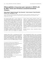
Figure 1
Repression of interleukin (IL)-1β-inducible matrix metalloproteinase (MMP)-3 and MMP-13 RNA expression by mithramycinRepression of interleukin (IL)-1β-inducible matrix metalloproteinase
(MMP)-3 and MMP-13 RNA expression by mithramycin. Quiescent
human chondrosarcoma (a), primary human chondrocytes (b) or bovine
chondrocytes (c) were pretreated with different concentrations of mith-
ramycin for 30 min, followed by additional treatment with IL-1β for 24
hours. The MMP-3, MMP-13 and 28S RNA levels were measured by
Northern hybridization, and MMP-13 protein levels were measured by
Western blot analysis. For protein gels, 3 µg (a) or 4 µg (b, c) of pro-
tein was applied to each lane. The resulting autoradiograms indicating
the respective gene products are shown.
Arthritis Research & Therapy Vol 7 No 4 Liacini et al.
R780
bovine cartilage explants were maintained in low-serum
(0.01%) medium and exposed to mithramycin (150 nM) and
IL-1β for 15 days with changes of reagents every 2 days.
Human cartilage had somewhat elevated constitutive levels of
MMP-3 and MMP-13. Mithramycin drastically reduced the
secreted basal and IL-1-induced MMP-3 and MMP-13 protein
levels in human cartilage (Fig. 4a) and MMP-13 in bovine car-
tilage (Fig. 4b) as measured by Western blotting of the condi-
tioned media. MMP-3 levels were too low to be measurable in
Figure 2
Decrease in interleukin (IL)-17-inducible matrix metalloproteinase (MMP)-3 and MMP-13 gene expression by mithramycinDecrease in interleukin (IL)-17-inducible matrix metalloproteinase
(MMP)-3 and MMP-13 gene expression by mithramycin. Quiescent
bovine chondrocytes (a) or primary human chondrocytes (b) were pre-
treated with different doses of mithramycin for 30 min and treated fur-
ther with IL-17 for 24 hours. The MMP-3, MMP-13 and 28S RNA levels
were measured by Northern hybridization, and MMP-13 protein levels
were measured by Western blot analysis. For protein gels, 4 µg of pro-
tein was applied to each lane. The resulting autoradiograms indicating
the respective gene products are shown.
Figure 3
Downregulation of tumor necrosis factor (TNF)-α-inducible matrix met-
alloproteinase (MMP)-3 and MMP-13 RNA expression by mithramycinDownregulation of tumor necrosis factor (TNF)-α-inducible matrix met-
alloproteinase (MMP)-3 and MMP-13 RNA expression by mithramycin.
Human SW1353 condrosarcoma cells (a), primary human femoral
head chondrocytes (b) and bovine chondrocytes (c) were pre-exposed
to the indicated concentrations of mithramycin for 30 min, followed by
additional treatment with TNF-α for 24 hours. The MMP-3, MMP-13
and 28S RNA levels were measured by Northern hybridization, and
MMP-3 protein levels were measured by Western blot analysis. For pro-
tein gels, 3 µg (a) or 4 µg (b) of protein was applied to each lane. The
resulting autoradiograms indicating the respective products are shown.
Available online />R781
bovine explants. Therefore mithramycin diminishes IL-1-stimu-
lated MMP production in cartilage explants.
Mithramycin does not affect the phosphorylation of ERK,
p38 and JNK
Because MAPKs are important mediators of proinflammatory
cytokine signal transduction [25], we investigated whether
mithramycin affected these signaling cascades. As reported
previously [25], TNF-α induced the phosphorylation of the
ERK, p38 and JNK subclasses of MAPKs without affecting
their total protein levels. Mithramycin did not significantly influ-
ence their phosphorylation levels (Fig. 5).
Discussion
We have shown here that mithramycin downregulates basal
and proinflammatory cytokine-stimulated MMP-3 and MMP-13
gene expression in chondrocytes and cartilage. This inhibition
might be via multiple mechanisms. Sp1 is a ubiquitous tran-
scription factor generally associated with the constitutive
expression of genes. However, serum and growth-promoting
conditions can stimulate its phosphorylation at specific car-
boxy-terminal serine residues and can affect the expression of
several genes [15,20,26]. Mithramycin is a GC-specific DNA-
Figure 4
Downregulation of interleukin (IL)-1β-inducible matrix metalloproteinase (MMP) protein expression by mithramycin in cartilage explantsDownregulation of interleukin (IL)-1β-inducible matrix metalloproteinase
(MMP) protein expression by mithramycin in cartilage explants. Human
(a) or bovine (b) cartilage explants maintained in DMEM with 0.01%
serum were either treated with mithramycin and IL-1β vehicles (water
and PBS containing 0.1% BSA) or exposed to mithramycin (150 nM)
and IL-1β (33 ng/ml) for 15 days, with renewal of the reagents every 2
days. The secreted media were concentrated by precipitation, and
equal amounts of protein (human, 16 µg per lane; bovine, 20 µg per
lane) were subjected to Western blotting. The MMP-3 and MMP-13
protein bands are shown.
Figure 5
Impact of mithramycin on interleukin (IL)-1β-induced phosphorylation of mitogen-activated protein kinasesImpact of mithramycin on interleukin (IL)-1β-induced phosphorylation of
mitogen-activated protein kinases. Primary human chondrocytes were
pretreated with the indicated doses of mithramycin for 30 min and then
stimulated with IL-1β for 20 min. Protein extracts (20 µg per lane) were
analyzed by Western blotting with phosphorylation-specific and total
antibodies. The resulting bands are shown. ERK, extracellular signal-
regulated kinase; JNK, c-Jun N-terminal kinase.
Arthritis Research & Therapy Vol 7 No 4 Liacini et al.
R782
binding drug, which prevents the binding of Sp1 to its cognate
DNA [19]. MMP-3 and MMP-13 induction by the three major
inflammatory cytokines and inhibition by mithramycin imply that
interference with Sp1 binding might be one of the possible
mechanisms. The putative Sp1 site in the MMP-13 promoter
[16] might be the target of mithramycin. Because no obvious
Sp1-binding site has been found in the MMP-3 promoter [13],
the mechanism of MMP-3 inhibition is not known. Suppression
by mithramycin might also involve indirect mechanisms. These
could include blocking the transcription of other Sp1-respon-
sive MMP regulatory genes such as ets-1, which has Sp1-
binding sites in its promoter [27]. Analogously to our results, a
requirement for Sp1 activity was demonstrated for the induc-
tion of monocyte chemoattractant protein-1 (MCP-1) by TNF-
α, and a possible interaction between Sp1 and NF-κB was
suggested [28]. Another possibility is that TNF-α-induced c-
Jun (a component of AP-1) might superactivate Sp1, and their
physical and functional interaction [29] might upregulate MMP
promoters. An interaction of Sp1 and c-Jun has also been
observed in the gene encoding atrial natriuretic factor [30].
ERK2 was shown to phosphorylate Sp1 [31]. IL-1 can
increase the phosphorylation and activity of Sp1 in synovial
fibroblasts [32]. However, in our experience, mithramycin had
no effect on the IL-1-induced activation of ERK1/2, p38 and
JNK MAPKs. Further, a calcium-influx-reducing agent (bis-(o-
aminophenoxy)ethane-N, N, N', N' -tetra-acetic acid ace-
toxymethyl ester (BAPTA-AM)) did not mimic the inhibition of
MMP expression by mithramycin (results not shown). Thus,
inhibition by mithramycin does not seem to involve MAPKs or
a decrease in calcium concentration. Mithramycin might work
through the aforementioned mechanisms or by interfering with
Sp1/AP-1, ets-1/Sp1 and Sp1/NF-κB interactions, which are
important regulators of MMPs. These hypotheses will be
tested in future.
The inhibition of MMP gene expression by mithramycin is not
unique to this antibiotic. Interestingly, a tetracycline analogue,
doxycycline, downregulated the TNF-α-induced expression of
MMP-13 RNA in human chondrocytes [33]. Similarly, tetracy-
cline also reduced the IL-1-induced accumulation of strome-
lysin mRNA [34] as well as that of MMP-1 and MMP-3 in
bovine chondrocytes [35]. Subsequent studies revealed that
inhibition occurred by decreasing IL-1 and increasing trans-
forming growth factor-β and its receptors, which could down-
regulate MMP gene expression [36]. It is not known whether
mithramycin works through similar mechanisms. Mithramycin
also has an interesting property of blocking bone resorption
[18], which could be through the suppression of MMP gene
expression. Indeed, osteoblast-derived interstitial collagenase
initiates bone resorption by the generation of collagen frag-
ments, which in turn activate bone-resorbing osteoclasts [37].
Thus, the ability of mithramycin to block the resorption of bone
and cartilage (as implied here) can be advantageous in treat-
ing arthritis, in which both tissues are damaged by MMPs.
Alternatively, it might work through multiple mechanisms attrib-
uted to bisphosphonates, which also prevent cartilage and
bone loss and might have utility in treating arthritis [38,39].
Mithramycin is known to have several side effects in patients,
including bleeding in the stomach [17], so its benefits in arthri-
tis in vivo are questionable, requiring the development of safer
and more specific analogues.
Conclusion
We have shown that the upregulation of MMP-3 and MMP-13
gene expression by IL-1, IL-17 and TNF-α can be inhibited by
mithramycin. The mechanisms of inhibition remain to be deci-
phered but do not seem to involve MAPKs. Multiple
mechanisms of action similar to those of bisphosphonate may
be operative. It is worth exploring whether this knowledge
could lead to the development of novel therapies for blocking
tissue damage in arthritis.
Competing interests
The author(s) declare that they have no competing interests.
Authors' contributions
AL performed most of the tissue culture work and Western
blotting experiments. JS conducted several Northern blotting
and hybridization experiments. WQL cloned and tested the
MMP-13 probe. MZ designed the experimental plan, coordi-
nated the research and drafted the manuscript. All authors
read and approved the final manuscript.
Acknowledgements
This work was supported by the Canadian Institutes of Health Research,
Arthritis Society and the Canadian Arthritis Network. We thank Dr Fara-
maze Dehnade, Dr Julio Fernandes and Dr Nicolas Duval for human car-
tilage, and Ms Anna Chelchowska for preparing the figures.
References
1. Poole AR: An introduction to the pathophysiology of
osteoarthritis. Front Biosci 1999, 4:D662-D670.
2. Goldring MB: The role of the chondrocyte in osteoarthritis.
Arthritis Rheum 2000, 43:1916-1926.
3. Nagase H, Woessner JF Jr: Matrix metalloproteinases. J Biol
Chem 1999, 274:21491-21494.
4. Caterson B, Flannery CR, Hughes CE, Little CB: Mechanisms
involved in cartilage proteoglycan catabolism. Matrix Biol 2000,
19:333-344.
5. Billinghurst RC, Dahlberg L, Ionescu M, Reiner A, Bourne R,
Rorabeck C, Mitchell P, Hambor J, Diekmann O, Tschesche H, et
al.: Enhanced cleavage of type II collagen by collagenases in
osteoarthritic articular cartilage. J Clin Invest 1997,
99:1534-1545.
6. Konttinen YT, Ainola M, Valleala H, Ma J, Ida H, Mandelin J, Kinne
RW, Santavirta S, Sorsa T, Lopez-Otin C, et al.: Analysis of 16 dif-
ferent matrix metalloproteinases (MMP-1 to MMP-20) in the
synovial membrane: different profiles in trauma and rheuma-
toid arthritis. Ann Rheum Dis 1999, 58:691-697.
7. Reboul P, Pelletier JP, Tardif G, Cloutier JM, Martel-Pelletier J: The
new collagenase, collagenase-3, is expressed and synthe-
sized by human chondrocytes but not by synoviocytes. A role
in osteoarthritis. J Clin Invest 1996, 97:2011-2019.
8. Neuhold LA, Killar L, Zhao W, Sung ML, Warner L, Kulik J, Turner
J, Wu W, Billinghurst C, Meijers T, et al.: Postnatal expression in
hyaline cartilage of constitutively active human collagenase-3
(MMP-13) induces osteoarthritis in mice. J Clin Invest 2001,
107:35-44.
Available online />R783
9. Tetlow LC, Adlam DJ, Woolley DE: Matrix metalloproteinase and
proinflammatory cytokine production by chondrocytes of
human osteoarthritic cartilage: associations with degenera-
tive changes. Arthritis Rheum 2001, 44:585-594.
10. Westacott CI, Sharif M: Cytokines in osteoarthritis: mediators
or markers of joint destruction? Semin Arthritis Rheum 1996,
25:254-272.
11. Shalom-Barak T, Quach J, Lotz M: Interleukin-17-induced gene
expression in articular chondrocytes is associated with activa-
tion of mitogen-activated protein kinases and NF-κB. J Biol
Chem 1998, 273:27467-27473.
12. Bigg HF, Rowan AD: The inhibition of metalloproteinases as a
therapeutic target in rheumatoid arthritis and osteoarthritis.
Curr Opin Pharmacol 2001, 1:314-320.
13. Buttice G, Quinones S, Kurkinen M: The AP-1 site is required for
basal expression but is not necessary for TPA-response of the
human stromelysin gene. Nucleic Acids Res 1991,
19:3723-3731.
14. Pendas AM, Balbin M, Llano E, Jimenez MG, Lopez-Otin C: Struc-
tural analysis and promoter characterization of the human col-
lagenase-3 gene (MMP13). Genomics 1997, 40:222-233.
15. Black AR, Black JD, Azizkhan-Clifford J: Sp1 and kruppel-like fac-
tor family of transcription factors in cell growth regulation and
cancer. J Cell Physiol 2001, 188:143-160.
16. Wu N, Opalenik S, Liu J, Jansen ED, Giro MG, Davidson JM: Real-
time visualization of MMP-13 promoter activity in transgenic
mice. Matrix Biol 2002, 21:149-161.
17. Zojer N, Keck AV, Pecherstorfer M: Comparative tolerability of
drug therapies for hypercalcaemia of malignancy. Drug Saf
1999, 21:389-406.
18. Kiang DT: Effect of mithramycin on bone beta-glucuronidase
and resorption. Calcif Tissue Res 1978, 26:209-213.
19. Blume SW, Snyder RC, Ray R, Thomas S, Koller CA, Miller DM:
Mithramycin inhibits SP1 binding and selectively inhibits tran-
scriptional activity of the dihydrofolate reductase gene in vitro
and in vivo. J Clin Invest 1991, 88:1613-1621.
20. Ihn H, Tamaki K: Increased phosphorylation of transcription
factor Sp1 in scleroderma fibroblasts: association with
increased expression of the type I collagen gene. Arthritis
Rheum 2000, 43:2240-2247.
21. Chomczynski P, Sacchi N: Single-step method of RNA isolation
by acid guanidinium thiocyanate-phenol-chloroform
extraction. Anal Biochem 1987, 162:156-159.
22. Shlopov BV, Lie WR, Mainardi CL, Cole AA, Chubinskaya S, Hasty
KA: Osteoarthritic lesions: involvement of three different
collagenases. Arthritis Rheum 1997, 40:2065-2074.
23. Freije JM, Diez-Itza I, Balbin M, Sanchez LM, Blasco R, Tolivia J,
Lopez-Otin C: Molecular cloning and expression of colla-
genase-3, a novel human matrix metalloproteinase produced
by breast carcinomas. J Biol Chem 1994, 269:16766-16773.
24. Jovanovic DV, Martel-Pelletier J, Di Battista JA, Mineau F, Jolicoeur
FC, Benderdour M, Pelletier JP: Stimulation of 92-kd gelatinase
(matrix metalloproteinase 9) production by interleukin-17 in
human monocyte/macrophages: a possible role in rheuma-
toid arthritis. Arthritis Rheum 2000, 43:1134-1144.
25. Liacini A, Sylvester J, Li WQ, Huang W, Dehnade F, Ahmad M,
Zafarullah M: Induction of matrix metalloproteinase-13 gene
expression by TNF-α is mediated by MAP kinases, AP-1, and
NF-κB transcription factors in articular chondrocytes. Exp Cell
Res 2003, 288:208-217.
26. Black AR, Jensen D, Lin SY, Azizkhan JC: Growth/cell cycle reg-
ulation of Sp1 phosphorylation. J Biol Chem 1999,
274:1207-1215.
27. Oka T, Rairkar A, Chen JH: Structural and functional analysis of
the regulatory sequences of the ets-1 gene. Oncogene 1991,
6:2077-2083.
28. Ping D, Boekhoudt G, Zhang F, Morris A, Philipsen S, Warren ST,
Boss JM: Sp1binding is critical for promoter assembly and
activation of the MCP-1 gene by tumor necrosis factor. J Biol
Chem 2000, 275:1708-1714.
29. Kardassis D, Papakosta P, Pardali K, Moustakas A: c-Jun transac-
tivates the promoter of the human p21(WAF1/Cip1) gene by
acting as a superactivator of the ubiquitous transcription fac-
tor Sp1. J Biol Chem 1999, 274:29572-29581.
30. McDonough PM, Hanford DS, Sprenkle AB, Mellon NR, Glembot-
ski CC: Collaborative roles for c-Jun N-terminal kinase, c-Jun,
serum response factor, and Sp1 in calcium-regulated myocar-
dial gene expression. J Biol Chem 1997, 272:24046-24053.
31. Merchant JL, Du M, Todisco A: Sp1 phosphorylation by Erk 2
stimulates DNA binding. Biochem Biophys Res Commun 1999,
254:454-461.
32. Ray A, Schatten H, Ray BK: Activation of Sp1 and its functional
co-operation with serum amyloid A-activating sequence bind-
ing factor in synoviocyte cells trigger synergistic action of
interleukin-1 and interleukin-6 in serum amyloid A gene
expression. J Biol Chem 1999, 274:4300-4308.
33. Shlopov BV, Smith GN Jr, Cole AA, Hasty KA: Differential pat-
terns of response to doxycycline and transforming growth fac-
tor β1 in the down-regulation of collagenases in osteoarthritic
and normal human chondrocytes. Arthritis Rheum 1999,
42:719-727.
34. Jonat C, Chung FZ, Baragi VM: Transcriptional downregulation
of stromelysin by tetracycline. J Cell Biochem 1996,
60:341-347.
35. Sadowski T, Steinmeyer J: Effects of tetracyclines on the pro-
duction of matrix metalloproteinases and plasminogen activa-
tors as well as of their natural inhibitors, tissue inhibitor of
metalloproteinases-1 and plasminogen activator inhibitor-1.
Inflamm Res 2001, 50:175-182.
36. Shlopov BV, Stuart JM, Gumanovskaya ML, Hasty KA: Regulation
of cartilage collagenase by doxycycline. J Rheumatol 2001,
28:835-842.
37. Holliday LS, Welgus HG, Fliszar CJ, Veith GM, Jeffrey JJ, Gluck SL:
Initiation of osteoclast bone resorption by interstitial
collagenase. J Biol Chem 1997, 272:22053-22058.
38. Catterall JB, Cawston TE: Drugs in development: bisphospho-
nates and metalloproteinase inhibitors. Arthritis Res Ther 2003,
5:12-24.
39. Hayami T, Pickarski M, Wesolowski GA, McLane J, Bone A, Deste-
fano J, Rodan GA, Duong le T: The role of subchondral bone
remodeling in osteoarthritis: reduction of cartilage degenera-
tion and prevention of osteophyte formation by alendronate in
the rat anterior cruciate ligament transection model. Arthritis
Rheum 2004, 50:1193-1206.