Báo cáo y học: "The differential contribution of tumour necrosis factor to thermal and mechanical hyperalgesia during chronic inflammation" pps
Bạn đang xem bản rút gọn của tài liệu. Xem và tải ngay bản đầy đủ của tài liệu tại đây (1.48 MB, 10 trang )
Open Access
Available online />R807
Vol 7 No 4
Research article
The differential contribution of tumour necrosis factor to thermal
and mechanical hyperalgesia during chronic inflammation
Julia J Inglis
1
, Ahuva Nissim
1
, Delphine M Lees
2
, Stephen P Hunt
3
, Yuti Chernajovsky
1
and
Bruce L Kidd
1
1
Bone and Joint Research Unit, Barts and The London School of Medicine and Dentistry, John Vane Science Centre, London, UK
2
Experimental Therapeutics, Barts and The London School of Medicine and Dentistry, John Vane Science Centre, London, UK
3
Department of Anatomy and Developmental Biology, University College London, London, UK
Corresponding author: Bruce L Kidd,
Received: 5 Oct 2004 Revisions requested: 3 Dec 2004 Revisions received: 21 Feb 2005 Accepted: 16 Mar 2005 Published: 12 Apr 2005
Arthritis Research & Therapy 2005, 7:R807-R816 (DOI 10.1186/ar1743)
This article is online at: />© 2005 Inglis et al.; licensee BioMed Central Ltd
This is an Open Access article distributed under the terms of the Creative Commons Attribution License ( />2.0), which permits unrestricted use, distribution, and reproduction in any medium, provided the original work is properly cited.
Abstract
Therapies directed against tumour necrosis factor (TNF) are
effective for the treatment of rheumatoid arthritis and reduce
pain scores in this condition. In this study, we sought to explore
mechanisms by which TNF contributes to inflammatory pain in
an experimental model of arthritis. The effects of an anti-TNF
agent, etanercept, on behavioural pain responses arising from
rat monoarthritis induced by complete Freund's adjuvant were
assessed and compared with expression of TNF receptors
(TNFRs) by dorsal root ganglion (DRG) cells at corresponding
time points. Etanercept had no effect on evoked pain responses
in normal animals but exerted a differential effect on the thermal
and mechanical hyperalgesia associated with rat arthritis
induced by complete Freund's adjuvant (CFA). Joint
inflammation was associated with increased TNFR1 and TNFR2
expression on DRG cells, which was maintained throughout the
time course of the model. TNFR1 expression was increased in
neuronal cells of the DRG bilaterally after arthritis induction. In
contrast, TNFR2 expression occurred exclusively on non-
neuronal cells of the macrophage–monocyte lineage, with cell
numbers increasing in a TNF-dependent fashion during CFA-
induced arthritis. A strong correlation was observed between
numbers of macrophages and the development of mechanical
hyperalgesia in CFA-induced arthritis. These results highlight
the potential for TNF to play a vital role in inflammatory
hyperalgesia, both by a direct action on neurons via TNFR1 and
by facilitating the accumulation of macrophages in the DRG via
a TNFR2-mediated pathway.
Introduction
Pain and disability are the principal clinical features associated
with chronic arthritis. Inflammation is associated with sensiti-
sation of specialised sensory neurons that comprise the noci-
ceptive (pain) pathway, leading to enhanced pain sensations
in response to both noxious and non-noxious stimuli (termed
hyperalgesia and allodynia, respectively) [1]. Neural sensitisa-
tion is thought to arise in response to the actions of mediators
on both peripheral and central nociceptive neurons. Whereas
acute inflammatory mediators such as prostaglandins and
bradykinin have been shown to exert important effects on neu-
ronal sensitisation in the short term, the longer-term influences
of these and other mediators on nociceptive neurons remain
less clear.
Therapies directed against tumour necrosis factor (TNF) have
proved highly effective in treating rheumatoid arthritis. In addi-
tion to exerting an anti-inflammatory effect and slowing the pro-
gression of rheumatoid arthritis, anti-TNF therapy produces a
profound and rapid analgesia [2]. The mechanism of this latter
action remains uncertain, but the action suggests an important
role for TNF in persistent inflammatory (neuroplastic) pain.
In acute situations, TNF has been reported to sensitize nocic-
eptive neurons indirectly via the induction of a proinflammatory
ATF-3 = activating transcription factor-3; CFA = complete Freund's adjuvant; DRG = dorsal root ganglion; ELISA = enzyme-linked immunosorbent
assay; FITC, fluorescein isothiocyanate; GFAP = glial fibrillary acidic protein; L4/L5 = fourth/fifth lumbar vertebrae; PBS = phosphate-buffered saline;
ScFv = single-chain variable fragment; TNF = tumour necrosis factor; TNFR(1/2) = tumour necrosis factor receptor (type 1/2).
Arthritis Research & Therapy Vol 7 No 4 Inglis et al.
R808
cytokine cascade involving IL-1β, IL-6, and IL-8, resulting in the
release of prostaglandins and other mediators from immune
cells [3-5]. Evidence for more direct TNF actions comes from
electrophysiological studies showing that low-dose subcuta-
neous injections of TNF-α induce ectopic activity in nocicep-
tive neurons within 2 minutes, with higher doses producing
significant mechanical and thermal hyperalgesia by 15 min-
utes [6-8]. Furthermore, application of TNF-α enhances cal-
cium currents and increases neuron sensitivity to the
neurotoxin capsaicin in cultures of sensory neurons [9-11].
TNF acts via two receptors, including the p55 TNF receptor
type 1 (TNFR1) and the p75 TNF receptor 2 (TNFR2). Both
receptors have been reported as being present within the rat
dorsal root ganglion (DRG), although the cellular distribution
remains controversial [11-13]. Recently, increased neuronal
TNFR1 expression was reported in association with intraperi-
toneal lipopolysaccharide [13], suggesting a direct TNF effect
on nociceptive pathways via TNFR1. Neuronal expression of
TNFR2 has been reported by some investigators [12,14,15]
but not others [13]. While these studies have been performed
in normal animals or within hours of the onset of inflammation,
the situation during more chronic phases of inflammatory dis-
ease remains unclear.
The antihyperalgesic actions of an anti-TNF drug, etanercept,
have been investigated in an experimental neuropathic model
[16], but there are no reports of the antihyperalgesic effects of
these drugs in inflammatory models. Nociceptive mechanisms
in neuropathic conditions differ from those present in inflam-
matory disorders. Therefore, our aim was to assess the effects
of etanercept on various behavioural pain measures in an
experimental model of persistent arthritis and to compare
these with the sequential expression and cellular distribution
of TNF receptors by DRG cells, particularly during the later
and clinically more relevant phases of inflammatory disease.
Materials and methods
Animals
Adult (180 to 250 g) male Wistar Rats were kept in groups of
between 3 and 5 animals in cages maintained at 20°C with a
12-hour light/dark cycle and food and water ad libitum. All
experimental procedures were approved by the UK Home
Office and followed guidelines issued by the International
Association for the Study of Pain. Inflammation was induced
by a single intraplantar injection (100 µl) of complete Freund's
adjuvant (CFA) (Becton Dickinson, Franklin Lakes, NJ, USA)
into the right hind footpad of each animal, prepared as a 10-
mg/ml suspension of heat-attenuated Mycobacterium tuber-
culosis in sterile paraffin oil (Sigma, St Lois, MO, USA). For
subsequent in situ hybridisation and real-time PCR analysis,
animals were killed with CO
2
, and for immunohistochemical
analysis they were killed with an overdose of phenobarbitone,
followed by intracardiac perfusion with 250 ml saline and 500
ml periodate–lysine–paraformaldehyde fixative [17]. L4/L5
DRG tissues were removed for assessment.
Drugs
Etanercept (Wyeth Pharmaceuticals, Madison, WI, USA) (0.5
mg/kg reconstituted in 0.5 ml sterile water) was administered
subcutaneously on alternate days starting either 1 day before
or 3 days after the induction of inflammation with CFA. Control
animals received denatured etanercept that had previously
been heated to 95°C for 10 min.
Behavioural assessments
Footpad diameter was assessed with calipers before and 3
and 7 days after CFA injection (n = 4 each time). Behavioural
pain measures were assessed at the same time points. Nocic-
eptive thresholds to mechanical stimulation were determined
using von Frey hairs of increasing gauge (0.6 to 12.6 g), as
previously described [18]. Animals were placed in boxes of
wire mesh and von Frey hairs were applied to the plantar sur-
face of the hind paw. The lowest-weight von Frey hair to evoke
a withdrawal from three consecutive applications was consid-
ered to indicate the threshold. Thermal hyperalgesia was
assessed with the Hargreaves algometer test [19]. Animals
were placed in a Perspex (methyl methacrylate) box and an
increasing thermal stimulus was delivered to the plantar sur-
face of the hind paw. The time interval to lifting of the paw was
recorded. All assessments were performed with the assessor
blinded with respect to treatment and were repeated on at
least two separate occasions.
In situ mRNA hybridisation
Primers were designed against fragments of the rat TNFR1
and TNFR2 cDNA [20,21] (Invitrogen, San Diego, CA, USA).
TNFR1 (5'-CGG AAT TCC AAA GAG GTG GAG GGT GAA
GGA-3' (bp 999 to 1020), 5'-CCA TCG ATC AGT GTC AAG
CCG TTG TTG CTG-3' (bp 1297 to 1318)); TNFR2 (5'-CGG
AAT TCC CCA GGA TGC AGT AGG CCT TGA-3' (bp 11 to
32), 5'-CCA TCG ATC AGA CGT TCA CGA TGC AGG
TGA-3' (bp 230 to 253)). 320- and 242-bp fragments of the
TNFR1 and TNFR2 receptors from rat brain and monocyte
cDNA, respectively (gifts from Adrian Bristow, National Insti-
tute for Biological Standards and Control, UK) were cloned
into PCR2.1, using a TA cloning kit (Invitrogen). Plasmids were
linearised and were transcribed with DIG-UTP RNA mix
(Roche, Basel, Switzerland).
In situ mRNA hybridisation was performed on 10 µM sections
of DRG from naive rats 3 and 7 days after the induction of
inflammation with CFA [22]. Labelling was detected with
sheep anti-DIG-AP antibody (Roche), and NBT/BCIP (Roche).
Real-time RT-PCR
Forward and reverse primers were designed for the TNFR1
and TNFR2 mRNA using Primer Express software (Applied
Biosystems, Foster City, CA, USA). The primers chosen for
Available online />R809
TNFR1 and TNFR2 (TNFR1: 5'-CTC TTG GTG ACC GGG
AGA AG-3' (bp 98-117), 5'-GGT TCC TTT GTG GCA CTT
GGT-3' (bp 203-183); and TNFR2: 5'-CAT CCC TGT GTC
CTT GGG-3' (bp 709–808), 5'-CCC GTG ATG CTT GGT
TCA-3' (bp 839-820)) gave products of 101 and 51 bp,
respectively.
Total RNA was extracted from frozen L4/L5 DRGs using Qia-
gen RNeasy mini-kit with on-column DNase digestion (Qiagen,
Hilden, Germany), and reverse transcription was performed
using a Promega Reverse Transcription kit according to the
manufacturer's instructions (Promega, Madison, WI, USA).
Real-time PCR was carried out with SYBR Green PCR mas-
termix (Applied Biosystems) containing AmpliTaq Gold [23].
Taqman real-time PCR was performed on 10 ng of each sam-
ple and the standard curve (5 to 20 ng) for 18S RNA [23]
(Applied Biosystems) in a 20-µl reaction volume. The TNFR
mRNA level was expressed as a ratio to 18S RNA.
For cloning and expression of the rat TNFR2, total RNA was
isolated from rat spleen using a Qiagen RNeasy mini-kit with
on-column DNAse digestion (Qiagen). RNA was reverse-tran-
scribed using Promega Reverse Transcription kit (Promega).
Primers were generated to amplify the full coding region of the
receptor, according to the corresponding cDNA sequence in
the mouse [24] (Invitrogen) (5'-CTG GGT ACC ACC ATG
GCG CCC GCC GCC CTC-3', 5'-GGC CAC TTT GAC TGC
AAT CT-3'). The 1384-bp fragment was cloned into PCDNA6
His B (Invitrogen). TNFR2-PCDNA6, or empty vector, was
transfected into human embryonic kidney 293T cells using a
CaCl
2
–glycerol shock protocol described previously [25]. The
cloned receptor was sequenced and submitted to GenBank
(17-07-2003, NCBI Accession number AY344841).
Production and characterisation of an anti-TNFR2
single-chain variable fragment (ScFv)
Selection was carried out as described by White and col-
leagues [26], with minor modifications. 293T cells were
seeded in 6-well plates at 5 × 10
5
. One well per plate was
transfected with TNFR2, while two were left untransfected.
Forty-eight hours after transfection, 500 µl Tomlinson J-library
phage [27], prepared as described at -
cpe.cam.ac.uk, (2 × 10
11
TU) was depleted on untransfected
293T cells, and unbound phage were collected. Unbound
phage was then applied to a transfected culture and bound
phage was eluted by incubation with 500 µl 100 mM triethyl-
amine, pH 12. Three rounds of selection were performed. After
the final round, phage was infected into HB2151 bacteria, and
monoclonal ScFv ELISA was performed against TNFR2-trans-
fected cell lysate and vector-transfected lysate. A strongly
binding clone was produced.
To ensure specificity, double immunohistochemistry was per-
formed on TNFR2-transfected 293T cells, first with anti-His
FITC (Qiagen) in accordance with the manufacturer's instruc-
tions. Subsequently, cells were stained with ScFv and
detected using anti-myc-CY-3 antibody (Sigma, St Louis, MO,
USA). To further assess specificity, immunoprecipitation was
performed against 293T cells expressing His-tagged TNFR1,
or TNFR2 using the produced ScFv, and an anti-His antibody
(Qiagen). ScFv or mouse anti-His antibody (5 µg) was mixed
with 20 µl 50% (w/v) Protein A beads (Amersham, Little Chal-
font, UK) and 100 µg TNFR1- or TNFR2-transfected 293T
lysate. Protease cocktail inhibitors were added, and solutions
were incubated overnight with agitation at 4°C. Protein A
beads were centrifuged and washed three times with 1 ml
PBS to remove non-bound protein. Beads were then resus-
pended in 30 µl SDS loading dye and heated for 10 min at
95°C. Samples were then run on a 10% SDS gel and probed
with an anti-His antibody.
Immunohistochemistry and histology
L4/L5 DRGs were removed and cryoprotected overnight in a
30% sucrose solution. 10 µM sections of the DRGs were
stained with mouse anti-ED1 (Serotec, Oxford, UK), rabbit
anti-ATF-3 (Santa Cruz, Santa Cruz, CA, USA), rabbit anti-
GFAP (Dako Cytomation, Glostrup, Denmark), guinea pig anti
VR-1 (Chemicon, Temecula, CA, USA), or human antirat
TNFR2 ScFv. Detection was performed with fluorescently
labelled conjugates (Jackson Immunolabs, West Grove, PA,
USA). An anti-His Alexa Fluor 488 (Qiagen) was used for
detection of ScFv binding. ED-1-positive cells adjacent to neu-
ronal somata were counted, and the results were expressed as
the ratio of the number of positive cells to the number of neu-
ronal somata [28]. Macrophages with a nucleus were
counted; hence measurements are conservative. Joints were
decalcified, embedded in paraffin wax, sectioned, and stained
with haematoxylin and eosin. Joints were assessed for inflam-
matory infiltrate, bone necrosis, and cartilage degradation
[17]. Joints were scored from 0, indicating no damage, to 4,
indicating severe damage or infiltration. Assessments were
performed in a blinded fashion by a trained investigator (BLK).
Data analysis
Statistical analysis was performed using one-way analysis of
variance with post hoc Bonferroni multiple range testing and
unpaired t-test for comparing two means. Linear regression
and Pearson's correlation were performed to assess the rela-
tion between macrophage numbers and mechanical
hyperalgesia.
Results
Characteristics of CFA-induced arthritis
Administration of CFA produced a twofold increase in paw
diameter in control arthritic animals treated with denatured
etanercept (Fig. 1a) as well as a reduction in both the thermal
withdrawal latency (Fig. 1b) and the mechanical withdrawal
threshold (Fig. 1c). Histological assessment demonstrated a
pronounced inflammatory infiltrate with modest bone necrosis
and cartilage loss (Fig. 1d).
Arthritis Research & Therapy Vol 7 No 4 Inglis et al.
R810
Effects of TNF antagonism on CFA-induced arthritis
The effects of the TNF antagonist etanercept on inflammation
and behavioural pain measurements during CFA arthritis were
assessed. Etanercept produced a modest decrease in paw
swelling when given either before or after the onset of inflam-
mation (Fig. 1a). No gross histological differences were seen
between etanercept-treated and control-treated animals (Fig.
1d).
Treatment with etanercept before the onset of inflammation
had no effect on thermal withdrawal latencies in naive animals
or those tested 3 days after CFA injection, but abolished ther-
mal hyperalgesia at 7 days (Fig. 1b). It was notable that treat-
ment with etanercept after the onset of inflammation also
abolished thermal hyperalgesia at the 7-day time point.
Mechanical hyperalgesia in naive animals was unaffected by
etanercept. When administered before the onset of inflamma-
tion, etanercept significantly attenuated mechanical hyperal-
gesia such that the withdrawal threshold increased from 12 g
to 23 g at 3 days and to 39 g at 7 days (P < 0.05). In contrast,
little effect was observed on mechanical hyperalgesia when
etanercept treatment was commenced 3 days after injection of
CFA (Fig. 1c).
TNF receptor mRNA expression in rat dorsal root ganglia
during CFA arthritis
In order to investigate changes in TNF receptor expression in
response to inflammation, we performed real-time RT-PCR
and in situ mRNA hybridisation using gene-specific primers on
DRG tissues taken from normal animals and at 3- and 7-day
time points after induction of CFA arthritis. Real-time RT-PCR
showed a modest expression of TNFR1 mRNA in naive tis-
sues, with a threefold increase in TNFR1 mRNA in both the
ipsilateral and contralateral DRG at 3 and 7 days after induc-
tion of inflammation (Fig. 2a). In situ hybridisation failed to
detect TNFR1 mRNA in the naive rat (Fig. 2b) but identified
TNFR1 mRNA in DRG cells at 3 and 7 days (Fig. 2c).
Expression of TNFR2 mRNA measured by real-time RT-PCR
was increased after the induction of inflammation (Fig. 3a), ris-
ing to four times the initial level by 7 days after induction of
inflammation. In contrast to TNFR1, TNFR2 mRNA was pre-
dominantly increased in the ipsilateral DRG. In situ hybridisa-
Figure 1
Etanercept treatment attenuates mechanical and thermal hyperalgesia, with little effect on swelling and histological damageEtanercept treatment attenuates mechanical and thermal hyperalgesia, with little effect on swelling and histological damage. (a) Prearthritic etaner-
cept treatment in rats with arthritis induced by complete Freund's adjuvant (CFA) reduced paw swelling significantly, by 10%, until 4 days after
inflammation induction (n = 4). Thermal (b) and mechanical (c) hyperalgesia were assessed throughout the study. (b)Both prearthritic and postar-
thritic etanercept therapy abolished thermal hyperalgesia 7 days after inflammation (n = 4). (c) Prearthritic etanercept treatment reduced mechanical
hyperalgesia significantly throughout the period studied, and postarthritic treatment reduced hyperalgesia to a lesser extent. (d) Joints were stained
with haematoxylin and eosin and were assessed on a 4-point scale (where 0 = absent, 1 = mild, 2 = moderate, 3 = severe) for severity of inflamma-
tory infiltrate, bone necrosis, and cartilage damage. CFA induced inflammatory infiltration 3 days (not shown) and 7 days (d) after CFA, while bone
and cartilage were largely preserved. We found no reduction in histological score with either etanercept treatment regime. *P < 0.05; ***P < 0.005
in comparison with controls.
Available online />R811
tion did not detect TNFR2 mRNA in the DRGs of naive animals
(Fig. 3b) but identified ipsilateral expression of TNFR2 mRNA
in small, non-neuronal cells 3 and 7 days after induction of
inflammation (Fig. 3c,d).
Development of antibody against TNFR2 by subtractive
phage display
The cellular distribution of TNFR2 in DRG tissues remains
controversial, and in the absence of a specific antibody to
TNFR2 for immunohistochemical use, studies to date have
mostly relied on identifying TNFR2 mRNA in DRG sections or
ganglia extracts. In order to overcome this problem, we pro-
duced a specific anti-TNFR2 ScFv for use in immunohisto-
chemical studies in the rat.
TNFR1 and TNFR2 were cloned with a histidine tag and were
then used to transfect 293T cells. Subsequently, phage dis-
play human antibody library was used to produce a ScFv
against TNFR2 employing 293T cells transfected with TNFR2.
Specificity of the ScFv was confirmed through ELISA (9.3-fold
increased binding to TNFR2 relative to TNFR1). The ScFv was
shown to immunoprecipitate TNFR2 but not TNFR1 (Fig. 4a).
Specificity was also tested using immunohistochemistry
against TNFR2-transfected 293T cells. An anti-His-FITC anti-
body was used to detect cells expressing the TNFR2 (Fig. 4b),
and colocalisation was shown by double immunohistochemis-
try using the ScFv and an anti myc-CY-3 antibody (Fig. 4c).
Characterisation of TNFR2-expressing DRG cells
The ScFv TNFR2 antibody described in the previous section
was used in triple immunohistochemical studies to character-
ise TNFR2-expressing DRG cells in naive and arthritic rats. A
monoclonal antibody against rat ED1 antigen was used to rec-
ognise cells of macrophage–monocyte lineage, and an anti-
body against glial fibrillary acidic protein (GFAP) was used to
identify glial cells.
Seven days after CFA injection, TNFR2 protein was detected
in small DRG cells (Fig. 4d). At all time points studied, TNFR2
colocalisation was observed exclusively with ED1-positive
cells (Fig. 4e), and no colocalisation was observed with
GFAP-positive cells (Fig. 4f).
Modest numbers of ED1-positive cells were observed in the
naive rat DRG. Significantly, there were 7- and 10-fold
increases in the ratio of ED1-positive cells to neuronal cells at,
respectively, 3 and 7 days after CFA injection in the ipsilateral
DRG (Fig. 5a). A less marked increase in ED1-positive cell
numbers was observed in the contralateral DRG, reaching sta-
tistical significance only at 7 days.
In order to assess for possible neuronal injury in the CFA
model, we used double immunohistochemistry with ED1 (Fig.
5b) and activating transcription factor-3 (ATF-3), a marker of
nerve damage (Fig. 5c.) At all assessed time points, little or no
ATF-3 expression was observed, with a maximum of two ATF-
3-positive cells per DRG section.
Figure 2
TNFR1 mRNA is increased following inflammation in the dorsal root ganglion (DRG)TNFR1 mRNA is increased following inflammation in the dorsal root
ganglion (DRG). (a) Real-time RT-PCR showed a threefold increase in
mRNA of tumour necrosis receptor type 1 (TNFR1) (expressed as the
ratio of TNFR1 mRNA to 18S mRNA) in the DRG following the induc-
tion of inflammation by complete Freund's adjuvant (CFA) (n = 4). The
increase was bilateral, being observed on both the ipsilateral (black
bars) and the contralateral (white bars) DRGs. (b) In situ mRNA hybrid-
isation showed no TNFR1 detection in the naive DRG. (c) In situ
hybridisation for TNFR1 in the ipsilateral DRG 7 days after inflammation
showed receptor expression in neuronal cells. *P < 0.05, **P < 0.01.
Arthritis Research & Therapy Vol 7 No 4 Inglis et al.
R812
Effects of etanercept on ED1-positive cell numbers in rat
DRG
Administration of etanercept before onset of CFA-induced
inflammation produced a 75% reduction in ED1-positive cell
numbers in the ipsilateral DRG in comparison with controls at
7 days (Fig. 5d). In contrast, treatment with etanercept after
the onset of inflammation had no effect on ED1-positive cell
numbers at the same time point (Fig. 5d). A highly significant
correlation between numbers of ED1-positive cells in the DRG
and development of mechanical hyperalgesia was observed 7
days after inflammation (R
2
= 0.677, P < 0.01.)
Discussion
This study has provided several novel findings. One is that
etanercept exerts a differential effect on the thermal and
mechanical hyperalgesia associated with rat CFA-induced
arthritis. Another is that experimental joint inflammation is
associated with increased TNF-receptor expression on DRG
cells that is maintained through the time course of the model.
We also found that DRG TNFR2 expression occurs exclu-
sively on non-neuronal cells of the macrophage–monocyte lin-
eage, with cell numbers increasing in a TNF-dependent
fashion during CFA arthritis. And, finally, we found a strong
correlation between numbers of macrophage–monocyte cells
and development of mechanical hyperalgesia in CFA arthritis.
CFA-induced arthritis has been used extensively in studies of
behavioural pain responses [29,30]. Three hours after induc-
tion of inflammation, TNF is significantly up-regulated in local
tissues, with the rise being maintained for a minimum of 5 days
[3]. Consistent with a nociceptive role for TNF in this and other
experimental models, antisera to TNF have been reported to
transiently reduce thermal and mechanical hyperalgesia early
in the course of CFA arthritis (within 24 hours) as well as after
intraplantar injection of carageenan and also lipopolysaccha-
ride, [31]. The effect of anti-TNF treatment at later time points
has not previously been reported; however, prior inflammation
(of rat knee) has been shown to be associated with an
increased sensitivity to TNF [32], suggesting the presence of
a dynamic, time-dependent process.
Etanercept had little or no effect on pain-related behaviour in
naive animals. This observation accords with the observation
that TNF administration into noninflamed tissues induces only
modest hyperalgesia [33]. It is also consistent with the present
finding of relatively low-grade TNF receptor expression in both
neuronal and non-neuronal DRG cells taken from animals with-
out inflammation.
In this study, the hyperalgesic response to thermal stimuli was
reduced by etanercept, irrespective of whether it was admin-
istered before or after the onset of inflammation. In contrast,
etanercept reduced mechanical hyperalgesia only when
administered before the onset of inflammation. These results
imply that although TNF plays a role in both the thermal and the
mechanical hyperalgesia that accompanies inflammation, dif-
ferent mechanisms may be operating.
After induction of CFA arthritis, expression of both TNFR1 and
TNFR2 within the DRG tissues was markedly increased. The
Figure 3
TNFR2 mRNA is increased following inflammation in the dorsal root ganglion (DRG)TNFR2 mRNA is increased following inflammation in the dorsal root ganglion (DRG). (a) Real-time RT-PCR showed an increase in mRNA of tumour
necrosis factor receptor type 2 (TNFR2) (expressed as the ratio of TNFR2 mRNA to 18S mRNA) in the DRG ipsilateral to inflammation (n = 4),
reaching five times its original level by 7 days. Contralateral TNFR2 levels increased significantly 7 days after inflammation. (b) In situ mRNA hybrid-
isation in the naive DRG detected no TNFR2 mRNA. (c) In situ hybridisation detected TNFR2 mRNA in small cells of the DRG surrounding neuronal
somata in the ipsilateral DRG 7 days after inflammation. (d) High-power magnification of TNFR2-labelled cells in the DRG, showing a perisomal dis-
tribution. *P < 0.05, **P < 0.01. CFA, complete Freund's adjuvant.
Available online />R813
observation that neuronal expression of neuronal TNFR1
increased with time confirms and extends previous observa-
tions made in neuropathic models [15,13]. It also accords with
the time-dependent effects of etanercept, which was effective
at reducing thermal hyperalgesia at 7 days but not at earlier
time points of the experimental model, when TNFR1 expres-
sion was less apparent. This evolving pattern mirrors that
observed after spinal ligation, which is associated with
increasing sensitivity of both injured and noninjured neurons to
TNF [34].
In contrast to TNFR1, we found no evidence for neuronal
expression of TNFR2. Previously, TNFR2 has variously been
reported either as being expressed on neuronal cells [11,12]
or as not being expressed [13]. In our study, we used a highly
specific antibody to TNFR2 protein, and although low-grade
neuronal TNFR2 expression may have been below the detec-
tion threshold for the immunocytochemical technique used, it
seems unlikely that functionally important expression was
present.
TNFR2 was, however, shown to be expressed by non-neuronal
cells of macrophage–monocyte lineage. The expression of
TNFR2 increased significantly during the time course of CFA
arthritis as a result of increased numbers of ED1-positive cells
in the DRG following inflammation. To our knowledge, macro-
phage invasion of the DRG has previously been reported only
in association with nerve injury [28]. In neuropathic models,
ATF-3, a member of the ATF/CREB family, is up-regulated in
damaged neurons [35]. Although its role after nerve damage
is not known, it is regarded as a unique neurochemical marker
of nerve injury. Little or no ATF-3 DRG expression was
Figure 4
Monitoring expression of TNFR2 with anti-TNFR2 single-chain variable fragment (ScFv)Monitoring expression of TNFR2 with anti-TNFR2 single-chain variable fragment (ScFv). (a) Immunoprecipitation of His-tagged tumour necrosis fac-
tor types 1 and 2 (TNFR1 and TNFR2) with anti-His antibody (left panel), and anti-TNFR2 single-chain variable fragment (ScFv) (right panel). The
ScFv precipitated TNFR2, at ~75 KDa, but not TNFR1, at ~55 KDa, while the anti-His antibody precipitated both receptors, indicating specificity of
the ScFv. Double immunohistochemistry against TNFR2-transfected 293T cells with anti-His antibody (b), and ScFv plus anti-myc-CY-3 (c) indi-
cates that the selected ScFv specifically binds toTNFR2. (d) Immunohistochemistry with anti-TNFR2 ScFv in the dorsal root ganglion 7 days after
inflammation ipsilateral to injection. TNFR2 colocalisation was observed with the macrophage marker, ED1 (e) but not with glial fibrillary acidic pro-
tein (GFAP) (f), indicating expression of TNFR2 by macrophages following inflammation.
Arthritis Research & Therapy Vol 7 No 4 Inglis et al.
R814
observed at any time point during CFA-induced inflammation;
this observation provides strong evidence that there is not sig-
nificant nerve injury in the CFA model.
A unique finding in the present study is that there was a highly
significant correlation between macrophage numbers and the
mechanical pain threshold 7 days after the onset of inflamma-
tion (Fig. 5e). Pretreatment with etanercept virtually abolished
CFA-related macrophage infiltration, while substantially reduc-
ing the development of mechanical hyperalgesia. Paradoxi-
cally, treatment after the onset of inflammation had no effect on
macrophage numbers and did not ameliorate mechanical
hyperalgesia.
The antihyperalgesic properties of etanercept observed in the
CFA model show striking similarities to those observed in
chronic constriction injury of peripheral nerves [16]. Etaner-
cept treatment that is started before the nerve is injured
reduces subsequent mechanical hyperalgesia dramatically,
while treatment started after injury has little effect. In addition,
both prophylactic and postconstriction treatment with etaner-
cept in the chronic-constriction-injury model reduces thermal
hyperalgesia only at later time points. The magnitude of the
infiltration of macrophages into the DRG after induction of
CFA arthritis was of a similar degree to that observed following
axotomy [28], leading us to question both the mechanism
leading to the infiltration and the resulting functional role that
DRG macrophages might play in hyperalgesic responses in
models of inflammatory disease.
Traditionally, macrophage infiltration into the DRG has been
thought to play a key role in degeneration and repair of the
damaged nervous system [36]. However, the apparent
absence of nerve damage in the CFA arthritis model leads to
speculation that DRG macrophage infiltration may play addi-
tional roles. Depletion of macrophages during neuropathic
conditions reduces hyperalgesia [37]. A study comparing
macrophage infiltration into the DRG in neuropathic models
[38] found low-level macrophage infiltration in models with
extensive nerve damage, whereas macrophage numbers were
highest in models with the greatest mechanical hyperalgesia.
The conclusion was that DRG macrophages played a more
important role in the genesis of hyperalgesia than in repair of
neuronal damage. It therefore seems likely that DRG macro-
phages play a role in hyperalgesic states associated both with
neuronal injury and with conditions associated with chronic
inflammation.
Figure 5
Prearthritic treatment with etanercept reduces postarthritic macrophage accumulation in the dorsal root ganglion (DRG)Prearthritic treatment with etanercept reduces postarthritic macrophage accumulation in the dorsal root ganglion (DRG). (a) Inflammation due to
complete Freund's adjuvant (CFA) induced a 10-fold increase in perineuronal macrophages (n = 4) by 7 days after inflammation. Infiltration of mac-
rophages was restricted to the ipsilateral side, except for 7 days after inflammation, when a threefold increase in perineuronal macrophages was
observed in the contralateral DRG. Nerve damage was assessed through double immunohistochemistry using the macrophage marker, ED1 (b) and
a neuropathic marker, ATF-3 (activating transcription factor-3) (c). A maximum of two ATF-3-positive cells was detected per DRG, indicating that lit-
tle nerve damage occurs in this model. (d) Prearthritic treatment with etanercept reduced macrophage numbers in the ipsilateral DRG by 75% 7
days after inflammation in comparison with controls treated with denatured etanercept (n = 4). No differences in macrophage numbers were
detected with etanercept treatment commencing 3 days after inflammation induction. (e) A significant correlation between macrophage numbers in
the DRG and mechanical hyperalgesia was observed 7 days after inflammation (n = 12). All treatment groups and controls were plotted. R
2
= 0.667,
P < 0.01. *P < 0.05,**P < 0.01, ***P < 0.005.
Available online />R815
Conclusion
This study has shown a differential effect of anti-TNF therapy
on thermal and mechanical inflammatory hyperalgesia. Inflam-
mation is associated with increased TNFR1 expression by
neuronal cells, potentially leading to direct activation of these
cells by TNF under inflammatory conditions. Inflammation was
associated with a TNFR2-dependent accumulation of macro-
phages into the DRG. Macrophage infiltration into the DRG
has previously only been reported following nerve injury and
has been linked to a role in tissue damage and repair. The cor-
relation between DRG macrophage numbers and mechanical
hyperalgesia suggests a much broader role for these cells in
the maintenance of chronic pain states. The analgesic efficacy
of anti-TNF therapies in human arthropathies attests to the
importance of TNF in the pathogenesis of chronic arthritic
pain. These results indicate that TNF makes a more direct con-
tribution to chronic inflammatory pain than has hitherto been
assumed.
In summary, present evidence suggests that under noninflam-
matory conditions, TNF-α acts on peripheral cells to induce a
proinflammatory cascade resulting in the release of additional
mediators to sensitise and activate nociceptive neurons. The
present study has shown that chronic inflammation is associ-
ated with up-regulation of TNFR1 on DRG neurons, thereby
providing an opportunity for direct interaction between TNF
and sensory neurons. This study has also shown the presence
of TNFR2-expressing infiltrating macrophages, providing a
second pathway by which TNF can modulate neuronal func-
tion in inflammatory as well as neuropathic conditions (see Fig.
6).
Competing interests
The author(s) declare that they have no competing interests.
Authors' contributions
JJI carried out the study and analysed the data. JJI, BLK, YC,
and SPH designed the study. AN contributed to the phage
display experimental design. YC contributed to the cloning
and expression of the TNFRs and riboprobes. DML assisted
with the real-time RT-PCR experiments. All authors read and
approved the final manuscript.
Acknowledgements
This work was supported by Barts and the London Joint Research
Board, the Arthritis Research Campaign, and the William Harvey
Research Foundation.
References
1. Millan MJ: The induction of pain: an integrative review. Prog
Neurobiol 1999, 57:1-164.
2. Feldmann M: Development of anti-TNF therapy for rheumatoid
arthritis. Nat Rev Immunol 2002, 2:364-371.
3. Woolf CJ, Allchorne A, Safieh-Garabedian B, Poole S: Cytokines,
nerve growth factor and inflammatory hyperalgesia: the contri-
bution of tumour necrosis factor alpha. Br J Pharmacol 1997,
121:417-424.
4. Ferreira SH, Lorenzetti BB, Cunha FQ, Poole S: Bradykinin
release of TNF-alpha plays a key role in the development of
inflammatory hyperalgesia. Agents Actions 1993, 38:C7-C9.
5. Cunha FQ, Lorenzetti BB, Poole S, Ferreira SH: Interleukin-8 as
a mediator of sympathetic pain. Br J Pharmacol 1991,
104:765-767.
6. Sorkin LS, Xiao WH, Wagner R, Myers RR: Tumour necrosis fac-
tor-alpha induces ectopic activity in nociceptive primary affer-
ent fibres. Neuroscience 1997, 81:255-262.
7. Junger H, Sorkin LS: Nociceptive and inflammatory effects of
subcutaneous TNFalpha. Pain 2000, 85:145-151.
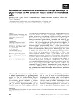
Figure 6
Actions of tumour necrosis factor (TNF) on hyperalgesia in health and in chronic inflammationActions of tumour necrosis factor (TNF) on hyperalgesia in health and in chronic inflammation. In the naive dorsal root ganglion (DRG), TNFα acts on
peripheral cells to induce a proinflammatory cascade resulting in the release of mediators, such as prostaglandins, that activate nociceptive neurons,
resulting in pain. After chronic inflammation, tumour necrosis factor receptor type 1(TNFR1) is up-regulated on DRG neurons, while TNFR2 is
expressed by infiltrating macrophages. TNF can directly modulate neuronal function and act on peripheral cells and DRG macrophages to induce
inflammatory mediators that can modulate neuronal function. This results in exaggerated pain.
Arthritis Research & Therapy Vol 7 No 4 Inglis et al.
R816
8. Opree A, Kress M: Involvement of the proinflammatory
cytokines tumor necrosis factor-alpha, IL-1 beta, and IL-6 but
not IL-8 in the development of heat hyperalgesia: effects on
heat-evoked calcitonin gene-related peptide release from rat
skin. J Neurosci 2000, 20:6289-6293.
9. Nicol GD, Lopshire JC, Pafford CM: Tumor necrosis factor
enhances the capsaicin sensitivity of rat sensory neurons. J
Neurosci 1997, 17:975-982.
10. Morioka N, Inoue A, Nakata Y: [Neural-immune interactions in
dorsal root ganglia] [in Japanese]. Nippon Yakurigaku Zasshi
2000, 115:219-227.
11. Pollock J, McFarlane SM, Connell MC, Zehavi U, Vandenabeele P,
MacEwan DJ, Scott RH: TNF-alpha receptors simultaneously
activate Ca2+ mobilisation and stress kinases in cultured sen-
sory neurones. Neuropharmacology 2002, 42:93-106.
12. Schafers M, Sorkin LS, Geis C, Shubayev VI: Spinal nerve liga-
tion induces transient upregulation of tumor necrosis factor
receptors 1 and 2 in injured and adjacent uninjured dorsal root
ganglia in the rat. Neurosci Lett 2003, 347:179-182.
13. Li Y, Ji A, Weihe E, Schafer MK: Cell-specific expression and
lipopolysaccharide-induced regulation of tumor necrosis fac-
tor alpha (TNFalpha) and TNF receptors in rat dorsal root
ganglion. J Neurosci 2004, 24:9623-9631.
14. Shubayev VI, Myers RR: Axonal transport of TNF-alpha in pain-
ful neuropathy: distribution of ligand tracer and TNF receptors.
J Neuroimmunol 2001, 114:48-56.
15. Parada CA, Yeh JJ, Joseph EK, Levine JD: Tumor necrosis factor
receptor type-1 in sensory neurons contributes to induction of
chronic enhancement of inflammatory hyperalgesia in rat. Eur
J Neurosci 2003, 17:1847-1852.
16. Sommer C, Schafers M, Marziniak M, Toyka KV: Etanercept
reduces hyperalgesia in experimental painful neuropathy. J
Peripher Nerv Syst 2001, 6:67-72.
17. Kidd BL, Inglis JJ, Vetsika K, Hood VC, De Felipe C, Bester H, Hunt
SP, Cruwys SC: Inhibition of inflammation and hyperalgesia in
NK-1 receptor knock-out mice. Neuroreport 2003,
14:2189-2192.
18. Tal M, Bennett GJ: Extra-territorial pain in rats with a peripheral
mononeuropathy: mechano-hyperalgesia and mechano-allo-
dynia in the territory of an uninjured nerve. Pain 1994,
57:375-382.
19. Hargreaves K, Dubner R, Brown F, Flores C, Joris J: A new and
sensitive method for measuring thermal nociception in cuta-
neous hyperalgesia. Pain 1988, 32:77-88.
20. Himmler A, Maurer-Fogy I, Kronke M, Scheurich P, Pfizenmaier K,
Lantz M, Olsson I, Hauptmann R, Stratowa C, Adolf GR: Molecu-
lar cloning and expression of human and rat tumor necrosis
factor receptor chain (P60) and its soluble derivative, tumor
necrosis factor-binding protein. DNA Cell Biol 1990,
9:705-715.
21. Bader T, Nettesheim P: Tumor necrosis factor-alpha modulates
the expression of its P60 receptor and several cytokines in rat
tracheal epithelial cells. J Immunol 1996, 157:3089-3096.
22. Meltzer JC, Sanders V, Grimm PC, Stern E, Rivier C, Lee S, Rennie
SL, Gietz RD, Hole AK, Watson PH: Production of digoxigenin-
labelled RNA probes and the detection of cytokine mRNA in rat
spleen and brain by in situ hybridization. Brain Res Brain Res
Protoc 1998, 2:339-351.
23. Lees DM, Khan NQ, Barker S, Corder R: Quantitative measure-
ment of mRNA levels by RT-PCR. studies of ECE-1 isoforms.
Methods Mol Biol 2002, 206:125-145.
24. Lewis M, Tartaglia LA, Lee A, Bennett GL, Rice GC, Wong GH,
Chen EY, Goeddel DV: Cloning and expression of cDNAs for
two distinct murine tumor necrosis factor receptors demon-
strate one receptor is species specific. Proc Natl Acad Sci USA
1991, 88:2830-2834.
25. DuBridge RB, Tang P, Hsia HC, Leong PM, Miller JH, Calos MP:
Analysis of mutation in human cells by using an Epstein-Barr
virus shuttle system. Mol Cell Biol 1987, 7:379-387.
26. White SJ, Nicklin SA, Sawamura T, Baker AH: Identification of
peptides that target the endothelial cell-specific Lox-1
receptor. Hypertension 2001, 37:449-455.
27. de Wildt RM, Mundy CR, Gorick BD, Tomlinson IM: Antibody
arrays for high-throughput screening of antibody-antigen
interactions. Nat Biotechnol 2000, 18:989-994.
28. Lu X, Richardson PM: Responses of macrophages in rat dorsal
root ganglia following peripheral nerve injury. J Neurocytol
1993, 22:334-341.
29. Pearson CM: Development of arthritis, periarthritis and perios-
titis in rats given adjuvant. Proc Soc Exp Biol Med 1956,
91:95-101.
30. Walker K, Fox AJ, Urban LA: Animal models for pain research.
Mol Med Today 1999, 5:319-321.
31. Ferreira SH, Lorenzetti BB, Poole S: Bradykinin initiates
cytokine-mediated inflammatory hyperalgesia. Br J Pharmacol
1993, 110:1227-1231.
32. Tonussi CR, Ferreira SH: Tumour necrosis factor-alpha medi-
ates carrageenin-induced knee-joint incapacitation and also
triggers overt nociception in previously inflamed rat knee-
joints. Pain 1999, 82:81-87.
33. Cunha FQ, Poole S, Lorenzetti BB, Ferreira SH: The pivotal role
of tumour necrosis factor alpha in the development of inflam-
matory hyperalgesia. Br J Pharmacol 1992, 107:660-664.
34. Schafers M, Lee DH, Brors D, Yaksh TL, Sorkin LS: Increased
sensitivity of injured and adjacent uninjured rat primary sen-
sory neurons to exogenous tumor necrosis factor-alpha after
spinal nerve ligation. J Neurosci 2003, 23:3028-3038.
35. Tsujino H, Kondo E, Fukuoka T, Dai Y, Tokunaga A, Miki K, Yone-
nobu K, Ochi T, Noguchi K: Activating transcription factor 3
(ATF3) induction by axotomy in sensory and motoneurons: a
novel neuronal marker of nerve injury. Mol Cell Neurosci 2000,
15:170-182.
36. Zimmermann M: Pathobiology of neuropathic pain. Eur J
Pharmacol 2001, 429:23-37.
37. Liu T, van Rooijen N, Tracey DJ: Depletion of macrophages
reduces axonal degeneration and hyperalgesia following
nerve injury. Pain 2000, 86:25-32.
38. Cui JG, Holmin S, Mathiesen T, Meyerson BA, Linderoth B: Possi-
ble role of inflammatory mediators in tactile hypersensitivity in
rat models of mononeuropathy. Pain 2000, 88:239-248.