Báo cáo y học: "Accelerated development of arthritis in mice lacking endothelial selectins" pdf
Bạn đang xem bản rút gọn của tài liệu. Xem và tải ngay bản đầy đủ của tài liệu tại đây (766.47 KB, 12 trang )
Open Access
Available online />R959
Vol 7 No 5
Research article
Accelerated development of arthritis in mice lacking endothelial
selectins
Jeffrey H Ruth
1,2
, M Asif Amin
1,2
, James M Woods
1,3
, Xiaodong He
4
, Sharon Samuel
4
, Nengjun Yi
5
,
Christian S Haas
1,2
, Alisa E Koch
1,2,6,7
* and Daniel C Bullard
4
*
1
Northwestern University Feinberg School of Medicine, Chicago, Illinois, USA
2
University of Michigan Medical School, Ann Arbor, Michigan, USA
3
Midwestern University, Downers Grove, Illinois, USA
4
Department of Genetics, University of Alabama at Birmingham, Birmingham, Alabama, USA
5
Department of Biostatistics, University of Alabama at Birmingham, Birmingham, Alabama, USA
6
Veteran's Administration Chicago Health Care Medical Center, Lakeside Division, Chicago, Illinois, USA
7
Ann Arbor Veteran's Administration, Ann Arbor, Michigan, USA
* Contributed equally
Corresponding author: Alisa E Koch,
Received: 10 Nov 2004 Revisions requested: 7 Dec 2004 Revisions received: 12 May 2005 Accepted: 17 May 2005 Published: 15 Jun 2005
Arthritis Research & Therapy 2005, 7:R959-R970 (DOI 10.1186/ar1770)
This article is online at: />© 2005 Ruth et al.; licensee BioMed Central Ltd.
This is an Open Access article distributed under the terms of the Creative Commons Attribution License ( />2.0), which permits unrestricted use, distribution, and reproduction in any medium, provided the original work is properly cited.
Abstract
The selectins, along with very late antigen-4 and CD44, have
been implicated in mediating leukocyte rolling interactions that
lead to joint recruitment and inflammation during the
pathogenesis of rheumatoid arthritis. Previously, we showed
that P-selectin deficiency in mice resulted in accelerated onset
of joint inflammation in the murine collagen-immunized arthritis
model. Here, we report that mice deficient either in E-selectin or
in E-selectin and P-selectin (E/P-selectin mutant) also exhibit
accelerated development of arthritis compared with wild type
mice in the CIA model, suggesting that these adhesion
molecules perform overlapping functions in regulating joint
disease. Analyses of cytokine and chemokine expression in joint
tissue from E/P-selectin mutant mice before the onset of joint
swelling revealed significantly higher joint levels of macrophage
inflammatory protein-1α and IL-1β compared to wild-type mice.
IL-1β remained significantly increased in E/P-selectin mutant
joint tissue during the early and chronic phases of arthritis.
Overall, these data illustrate the novel finding that E-selectin and
P-selectin expression can significantly influence cytokine and
chemokine production in joint tissue, and suggest that these
adhesion molecules play important regulatory roles in the
development of arthritis in E/P-selectin mutant mice.
Introduction
Leukocyte recruitment from the vasculature into tissue in
response to an inflammatory stimulus is a regulated process
that requires both adhesion proteins and chemoattractant/
activating molecules [1]. Leukocyte emigration occurs prima-
rily from the postcapillary venules and involves a cascade of
events including leukocyte rolling, firm adhesion and activa-
tion, transendothelial migration, and migration into tissue. Roll-
ing is mediated principally by the selectins (E-, L-, and P-
selectin) and their ligands, although other adhesion molecules
such as α
4
integrins, vascular cell adhesion molecule-1, and
CD44 can mediate rolling of certain leukocyte subtypes [2-4].
The selectins share a common structure characterized by an
amino-terminal calcium-dependent lectin binding domain, an
epidermal growth factor-like domain, a series of repeats with
similarities to complement binding proteins, a transmembrane
segment, and a short cytoplasmic tail [2]. L-selectin is
expressed on the majority of leukocytes and is shed from the
cell surface following activation, whereas P-selectin and E-
selectin are expressed on endothelial cells following activation
by various inflammatory mediators. Unlike the other selectins,
P-selectin is also found on activated platelets.
Much of the early information about the roles of selectins in ini-
tiating leukocyte rolling and recruitment has come from in vitro
or in vivo studies using function-blocking monoclonal
CIA = collagen-immunized arthritis; ELISA = enzyme-linked immunosorbent assay; IL = interleukin; IL-1ra = interleukin-1 receptor antagonist; LN =
lymph node; MIP = macrophage inflammatory protein; PBS = phosphate-buffered saline; RA = rheumatoid arthritis; TNF = tumor necrosis factor.
Arthritis Research & Therapy Vol 7 No 5 Ruth et al.
R960
antibodies or other inhibitors. During the past 10 years, tar-
geted mutations in the genes that encode these proteins have
been generated in mice. Collectively, these studies suggest
that the selectins, particularly E-selectin and P-selectin, play
important and overlapping roles in leukocyte rolling. However,
the majority of these studies have focused on their contribu-
tions in neutrophil-dependent, short-term inflammatory mod-
els, and less is known about their roles in the development of
chronic inflammatory diseases [5].
Rheumatoid arthritis (RA) is a systemic immune disorder char-
acterized by polyarticular joint inflammation and destruction
[6]. Increased expression of E-selectin and P-selectin has
been observed in inflamed joints from RA patients, with several
studies showing significant elevations in soluble selectins in
the serum of patients with active disease [7-10]. In addition,
several anti-inflammatory drugs have been shown to decrease
the expression of E-selectin and P-selectin, as well as that of
other adhesion molecules, in joint tissue from patients under-
going remission [7]. These findings suggest that the selectins
may play an important role in the initiation and/or progression
of joint inflammation during RA. However, investigations in ani-
mal models have provided inconsistent results concerning the
role of selectins in the development of arthritis. For example,
antibodies to E-selectin but not P-selectin inhibited adjuvant-
induced arthritis in rats [11], whereas Staphylococcus-
induced arthritis was diminished in P-selectin mutant mice and
in mice treated with antibodies to L-selectin [12]. Previously,
we reported that P-selectin mutant mice exhibited accelerated
development of joint inflammation in the collagen-immunized
arthritis (CIA) model compared with wild-type mice [13]. The
severity of arthritis and the circulating levels of anti-type II col-
lagen antibodies were also increased in P-selectin mutant
mice. Further investigations suggested that this effect may
result from alterations in leukocyte trafficking and/or cytokine
production in lymphoid organs or joint tissue in these mice dur-
ing the initiation of CIA [13].
In the present study, we analyzed both E-selectin mutant and
E/P-selectin mutant mice in the CIA model. We observed that
each of these mutants showed faster onset and more severe
arthritis compared to wild-type mice, similar to our earlier stud-
ies of P-selectin deficient mice in the CIA model [13]. E/P-
selectin mutants, along with E-selectin mutant mice, exhibited
the most severe arthritis phenotype. Analyses of both cytokine
and chemokine levels in joints from E/P-selectin mutant mice
undergoing arthritis revealed elevated production of macro-
phage inflammatory protein (MIP)-1α and IL-1β compared with
wild-type mice. Elevated joint IL-1β levels were consistently
observed in the E/P-selectin mutants compared with wild-type
mice throughout the course of the disease compared with
nonimmunized mice.
Materials and methods
Mice
Mice with null mutations for E-selectin and E/P-selectin were
generated by gene targeting in 129/Sv embryonic stem cells,
as described previously [14]. The selectin mutations were
then backcrossed into the CIA susceptible DBA-1/J strain
(Jackson Laboratory, Bar Harbor, ME, USA) for five genera-
tions (N5). Inbred DBA-1/J mice were used as controls for all
experiments. E/P-selectin mutant mice were kept on antibiot-
ics (neomycin sulfate, tetracycline, and trimethoprim) until 2
weeks before the induction of CIA in order to inhibit the devel-
opment of mucocutaneous infections, which commonly occur
in these mice [15]. All mice were kept in pathogen-free condi-
tions with regular health surveillance.
Ethical use of animals
The University of Michigan's Unit for Laboratory Animal Medi-
cine is an AAALAC (Association for Assessment and Accred-
itation of Laboratory Animal Care) accredited facility.
Treatment of animals in this study was approved by ethics
committees located initially at Northwestern University and
later at the University of Michigan. Animal care at the Unit for
Laboratory Animal Medicine is supervised by a veterinarian
and operates in accordance with federal regulations. Mice
were housed in sterile rodent micro-isolator caging with fil-
tered cage tops in a specific pathogen-free environment to
prevent infection. All efforts were made to reduce stress or dis-
comfort in the animals used in these studies.
CIA induction and scoring for CIA development
Chick type II collagen (Chondrex; LLC, Redman, WA, USA)
was dissolved in 0.01 N acetic acid overnight at 4°C, and then
emulsified with an equal volume of complete Freund's adjuvant
(Difco, Detroit, MI, USA) to give a final concentration of 1 mg/
ml. Male and female mice aged 8–12 weeks were injected
intradermally at the base of the tail with 100 µl emulsified col-
lagen on day 0 [13]. Arthritis was evaluated on a daily basis for
45 days, and then every other day until day 90 by an observer
who was blinded to genotype. For these studies we used a
scoring system identical to that used in our previously pub-
lished CIA studies involving P-selectin and intercellular adhe-
sion molecule-1 mutant mice [13,15], based on the
methodology of Wooley and others [16-18]. Briefly, each joint
was inspected and assessed for the severity of swelling using
scores of 0 (normal appearance), 1 (mild), 2 (moderate), and
3 (severe), yielding a maximum score of 12 for each mouse.
Swollen digits were noted but paws were only considered
arthritic when the entire paw was inflamed for 2 consecutive
days. The day of onset of arthritis was recorded for each
mouse. During the chronic phase of the arthritis, paws and dig-
its were inspected for distortion and manipulated to identify
loss of flexion (ankylosis). Joints received an additional score
of 3 for the presence of distortion and 3 for the presence of
ankylosis (maximum additional score of 6 per joint). Daily
severity scores were obtained from the addition of
Available online />R961
inflammation, distortion, and ankylosis scores. Scores for each
mouse were used to derive a mean arthritis severity score for
the group.
Homogenization of joint tissue
E/P-selectin and wild-type mice were killed and both joints and
serum collected at three periods during the course of disease:
pre-arthritis (pre-CIA; days 16, 22, and 28), when no overt
signs of arthritis were present; immediate post-arthritis onset
(early-CIA), when mice were killed within 2 days of receiving a
score of 3 for any joint; and chronic-CIA (day 80). Only hind
joints were used in the study. Joints were removed directly
below the hairline and snap frozen in liquid nitrogen. Joints
were stored at -80°C before processing. Each joint was
thawed on ice, weighed, and quickly homogenized on ice in 1–
2 ml phosphate-buffered saline (PBS) containing a tablet of
proteinase inhibitors (10 ml PBS/tablet; Boehringer Man-
nheim, Indianapolis, IN, USA). Homogenized tissues were cen-
trifuged at 2000 g at 4°C for 10 min. Supernatants were
stored at -80°C until analysis with ELISA.
ELISA measurement of cytokines and chemokines in
joint homogenate
ELISA reagents for cytokine measurement were obtained from
R&D Systems (Minneapolis, MN, USA). Mouse cytokines were
measured in joint homogenates, lymph node (LN) superna-
tants, and serum by ELISA using 96-well polystyrene plates, in
accordance with the manufacturer's instructions. All homoge-
nates were normalized to total protein and are presented as
picogram or nanogram of cytokine per milligram of protein.
ELISA measurement of anti-collagen antibodies
Mouse serum from E/P-selectin mutant, E-selectin mutant, and
wild-type mice at 16 and 28 days after collagen immunization
was analyzed for anti-collagen antibodies using a mouse IgG
anti-type II collagen antibody assay kit, in accordance with the
manufacturer's instructions (Chondrex, Inc., Redmond, WA,
USA). ELISA samples were developed using a TMB substrate
system and plates were read at 450 nm using an ELISA plate
reader.
Immunohistology
Frozen tissue (NL mouse and mouse CIA joints) were cut
(approximately 7 µm) and immunoperoxidase stained using an
avidin–biotin technique (Vector Laboratories, Burlingame, CA,
USA) with all subsequent incubations being performed at
37°C in a humidified chamber. Slides were fixed in cold ace-
tone for 20 min and then treated with 3% peroxidase in 0.1
mol/l Tris for 5 min to block endogenous peroxidase activity.
Tissues were further blocked with 3% horse serum (in PBS)
for 1 hour, and then incubated with goat anti-mouse IL-1β IgG
(goat anti-mouse IL-1β; R&D Systems, Minneapolis, MN, USA)
or goat anti-mouse MIP-1α IgG (goat anti-mouse/rat MIP-1α;
Research Diagnostics Inc., Flanders, NJ, USA) or purified non-
specific goat IgG (Coulter, Miami, FL, USA). Tissue was
washed twice in PBS, and a 1:200 dilution (in PBS) of anti-rat
biotinylated antibody (Vector Laboratories) was added to the
tissue sections and incubated for an additional 20 min. After a
final washing (twice in PBS), slides were developed with a
diaminobenzidine tetrahydrochloride substrate (Vector Labo-
ratories) for 2 min at room temperature, rinsed in tap water,
counter-stained with Harris' hematoxylin, and dipped in satu-
rated lithium carbonate solution for bluing. Slides were exam-
ined for cellular immunoreactivity. Positive cells were identified
by morphology.
Isolation and culture of regional lymph nodes, spleen
cells, preparation of supernatants, and serum collection
Draining inguinal LNs and spleen cells were collected at the
time of joint harvest at pre-CIA (days 16, 22, and 28 after
immunization), early-CIA (within 2 days of receiving a score of
3 for any joint), and chronic-CIA (80 days), and teased into sin-
gle cell suspensions. Serum was collected from all mice at all
time points, including day 0, pre-CIA, early-CIA, and chronic-
CIA time points. LN cells were washed, counted, plated, and
then cultured in RPMI containing 10% fetal bovine serum at 3
× 10
6
cells/ml in the presence or absence of 5 µg/ml chick
type II collagen for 36 hours. After incubation, supernatants
were collected by centrifugation and stored at -80°C. LN
supernatants were then thawed and measured for cytokine
levels. Data were combined for all time points and presented
as wild-type or E/P-selectin LN cultures with or without stimu-
lus. LN supernatant cytokine concentrations were normalized
to pg/3 × 10
6
cells. Serum was collected when the animal was
killed and measured for cytokine production, as described
above.
Statistical analysis
Three nonparametric statistical tests – Savage, Wilcoxon, and
Kruskal–Wallis – were used to compare the incidence and
severity of arthritis. These were calculated using SAS proce-
dure NPAR1WAY (SAS Institute Inc., Cary, NC, USA). Similar
statistical significance values were obtained with all three
tests. Statistically significant differences in cytokine levels
between groups was analyzed using the Student's independ-
ent t-test, with a threshold of P < 0.05.
Results
Accelerated development and increased severity of CIA
in E- and E/P-selectin mutant mice
In three separate experiments, CIA was induced in E-selectin
DBA/1J single mutant, E/P-selectin DBA/1J double mutant,
and wild-type DBA/1J mice. Similar to our previously reported
findings in P-selectin mutant mice [13], we observed a signifi-
cant acceleration in the development of arthritis in both mutant
groups (Fig. 1). The day of onset (mean ± standard error) was
45.2 ± 13.9 days for wild-type mice, 36.2 ± 11.0 days for E-
selectin mutants, and 33.2 ± 11.4 days for E/P-selectin
mutant mice (P < 0.01 for both E-selectin mutant and E/P-
selectin mutant versus wild-type, as determined using the
Arthritis Research & Therapy Vol 7 No 5 Ruth et al.
R962
Kruskal–Wallis test). The E-/P-selectin mutants exhibited the
most pronounced acceleratory effect, although the average
day of onset was not significantly different compared with E-
selectin mutants (P = 0.52). The incidence of arthritis was also
increased in E/P-selectin mutant mice, with 100% showing
signs of joint inflammation compared with 88% and 85% for
E-selectin mutants and wild-type mice, respectively (Fig. 1).
Additionally, both the E-selectin and E/P-selectin mutants had
significantly higher average daily severity scores than did wild-
type mice (Fig. 2). However, the severity scores were not sig-
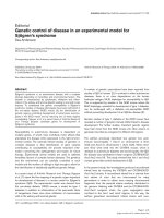
Figure 1
Incidence of CIA in E-selectin mutant, E/P-selectin mutant, and wild-type miceIncidence of CIA in E-selectin mutant, E/P-selectin mutant, and wild-type mice. E-selectin mutant (n = 56), E/P-selectin mutant (n = 54), and wild-
type mice (n = 48) were immunized with chick type II collagen and assessed for the development of arthritis for 90 days by a blinded observer. The
results are presented as the mean of three separate CIA experiments. The cumulative number of mice in each group is given as a percentage. CIA,
collagen-immunized arthritis; wt, wild-type.
Figure 2
Arthritis severity in E-selectin mutant, E/P-selectin mutant, and wild-type miceArthritis severity in E-selectin mutant, E/P-selectin mutant, and wild-type mice. Arthritic paws were evaluated for the severity of inflammation and the
presence of distortion and ankylosis as described in the Materials and method section. Daily severity scores were calculated from the combined
inflammation, distortion, and ankylosis scores. Data are shown from three separate CIA experiments, and the mean arthritis severity is shown for
each genotype. P < 0.05 for both E-selectin and E/P-selectin mutant versus wild type mice. wt, wild-type.
Available online />R963
nificantly different between E-selectin and E/P-selectin mutant
mice (P = 0.23 for E-selectin versus E/P-selectin).
Increased joint expression of IL-1β and MIP-1α in
collagen-immunized E-/P-selectin mutant mice prior to
the onset of arthritis
Altered cytokine production in synovial tissue appears to cor-
relate with RA pathology [8]. Consequently, we investigated
the hypothesis that mice that lack selectin molecules develop
accelerated CIA as a result of altered cytokine expression.
Because E/P-selectin mutant mice showed the strongest
acceleration effect, we initially analyzed five different proin-
flammatory cytokine and chemokines levels in the joints of
these mice before clinical signs of arthritis became obvious.
Figure 3a shows the mean levels of MIP-1α and IL-1β expres-
sion in the joints of collagen-immunized pre-arthritic mice. Lev-
els of both MIP-1α and IL-1β in the joint were elevated in E/P-
selectin mutant mice. Table 1 shows the levels of the type 1
proinflammatory cytokines IL-12, tumor necrosis factor (TNF)-
α, and MIP-2 in E/P-selectin mutants. In contrast to MIP-1α
and IL-1β, we found no differences in these cytokines between
immunized and/or nonimmunized wild-type and E/P-selectin
mutant mice, indicating that IL-12, TNF-α, and MIP-2 do not
directly correlate with the incidence or severity of CIA. We
also measured joint levels of MIP-1α in nonimmunized wild-
type and E/P-selectin mutant mice (data not shown) and
observed no significant differences in basal levels of MIP-1α
expression (values expressed as mean ± standard error; wild-
type: 4.1 ± 0.5; E/P-selectin mutant: 4.1 ± 0.6). Serum was
also collected from pre-arthritic mice. E-/P-selectin mutant
mice had increased levels of IL-1β 22 days after immunization
compared with wild-type mice (values expressed as mean ±
standard error; wild-type: 54.5 ± 1.8 pg/ml; E/P-selectin
mutant: 74.5 ± 6.4 pg/ml; P < 0.05, data not shown). There
was a trend toward higher serum IL-1β in E/P-selectin mutants
at days 16 and 28, but this was not statistically significant
(data not shown).
Elevated production of IL-1β in joint tissue of mice with
early arthritis
We next analyzed cytokine levels in the joints of wild-type and
E/P-selectin mutant mice showing the first signs of paw inflam-
mation. Early-CIA E/P-selectin mutant mice exhibited signifi-
cantly elevated levels of IL-1β but not of MIP-1α (Fig. 3b) or
MIP-2 (Table 1) in their joint homogenates compared with
wild-type mice. These findings indicate that in early-onset
arthritic joints, expression of E-selectin and P-selectin can sig-
nificantly influence the production of IL-1β but not of MIP-1α
and MIP-2. As shown in Table 1, we found no differences
between wild-type and E/P-selectin mutant mice in concentra-
tions of IL-12 or MIP-2 within the joint, although we did
observe increases in MIP-2 in the early-CIA joint tissue
compared with the pre-arthritic samples. Hence, MIP-2
appears to be important in the development of CIA but it does
not correlate with the acceleration in inflammation seen in E/P-
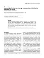
Figure 3
Cytokine expression in joint homogenates collected from pre-onset CIA miceCytokine expression in joint homogenates collected from pre-onset CIA
mice. (a) Collagen-immunized E/P-selectin mutant mice exhibit
increased MIP-1α and IL-1β in joint homogenates compared with
immunized wild-type mice. (b) Joints from E-/P-selectin mutant mice
during the early stages of arthritis show increased IL-1β expression
compared with wild-type mice. (c) Chronic (day 80 collagen-immu-
nized) E/P-selectin mutant mice show elevated levels of IL-1β and MIP-
1α in their joint tissues compared with wild-type mice. Values are
expressed as mean ± standard error of the mean. *P < 0.05. CIA, colla-
gen-immunized arthritis; MIP, macrophage inflammatory protein; n,
number of joints; wt, wild-type.
Arthritis Research & Therapy Vol 7 No 5 Ruth et al.
R964
Table 1
Cytokine levels
Cytokine Genotype Cytokine level P (versus E/P-selectin mutant) Number of observations
IL-12 Wild-type
Nonimmunized 2.2 ± 0.2 pg/mg >0.05 12
Pre-onset ND ND 0
Post-onset 1.6 ± 0.2 pg/mg >0.05 8
Chronic 2.6 ± 0.6 pg/mg >0.05 8
E/P-selectin mutant
Nonimmunized 5.7 ± 3.1 pg/mg 10
Pre-onset ND 0
Post-onset 1.7 ± 0.2 pg/mg 8
Chronic 2.3 ± 0.5 pg/mg 8
TNF-α
a
Wild-type
Nonimmunized <1 pg/mg NS 10
Pre-onset <1 pg/mg 13
Post-onset <1 pg/mg 8
Chronic <1 pg/mg 8
E/P-selectin mutant
Nonimmunized <1 pg/mg 10
Pre-onset <1 pg/mg 11
Post-onset <1 pg/mg 7
Chronic <1 pg/mg 8
MIP-2 Wild-type
Nonimmunized 2.2 ± 0.4 pg/mg >0.05 12
Pre-onset 1.9 ± 0.9 pg/mg >0.05 12
Post-onset 5.3 ± 0.9 pg/mg >0.05 8
Chronic 1.4 ± 0.1 pg/mg >0.05 10
E/P-selectin mutant
Nonimmunized 1.6 ± 0.2 pg/mg 10
Pre-onset 2.4 ± 1.8 pg/mg 9
Post-onset 5.2 ± 1.6 pg/mg 8
Chronic 1.7 ± 0.5 pg/mg 9
Cytokines were not elevated in murine joint homogenates from nonimmunized E/P-selectin mutant mice, or from pre-CIA onset, post-CIA onset,
and chronic-CIA (day 80) E/P-selectin mutant mice, compared with wild-type mice. Values for TNF-α are below the sensitivity of the assay. ELISA
assay sensitivities were 2.5 pg/ml for IL-12, 5 pg/ml for TNF-α and IL-1β, and 1.5 pg/ml for MIP-2 and MIP-1α. Pre-onset group corresponds to
combined values for day 16, day 22, and day 28 groups. All values are normalized to milligrams of total protein. CIA, collagen-immunized arthritis;
MIP, macrophage inflammatory protein; ND, no data.
Available online />R965
selectin mutant mice. TNF-α levels in these joints were below
the limit of detection.
Elevated production of IL-1β and MIP-1α in chronically
inflamed joint tissue
In order to examine the potential influence of selectins on the
pattern of cytokine expression during the chronic phase of
CIA, cytokines were measured in the joints of mice with CIA at
day 80. Joint tissue (day 80) collected from E/P-selectin
mutant mice had elevated levels of IL-1β and MIP-1α but not
of MIP-2 compared with wild-type mice (Fig. 3c, Table 1). As
shown in Table 1, IL-12 and MIP-2 levels in nonimmunized
joints from both wild-type and E/P-selectin mutant mice were
similar to those in immunized mice, suggesting MIP-2 or IL-12
are not pivotal cytokines in chronic-CIA. Consistent with the
pre-CIA and early-CIA analyses, no significant concentrations
of TNF-α were detected in the joints of wild-type or E/P-selec-
tin mutant mice with chronic arthritis.
Elevated levels of lymph node IL-1β and IL-2 from
collagen-immunized E/P-selectin mutant mice
In order to determine whether the systemic immune response
is affected by deletion of these selectin molecules, we meas-
ured cytokine expression from collagen-stimulated and non-
stimulated draining inguinal LN cells obtained from immunized
and nonimmunized wild-type and E/P-selectin mutant mice. E/
P-selectin mutant mice had elevated IL-1β and IL-2 production
from inguinal LN cells compared with collagen-immunized
wild-type mice (Fig. 4), indicating that deleting selectin genes
can affect the systemic immune response during CIA develop-
ment. This is consistent with the joint cytokine measurements,
showing increased IL-1β production in pre-CIA onset, post-
CIA onset, and chronic-CIA E-/P-selectin mutant mice (Fig. 3).
Increased IL-1β production in serum and joints of
nonimmunized E/P-selectin mutant mice
To investigate the possibility that mice lacking selectin mole-
cules are predisposed to develop an accelerated inflammatory
response, we analyzed basal proinflammatory cytokine con-
centrations in the serum and joints of nonimmunized wild-type
and E/P-selectin mutant mice. We found increased serum lev-
els of IL-1β but not of MIP-1α (data not shown for MIP-1α) in
E/P-selectin mutant compared with wild-type mice (Fig. 5a). In
addition, there was approximately a twofold increase in the
concentration of IL-1β in joint tissue from nonimmunized E/P-
selectin mutant mice compared with wild-type mice (Fig. 5b).
We examined nonimmunized mouse serum for IL-1β to test
whether E/P-selectin mutant mice may be primed to elicit an
inflammatory response to administered antigen by overex-
pressing basal levels of IL-1β. As shown in Fig. 5a, nonimmu-
nized E/P-selectin mutant mice had significantly elevated
levels of IL-1β in their serum compared with wild-type mice. In
order to rule out the possibility that the elevated serum IL-1β
levels in E/P-selectin mice were due to infections, we kept
nonimmunized E-/P-selectin mutant mice on antibiotics for 2
weeks and then examined serum IL-1β levels. As shown in Fig.
5a, serum IL-1β levels were not lowered by antibiotic
treatment but actually increased. This shows that the elevated
IL-1β in the serum of E/P-selectin mutant mice is not due to
infection.
Serum and joint IL-1 receptor antagonist protein levels
are not elevated in E/P-selectin mutant compared with
wild-type mice
We examined the serum of nonimmunized and post-CIA onset
E/P-selectin mutant and wild-type mice for IL-1 receptor
antagonist (IL-1ra) expression by ELISA. We found that both
wild-type and selectin mutant mice had approximately 2 ng/ml
IL-1ra in their serum, but we did not find any statistically signif-
icant differences between wild-type and selectin mutant mice.
Similar results were obtained from the serum of post-CIA
onset mice (Fig. 6a). We also examined the joints of wild-type
and E/P-selectin mutant mice for IL-1ra expression normalized
to total protein levels. Figure 6b shows that IL-1ra was
detected in higher amounts in whole joint homogenates than
in serum of both nonimmunized wild-type and E/P-selectin
mutant mice, but we did not find differences in IL-1ra expres-
sion between nonimmunized wild-type and E/P-selectin joint
homogenates, suggesting selectin deficiency does not signifi-
cantly alter IL-1ra expression in the serum or joints.
Mouse serum IgG anti-collagen antibody concentration
after collagen immunization is not increased in selectin
mutant compared with wild-type mice in pre-CIA onset
mice
To determine whether selectin mutant mice may have altered
responses to collagen immunization compared with wild-type
mice, we examined murine serum anti-collagen antibody
concentrations at days 16 and 28 post-CIA immunization in
wild-type, E-selectin mutant, and E/P-selectin mutant mice not
showing clinical signs of arthritis. As shown in Fig. 7, wild-type
and selectin mutant mice had elevated serum levels of anti-col-
lagen IgG at day 28 compared with day 16 mice, but we did
not find significant differences in anti-collagen antibody con-
centrations between wild-type, E-selectin, and E/P-selectin
mutant mice. This suggests that selectin deficiency does not
alter responses to collagen during the initiation of arthritis.
Immunohistochemistry for IL-1β and MIP-1α in synovial
tissue of E/P-selectin mutant mice with CIA
To determine the cell types responsible for elevated IL-1β and
MIP-1α in the joints of E/P-selectin deficient mice, we per-
formed immunohistology for both cytokines using goat anti-
mouse IL-1β or goat anti-mouse/rat MIP-1α. The staining
clearly shows immunopositivity on macrophages (arrows),
whereas ST endothelial cells (arrowheads) remained negative
(Fig. 8).
Arthritis Research & Therapy Vol 7 No 5 Ruth et al.
R966
Discussion
Studies in both RA patients and in animal models of arthritis
strongly implicate adhesion molecules as critical mediators in
both the initiation and maintenance of joint inflammation [7].
Multiple adhesion molecule pathways appear to mediate leu-
kocyte interactions, particularly rolling, within the synovial vas-
culature. Support for this model comes from studies showing
loss or inhibition of CD44 and VLA-4 (very late antigen-4)
interactions with their ligands that can reduce leukocyte rolling
interactions and inhibit joint inflammation in animal models of
arthritis [19,20]. However, conflicting results have been pub-
lished with regard to the roles of E-selectin and P-selectin in
the development of joint inflammation in different animal mod-
els [11-13,21]. Our previous and current studies in the CIA
model show that leukocyte rolling via E-selectin and P-selectin
is not required for the initiation of arthritis, and that loss of one
or both of these adhesion molecules significantly accelerates
the development and severity of joint inflammation. In the
present study there was no significant difference in day of
onset, number of mice showing arthritis, or severity between
the single E-selectin and the E/P-selectin mutant mice but,
overall, selectin mutant mice experienced accelerated CIA
development compared with wild-type mice. This may be
explained by a limit on how quickly a mouse can actually
develop CIA, even with loss of both E-selectin and P-selectin.
These factors may include the time for immunization to occur,
loss of tolerance to collagen, and immune complex deposition
in the joints. Also, once destructive processes such as bone
erosion start in the joint, individual scores do not change
significantly.
Accelerated development and/or increased severity of CIA in
mice have previously been reported where cytokine expres-
sion or activity has been altered [17,18,22,23]. Therefore, we
investigated whether the loss of E-selectin or P-selectin
affected the expression levels of five different cytokines and
chemokines that are implicated in the development of CIA.
Our analyses in E/P-selectin mutant mice suggested that
these adhesion molecules may influence the development of
CIA by altering the production of MIP-1α and IL-1β. E/P-selec-
tin mutant mice also produced significantly more IL-1β in their
joints and serum than did wild-type mice at all of the time
points examined, strongly implicating IL-1β in accelerated
development and increased severity of CIA. These studies
suggest that increased levels of IL-1β contribute to the
accelerated development of arthritis in double selectin defi-
cient mice.
Of significance, E/P-selectin mutant mice have elevated basal
levels of IL-1β in their hind joints compared with wild-type
mice, without collagen immunization. Therefore, mice lacking
both E-selectin and P-selectin are seemingly primed to mount
a robust inflammatory response in their joints. We also
observed increased MIP-1α levels in pre-CIA onset and
chronic-CIA mice, showing that selectins may also regulate
chemokine production. In order to rule out the possibility that
E/P-selectin mutant mice have enhanced basal levels of IL-1β
due to low-grade infections, we measured basal IL-1β levels in
nonimmunized E/P-selectin mutant mice kept on antibiotics for
2 weeks. We found that IL-1β levels were not decreased in
joints of E/P-selectin mutant mice, but actually increased.
Figure 4
IL-1β and IL-2 expression from LN cellsIL-1β and IL-2 expression from LN cells. IL-1β and IL-2 expression was increased in LN cell supernatants isolated from collagen-immunized E/P-
selectin mutant mice compared with immunized wild-type mice. Pre-onset, post-onset, and chronic arthritis time points were combined. LN superna-
tants were stimulated with 5 µg/ml chick II collagen. Values are expressed as mean ± standard error of the mean. *P < 0.05. LN, lymph node; n,
number of separate cultures; wt, wild-type.
Available online />R967
Therefore, it is likely that IL-1β is regulated differently in mice
that lack selectin molecules, and that these differences are not
due to infection.
We also measured basal levels of IL-1ra – the natural inhibitor
of IL-1β – in the joints and serum of both wild-type and E/P-
selectin mutant mice. We found substantial levels of IL-1ra in
both nonimmunized wild-type and nonimmunized E/P-selectin
mutant mouse joints and serum, but we did not find elevated
levels of IL-1ra in E/P-selectin mutant compared with wild-type
mice. We also measured serum of post-onset CIA wild-type
and E/P-selectin mutant mice for IL-1ra, and again observed
no significant differences between wild-type and selectin defi-
cient groups. These findings are in agreement with previous
reports that have shown TNF-α, but not IL-1β, to be a signifi-
cant in vivo and in vitro stimulus for murine IL-1ra production
[24]. Although TNF-α is a well established cytokine in RA
pathology, TNF-α was not detected in any mice at any of the
time points measured in the study. This cytokine may not be
necessary for the development of arthritis in this model,
because a study recently reported that severe CIA occurs in
TNF-α deficient mice [25]. It is currently unknown whether
similar mechanisms are at work in single selectin deficient
mice, and whether other inflammatory mediators may be
responsible for the accelerated CIA seen in these mice. We
also measured murine anti-collagen antibodies in the serum of
Figure 5
IL-1β expression in serum and joint tissue from non-collagen-immunized miceIL-1β expression in serum and joint tissue from non-collagen-immunized
mice. (a) IL-1β was significantly elevated in serum from nonimmunized
E/P-selectin mutant compared with nonimmunized wild-type mice.
Serum from E/P-selectin mutant mice kept on antibiotics for 2 weeks
also had elevated serum IL-1β levels, indicating that the basal overex-
pression of IL-1β in nonimmunized E/P-selectin mutant mice is not due
to bacterial infections. (b) IL-1β was also significantly increased in joint
homogenates from nonimmunized E/P-selectin mutant compared with
nonimmunized wild-type mice. Values are expressed as mean ± stand-
ard error of the mean. *P < 0.05. n, number of mice (panel (a) or
number of joints (or mice for serum; panel (b); wt, wild-type.
Figure 6
IL-1ra expression in serum and joint tissue in wild-type and E/P-selectin mutant miceIL-1ra expression in serum and joint tissue in wild-type and E/P-selectin
mutant mice. (a) IL-1ra was not significantly elevated in serum from
nonimmunized and post-onset CIA E/P-selectin mutant mice compared
with serum from wild-type mice. (b) IL-1ra levels were higher in the
joints compared with serum of nonimmunized wild-type and E/P-selec-
tin mutant and wild-type mice, but no difference in IL-1ra levels was
found in joint homogenates from nonimmunized E/P-selectin mutant
and nonimmunized wild-type mice. Values are expressed as mean ±
standard error of the mean. *P < 0.05. IL-1ra, IL-1 receptor antagonist;
n, number of mice (panel (a) or number of joints (or mice for serum;
panel (b); wt, wild-type.
Arthritis Research & Therapy Vol 7 No 5 Ruth et al.
R968
day 16 and day 28 pre-onset CIA wild-type, E-selectin, and E/
P-selectin mutant mice to determine whether anti-collagen
antibodies may be regulated differently in selectin deficient
and wild-type mice. We showed that anti-collagen antibody
levels were generally increased at day 28 compared with day
16 mice, but we observed no significant differences between
wild-type and E/P-selectin mutant mice, suggesting that anti-
collagen antibody is not a determining factor in the acceler-
ated CIA development seen in the E/P-selectin mutant mice.
Interestingly, we did not observe significant differences in the
cellular infiltrate by immunohistology in the joints of the wild-
type, E-selectin, and E/P-selectin mutant mice. In our hands,
the cell composition was similar between groups, but only the
severity and timing of the inflammatory response was altered
in mice lacking selectins, probably due to exaggerated and
early IL-1β production. This is concomitant with the elevated
blood leukocyte counts observed in P-selectin and E/P-selec-
tin mutant mice compared with wild-type mice, suggesting that
the elevated leukocytes in selectin deficient mice may be the
cause of accelerated CIA. However, E-selectin mutant mice
do not have elevated blood leukocyte counts, but they also
exhibit the accelerated CIA phenotype [14]. Therefore, the
increased cytokine production in the E/P-selectin mutant mice
cannot be fully explained by elevated white blood cell
numbers.
Further work is necessary to determine whether similar altera-
tions occur in other adhesion molecule deficient mice and to
investigate the possibility that these molecules regulate the
development of arthritis via a common pathway. An alternative
mechanism may involve the disruption of regulatory leukocyte
recruitment, including T regulatory cells, into the joints of
selectin deficient mice during the initial phases of arthritis. A
number of studies have shown that E-selectin and P-selectin
selectively mediate rolling of specific leukocyte subsets, such
as T-helper-1 cells, but not T-helper-2 cells [26], although their
contributions to T regulatory cell recruitment remain to be
defined. We are currently examining the roles of the selectins
in regulatory and effector T cell recruitment during the devel-
opment of arthritis in this model. It is also possible that the lack
of certain selectin molecules may result in an overexpression
of other adhesion molecules and/or selectins that may selec-
tively recruit IL-1β producing monocytes to the joints. This
would result in the observed elevated monokine production in
mice lacking E-selectin and P-selectin.
Several studies have identified IL-1β as a potent proinflamma-
tory cytokine in animal models of RA, and it is likely that this
cytokine contributes to the more severe disease phenotype
observed in the double mutants. For example, van den Berg
and coworkers [27] showed that a combination of antibodies
to IL-1α and IL-1β given before the onset of arthritis
completely prevented murine CIA. In addition, exogenously
administered IL-1β was found to potentiate both murine and
rat CIA [28,29]. In another study, IL-1β was shown to be criti-
cal for accelerated development of CIA, and expression of this
cytokine correlated with increased incidence and earlier dis-
Figure 7
Endothelial selectin deficiency does not influence anti-collagen antibody production in collagen immunized miceEndothelial selectin deficiency does not influence anti-collagen antibody production in collagen immunized mice. Mouse serum anti-collagen IgG
antibody concentration is not altered in single E-selectin and E/P-selectin mutant mice compared with wild-type mice at days 16 and 28 after colla-
gen immunization. Wild-type and selectin mutant mice were administered collagen, and then at days 16 and 28 mice were bled and serum was
measured for anti-collagen IgG. Although anti-collagen IgG was elevated in all the groups at day 28 compared with day 16, there were no differ-
ences in anti-collagen IgG between groups at the time points examined. Values are expressed as mean ± standard error of the mean. wt, wild-type.
Available online />R969
ease onset [30]. It was previously reported that mice overex-
pressing IL-1ra exhibited reduced incidence and severity of
CIA, whereas mice deficient in the IL-1ra gene showed accel-
erated development of arthritis with increased severity [31]. In
other studies, BALB/c mice lacking IL-1ra spontaneously
developed chronic inflammatory polyarthropathy, with joint
pathology closely resembling that of RA [32]. These findings
clearly underscore the importance of IL-1β in arthritis
development.
Our findings that MIP-1α levels were elevated in E/P-selectin
mutant mice are concordant with previous reports that MIP-1α
is proinflammatory in RA and CIA [33-35]. Increased MIP-1α
in the joints of pre-arthritic E/P-selectin mutant mice may also
contribute to increased IL-1β, because MIP-1α is chemotactic
for monocytes, which produce significant amounts of IL-1β
[36]. These findings are consistent with histology studies
showing that macrophages are the significant producers of
both MIP-1α and IL-1β in the joints of E/P-selectin mutant
mice with CIA. This suggests that macrophages in E/P-selec-
tin mutant mice may be altered in some way to overproduce
both MIP-1α and IL-1β, or that that these monocytes are spe-
cifically recruited to the joints of selectin deficient mice during
CIA development. Interestingly, we did not observe significant
expression of TNF-α in joint tissue in our study. It is possible
that TNF-α is present in CIA but is expressed only transiently
before the onset of disease. Previous studies of murine CIA
have indicated that TNF-α is expressed very early in the
disease, before IL-1β [37], and is tightly regulated, whereas IL-
1β expression is not [24].
Conclusion
We have observed accelerated development and increased
severity of CIA in P-selectin, E-selectin, and E/P-selectin
mutant mice, suggesting that these adhesion molecules play
important regulatory roles in the development of arthritis. In
addition, we found that E/P-selectin mutant mice show signif-
icantly increased expression of IL-1β in the joints and serum
under basal conditions, increased IL-1β and MIP-1α expres-
sion in joint tissue at several time points during the develop-
ment of CIA, and increased LN cell production of both IL-1β
and IL-2. This altered immune response may be due to built in
compensatory mechanisms or disrupted homeostasis, result-
ing in either monocyte over-recruitment or overstimulation of
tissue macrophages in E/P-selectin mutant mice. Alternatively,
these findings may suggest a novel function for these selectins
in regulating expression of cytokines and chemokines in joint
and other tissues during chronic inflammation.
Competing interests
The author(s) declare that they have no competing interests.
Authors' contributions
JHR, the first author, designed and developed all aspects of
the study. MAA and JMW assisted in the animal scoring and
cytokine measurements. XH assisted with the development
and generation of the selectin deficient mice, and performed
the anti-collagen ELISA. SS, of Dr Bullard's group, immunized
mice and conducted the scoring. NY performed the necessary
statistical analyses. CSH performed the immunohistology
work. AEK, a senior author, was instrumental in the design and
development of this project, and generously offered her exper-
tise. DCB, a co-senior author, developed, designed, and gen-
erated the selectin deficient mice, as well as supplied murine
joints and serum that made this study possible. DCB was also
responsible for generating CIA development in selectin defi-
cient mice, and offered valuable advice.
Acknowledgements
This work was supported by NIH grants AI40987 (AEK), HL58695
(AEK), AR48267 (AEK), and AR049907 (JHR) and a grant from the
Arthritis Foundation (DCB). Additional support included the Gallagher
Professorship for Arthritis Research, the Frederick GL Huetwell and Wil-
liam D Robinson, MD Professorship of Rheumatology, and funds from
the Veteran's Administration Research Service (AEK).
References
1. Springer TA: Traffic signals for lymphocyte recirculation and
leukocyte emigration: the multistep paradigm. Cell 1994,
76:301-314.
2. Patel KD, Cuvelier SL, Wiehler S: Selectins: critical mediators of
leukocyte recruitment. Semin Immunol 2002, 14:73-81.
3. Berlin C, Bargatze RF, Campbell JJ, von Andrian UH, Szabo MC,
Hasslen SR, Nelson RD, Berg EL, Erlandsen SL, Butcher EC:
alpha 4 integrins mediate lymphocyte attachment and rolling
under physiologic flow. Cell 1995, 80:413-422.
4. DeGrendele HC, Estess P, Picker LJ, Siegelman MH: CD44 and
its ligand hyaluronate mediate rolling under physiologic flow:
a novel lymphocyte-endothelial cell primary adhesion
pathway. J Exp Med 1996, 183:1119-1130.
5. Bullard DC: Adhesion molecules in inflammatory diseases:
insights from knockout mice. Immunol Res 2002, 26:27-33.
6. Szekanecz Z, Koch AE: Update on synovitis. Curr Rheumatol
Rep 2001, 3:53-63.
7. McMurray RW: Adhesion molecules in autoimmune disease.
Semin Arthritis Rheum 1996, 25:215-233.
8. Szekanecz Z, Szegedi G, Koch AE: Cellular adhesion molecules
in rheumatoid arthritis: regulation by cytokines and possible
clinical importance. J Investig Med 1996, 44:124-135.
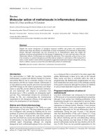
Figure 8
Macrophages stain positive for IL-1β and MIP-1α in the joints of nonim-munized E/P-selectin mutant miceMacrophages stain positive for IL-1β and MIP-1α in the joints of nonim-
munized E/P-selectin mutant mice. Immunohistochemistry for IL-1β and
MIP-1α in synovial tissue of E/P-selectin mutant CIA mice. Staining for
both cytokines showed clear immunopositivity on macrophages
(arrows), whereas ST endothelial cells (arrowheads) remained negative.
Original magnification: 400×. CIA, collagen-immunized arthritis; MIP,
macrophage inflammatory protein; wt, wild-type.
Arthritis Research & Therapy Vol 7 No 5 Ruth et al.
R970
9. Koch AE, Turkiewicz W, Harlow LA, Pope RM: Soluble E-selectin
in arthritis. Clin Immunol Immunopathol 1993, 69:29-35.
10. Hosaka S, Shah MR, Pope RM, Koch AE: Soluble forms of P-
selectin and intercellular adhesion molecule-3 in synovial
fluids. Clin Immunol Immunopathol 1996, 78:276-282.
11. Issekutz AC, Mu JY, Liu G, Melrose J, Berg EL: E-selectin, but not
P-selectin, is required for development of adjuvant-induced
arthritis in the rat. Arthritis Rheum 2001, 44:1428-1437.
12. Verdrengh M, Erlandsson-Harris H, Tarkowski A: Role of selectins
in experimental Staphylococcus aureus-induced arthritis. Eur J
Immunol 2000, 30:1606-1613.
13. Bullard DC, Mobley JM, Justen JM, Sly LM, Chosay JG, Dunn CJ,
Lindsey JR, Beaudet AL, Staite ND: Acceleration and increased
severity of collagen-induced arthritis in P-selectin mutant
mice. J Immunol 1999, 163:2844-2849.
14. Bullard DC, Kunkel EJ, Kubo H, Hicks MJ, Lorenzo I, Doyle NA,
Doerschuk CM, Ley K, Beaudet AL: Infectious susceptibility and
severe deficiency of leukocyte rolling and recruitment in E-
selectin and P-selectin double mutant mice. J Exp Med 1996,
183:2329-2336.
15. Bullard DC, Hurley LA, Lorenzo I, Sly LM, Beaudet AL, Staite ND:
Reduced susceptibility to collagen-induced arthritis in mice
deficient in intercellular adhesion molecule-1. J Immunol 1996,
157:3153-3158.
16. Schaefer CJ, Lawrence WD, Wooley PH: Influence of long term
silicone implantation on type II collagen induced arthritis in
mice. Ann Rheum Dis 1999, 58:503-509.
17. Vermeire K, Heremans H, Vandeputte M, Huang S, Billiau A, Mat-
thys P: Accelerated collagen-induced arthritis in IFN-gamma
receptor-deficient mice. J Immunol 1997, 158:5507-5513.
18. Alonzi T, Fattori E, Lazzaro D, Costa P, Probert L, Kollias G, De
Benedetti F, Poli V, Ciliberto G: Interleukin 6 is required for the
development of collagen-induced arthritis. J Exp Med 1998,
187:461-468.
19. Stoop R, Kotani H, McNeish JD, Otterness IG, Mikecz K:
Increased resistance to collagen-induced arthritis in CD44-
deficient DBA/1 mice. Arthritis Rheum 2001, 44:2922-2931.
20. Issekutz TB, Miyasaka M, Issekutz AC: Rat blood neutrophils
express very late antigen 4 and it mediates migration to
arthritic joint and dermal inflammation. J Exp Med 1996,
183:2175-2184.
21. Schimmer RC, Schrier DJ, Flory CM, Dykens J, Tung DK, Jacobson
PB, Friedl HP, Conroy MC, Schimmer BB, Ward PA: Streptococ-
cal cell wall-induced arthritis. Requirements for neutrophils, P-
selectin, intercellular adhesion molecule-1, and macrophage-
inflammatory protein-2. J Immunol 1997, 159:4103-4108.
22. Manoury-Schwartz B, Chiocchia G, Bessis N, Abehsira-Amar O,
Batteux F, Muller S, Huang S, Boissier MC, Fournier C: High sus-
ceptibility to collagen-induced arthritis in mice lacking IFN-
gamma receptors. J Immunol 1997, 158:5501-5506.
23. Joosten LA, Lubberts E, Durez P, Helsen MM, Jacobs MJ, Goldman
M, van den Berg WB: Role of interleukin-4 and interleukin-10 in
murine collagen-induced arthritis. Protective effect of inter-
leukin-4 and interleukin-10 treatment on cartilage destruction.
Arthritis Rheum 1997, 40:249-260.
24. Ruth JH, Bienkowski M, Warmington KS, Lincoln PM, Kunkel SL,
Chensue SW: IL-1 receptor antagonist (IL-1ra) expression,
function, and cytokine-mediated regulation during mycobacte-
rial and schistosomal antigen-elicited granuloma formation. J
Immunol 1996, 156:2503-2509.
25. Campbell IK, O'Donnell K, Lawlor KE, Wicks IP: Severe inflam-
matory arthritis and lymphadenopathy in the absence of TNF.
J Clin Invest 2001, 107:1519-1527.
26. Austrup F, Vestweber D, Borges E, Lohning M, Brauer R, Herz U,
Renz H, Hallmann R, Scheffold A, Radbruch A, Hamann A: P- and
E-selectin mediate recruitment of T-helper-1 but not T-helper-
2 cells into inflammed tissues. Nature 1997, 385:81-83.
27. van den Berg WB, Joosten LA, Helsen M, van de Loo FA: Amelio-
ration of established murine collagen-induced arthritis with
anti-IL-1 treatment. Clin Exp Immunol 1994, 95:237-243.
28. Hom JT, Gliszczynski VL, Cole HW, Bendele AM: Interleukin 1
mediated acceleration of type II collagen-induced arthritis:
effects of anti-inflammatory or anti-arthritic drugs. Agents
Actions 1991, 33:300-309.
29. Hom JT, Cole H, Estridge T, Gliszczynski VL: Interleukin-1
enhances the development of type II collagen-induced arthri-
tis only in susceptible and not in resistant mice. Clin Immunol
Immunopathol 1992, 62:56-65.
30. Killar LM, Dunn CJ: Interleukin-1 potentiates the development
of collagen-induced arthritis in mice. Clin Sci (Lond) 1989,
76:535-538.
31. Ma Y, Thornton S, Boivin GP, Hirsh D, Hirsch R, Hirsch E: Altered
susceptibility to collagen-induced arthritis in transgenic mice
with aberrant expression of interleukin-1 receptor antagonist.
Arthritis Rheum 1998, 41:1798-1805.
32. Horai R, Saijo S, Tanioka H, Nakae S, Sudo K, Okahara A, Ikuse T,
Asano M, Iwakura Y: Development of chronic inflammatory
arthropathy resembling rheumatoid arthritis in interleukin 1
receptor antagonist-deficient mice. J Exp Med 2000,
191:313-320.
33. Von Stebut E, Metz M, Milon G, Knop J, Maurer M: Early macro-
phage influx to sites of cutaneous granuloma formation is
dependent on MIP-1alpha /beta released from neutrophils
recruited by mast cell-derived TNFalpha. Blood 2003,
101:210-215.
34. Hatano Y, Kasama T, Iwabuchi H, Hanaoka R, Takeuchi HT, Jing L,
Mori Y, Kobayashi K, Negishi M, Ide H, Adachi M: Macrophage
inflammatory protein 1 alpha expression by synovial fluid neu-
trophils in rheumatoid arthritis. Ann Rheum Dis 1999,
58:297-302.
35. Kagari T, Doi H, Shimozato T: The importance of IL-1 beta and
TNF-alpha, and the noninvolvement of IL-6, in the develop-
ment of monoclonal antibody-induced arthritis. J Immunol
2002, 169:1459-1466.
36. Koch AE, Harlow LA, Haines GK, Amento EP, Unemori EN, Wong
WL, Pope RM, Ferrara N: Vascular endothelial growth factor. A
cytokine modulating endothelial function in rheumatoid
arthritis. J Immunol 1994, 152:4149-4156.
37. Marinova-Mutafchieva L, Williams RO, Mason LJ, Mauri C, Feld-
mann M, Maini RN: Dynamics of proinflammatory cytokine
expression in the joints of mice with collagen-induced arthritis
(CIA). Clin Exp Immunol 1997, 107:507-512.