Báo cáo y học: "Differential expression of chemokine receptors on peripheral blood B cells from patients with rheumatoid arthritis and systemic lupus erythematosus" ppsx
Bạn đang xem bản rút gọn của tài liệu. Xem và tải ngay bản đầy đủ của tài liệu tại đây (346.94 KB, 13 trang )
Available online />
Research article
Open Access
Vol 7 No 5
Differential expression of chemokine receptors on peripheral
blood B cells from patients with rheumatoid arthritis and systemic
lupus erythematosus
Maren Henneken1, Thomas Dörner2, Gerd-Rüdiger Burmester2 and Claudia Berek1
1Deutsches
Rheuma ForschungsZentrum, Berlin, Germany
of Rheumatology and Clinical Immunology, Charité, Humboldt University, Berlin, Germany
2Department
Corresponding author: Claudia Berek,
Received: 29 Nov 2004 Revisions requested: 22 Dec 2004 Revisions received: 17 May 2005 Accepted: 31 May 2005 Published: 22 Jun 2005
Arthritis Research & Therapy 2005, 7:R1001-R1013 (DOI 10.1186/ar1776)
This article is online at: />© 2005 Henneken et al.; licensee BioMed Central Ltd
This is an Open Access article distributed under the terms of the Creative Commons Attribution License ( />2.0), which permits unrestricted use, distribution, and reproduction in any medium, provided the original work is properly cited.
Abstract
Chemokines and their receptors are essential in the recruitment
and positioning of lymphocytes. To address the question of B
cell migration into the inflamed synovial tissue of patients with
rheumatoid arthritis (RA), peripheral blood naive B cells, memory
B cells and plasma cells were analyzed for cell surface
expression of the chemokine receptors CXCR3, CXCR4,
CXCR5, CCR5, CCR6, CCR7 and CCR9. For comparison, B
cells in the peripheral blood of patients with the autoimmune
disease systemic lupus erythematosus (SLE) or with the
degenerative disease osteoarthritis (OA) were analyzed.
Expression levels of chemokine receptors were measured by
flow cytometry and were compared between the different
patient groups and healthy individuals. The analysis of
chemokine receptor expression showed that the majority of
peripheral blood B cells is positive for CXCR3, CXCR4,
CXCR5, CCR6 and CCR7. Whereas a small fraction of B cells
were positive for CCR5, practically no expression of CCR9 was
found. In comparison with healthy individuals, in patients with RA
a significant fraction of B cells showed a decreased expression
of CXCR5 and CCR6 and increased levels of CXCR3. The
downregulation of CXCR5 correlated with an upregulation of
CXCR3. In patients with SLE, significant changes in CXCR5
expression were seen. The functionality of the chemokine
receptors CXCR3 and CXCR4 was demonstrated by
transmigration assays with the chemokines CXCL10 and
CXCL12, respectively. Our results suggest that chronic
inflammation leads to modulation of chemokine receptor
expression on peripheral blood B cells. However, differences
between patients with RA and patients with SLE point toward a
disease-specific regulation of receptor expression. These
differences may influence the migrational behavior of B cells.
Introduction
About 50 chemokines have been identified in humans and are
divided into four groups according to their cysteine motifs [4].
After activation and differentiation, cells of the lymphoid lineages dynamically change their expression profiles of chemokine receptors, which results in specific migration in response
to chemokines [5,6].
Rheumatoid arthritis (RA) is a complex autoimmune disease of
unknown etiology. It is characterized by chronic inflammation
of the synovial membrane and the formation of a pannus,
which leads to swollen joints and finally to joint destruction.
Inflammatory cells such as monocytes and neutrophils,
together with T and B cells, infiltrate the synovial membrane
[1]. Migration of lymphocytes from the blood to the synovial tissue is a multi-step process controlled in part by interactions
between chemokines and their receptors [2,3].
Under pathological conditions, such as RA, chemokines direct
lymphocytes into the chronically inflamed synovial tissue [7,8].
Both the residual synovial-lining cells and infiltrating leukocytes are the source of pro-inflammatory chemokines such as
CCL2, CCL3, CCL5, CCL20, CXCL9 and CXCL10, as well
BSA = bovine serum albumin; ELISA = enzyme-linked immunosorbent assay; FITC = fluorescein isothiocyanate; FACS = fluorescence-activated cell
sorting, mAb = monoclonal antibody; OA = osteoarthritis; PBMC = peripheral blood mononuclear cells; PE = phycoerythrin; RA = rheumatoid arthritis; RF = rheumatoid factor; SLE = systemic lupus erythematosus; TNF = tumor necrosis factor.
R1001
Arthritis Research & Therapy
Vol 7 No 5
Henneken et al.
as chemokines important for the homeostasis of lymphocytes,
such as CXCL12 and CXCL13 [2,9-13]. As a consequence
of the synovial inflammation, these chemokines are also found
in the synovial fluid.
Contradictory results have been reported for chemokine
receptor expression on peripheral blood T cells of patients
with RA [13,14] and even less is known about the expression
of chemokine receptors on B cells. The classical chemokine
receptor on B cells is CXCR5. Its ligand CXCL13 is a potent
B cell chemo-attractant molecule directing B cells into the follicles of secondary lymphoid organs [15]. In addition to
CXCR5, the chemokine receptors CXCR4 and CCR7 have
been implicated in B cell migration into the follicular structures,
and especially CXCR4 in directing plasma cells into the bone
marrow [6]. These chemokines and their corresponding chemokine receptors are also expressed in the inflamed synovial tissue of patients with RA. In particular, high expression of
CXCL13 was found when the synovial tissue contained large
aggregates of B cells, resembling the structure of germinal
centers [11,17]. These findings suggested that the chemokine
CXCL13 is involved in B cell trafficking into the inflamed tissue
and has a role in the formation of ectopic lymphoid tissue
[18,19].
CXCR3 is a chemokine receptor for the inflammatory chemokines CXCL9, CXCL10 and CXCL11. It has been described as
a marker for malignant B cells and is absent from the majority
of normal peripheral blood B cells [20,21]. The analysis of
patients with multiple sclerosis showed that CXCR3 becomes
upregulated on infiltration of the inflamed cerebrospinal fluid
[20].
To address the question of B cell migration, we analyzed
chemokine receptor expression on peripheral blood B cells.
Expression profiles of patients with RA were compared with
those from patients with systemic lupus erythematosus (SLE),
who – in contrast to patients with RA – usually show no or very
little B cell accumulation at the site of chronic inflammation
[22]. In addition, patients with osteoarthritis (OA) were
included into the study as a control cohort. OA is a non-inflammatory degenerative joint disease. Our data suggest that the
chronic inflammation in patients with RA and patients with SLE
leads to changes in the chemokine receptor expression pattern on peripheral B cells; however, the expression patterns of
chemokine receptors in RA and SLE are differentially
regulated.
criteria [23]. As control, blood samples from healthy blood
donors were analyzed. The demographic data and the treatment of patients are summarized in Table 1. The scientific ethics committee of Charité approved the study, and informed
consent was obtained.
Cell isolation and flow cytometry
Peripheral blood mononuclear cells (PBMC) were isolated by
gradient centrifugation with Ficoll (Amersham Biosciences)
and then stained (30 min at 4°C) with a biotin-coupled B cellspecific mAb against CD19 (clone SJ25-C1; Southern Biotechnology Associates), with the Cy5-labeled mAb against
CD27 (clone 2E4; gift from RA van Lier, Academic Medical
Center, Amsterdam) and with fluorescein isothiocyanate
(FITC)-labeled mAb specific for one of the chemokine receptors CXCR3 (clone 49801.111; R&D Systems), CXCR5
(clone 51505.111; R&D Systems), CCR5 (clone 45523.111;
R&D Systems), CCR6 (clone 53103.111; R&D Systems),
CCR7 (clone 3D12; a gift from M Lipp, Max Delbrück Center,
Berlin) or CCR9 (clone 112509.111; R&D Systems). Antibody against CXCR4 was labeled with phycoerythrin (PE;
clone 12G5; BD Pharmingen). Before incubation with streptavidin-PE or streptavidin-FITC (for CXCR4) (0.5 µg/ml;
Pharmingen), cells were washed twice in phosphate-buffered
saline/2% BSA/4 mM EDTA.
To determine the frequency of CD5+ B cells, PBMC were
stained as described above with biotin-CD19, Cy5-CD27 and
PE-labeled mAb against CD5 (clone UCHT2; BD Pharmingen). For the analysis of chemokine receptor expression,
CD5+ B cells were purified by magnetic cell sorting with
CD19-specific beads (Miltenyi). Isolated B cells were stained
as described above with PE-CD5 and Cy5-CD27 together
with the FITC-labeled mAb against the chemokine receptors
CXCR3 or CXCR5.
Propidium iodide (1 µg/ml; Sigma) was added immediately
before cytometric analysis for the exclusion of dead cells. Flow
cytometric analyses were performed by fluorescence-activated cell sorting (FACS) software (FACSCalibur and CellQuest; Becton Dickinson).
Methods
Patients and controls
Heparinized whole blood (9 ml) from patients with RA, patients
with SLE and patients with OA was obtained from the Departments of Rheumatology and Orthopedics (Charité, Humboldt
University, Berlin, Germany). Patients with RA were diagnosed
in accordance with the American College of Rheumatology
R1002
Measurement of CXCL10 concentrations
For further analysis, sera of controls and those of patients analyzed for chemokine receptor expression were stored at -20°C.
The concentration of CXCL10 was determined by using a sensitive ELISA test kit (HyCult Biotechnology). Samples were
tested in duplicate and values were compared with a standard
curve.
Transmigration assay
Cell migration was examined in Transwell™ inserts (Corning
Costar) with a diameter of 6.5 mm and 5 µm pores, by using
fibronectin-precoated membranes as described previously
[24]. In brief, 5 × 10⁵ PBMC were suspended in RPMI 1640
Available online />
Table 1
Demographics and medications of patients in the study
Parameter
RA
SLE
OA
Controls
Total numbers
26
11
13
21
Mean
55.1
42.5
71.2
42.6
Range
25–76
27–78
62–79
25–64
Female
21
10
8
15
Male
5
1
5
6
Mean
8.1
9.5
3.3
-
Range
0.1–37
2–25
0.5–10
-
Leukocytes (109/l)
9.5 ± 2.7
4.3 ± 1.1
7.8 ± 2.7
nd
CRP (mg/dl)
3.1 ± 3.0
1.1 ± 0.9
0.5 ± 0.6
nd
ESR (mm/h)
37.5 ± 30.5
34.9 ± 16.7
28.5 ± 19.4
nd
6
-
-
21
-
8a
-
Age (years)
Sex (n)
Duration of disease (years)
Clinical parameter (mean ± SD)
Treatment (n)
None
Corticosteroids and/or NSAID
7
13b
-
-
-
Predisolone
-
11c
-
-
Cyclophosphamide (i.v.)
-
6
-
-
Anti-TNF-α therapy
CRP, C-reactive protein; ESR, erythrocyte sedimentation rate; i.v., intravenous; nd, not determined; NSAID, non-steroidal anti-inflammatory drugs;
OA, osteoarthritis; RA, rheumatoid arthritis; SLE, systemic lupus erythematosus.
aTreatment with buprenorphine (1) or tramadol (1); btreatment with TNF-α-specific antibodies infliximab (6) or adalimumab (3) (mostly in
combination with methotrexate) or with the soluble TNF-α receptor etanercept (2); ctreatment with predisolone alone (2), in combination with
azathioprine (3), or in combination with cyclophosphamide (6).
medium (Life Technologies) without methyl red, supplemented
with 0.5% BSA. Cells were added to the upper well and chemokine dilution or assay medium to the lower compartment; they
were incubated for 90 min at 37°C under CO2-buffered conditions. Migrated cells from triplicate wells were pooled and
analyzed by flow cytometry. Optimal chemokine concentrations for migration were 50 nM for CXCL12 (R&D Systems)
and 100 nM for CXCL10 (R&D Systems).
Statistical analyses
Statistical analyses were performed with GraphPad Prism
software (Prism 3.0 software for windows; GraphPad). Frequencies of B cells were calculated with CellQuest software
(Becton Dickinson) and variations in chemokine receptor
expression on B cells within the analyzed group were compared in a hierarchic statistic analysis, using Kruskal–Wallis
and the nonparametric Mann–Whitney U test. Correlations
were determined by Spearman's product-moment correlation
for interval data (GraphPad software). P < 0.05 was considered significant.
Results
Chemokine receptor expression on peripheral blood B
cells of healthy controls
To analyze chemokine receptor expression on B cells of normal healthy volunteers PBMC were double-stained with antibodies specific for the B cell marker CD19 and for the
chemokine receptors CXCR3, CXCR4, CXCR5, CCR5,
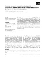
CCR6, CCR7 or CCR9. Data from a representative experiment are depicted in Fig. 1. The cytometric analysis of healthy
controls showed that 93.9% of the B cells were positive for
CXCR3, 81.6% for CXCR4, 97.6% for CXCR5, 76.7% for
CCR6 and 94.3% for CCR7. Most B cells were negative for
the receptors CCR5 (only 5.6% positive) and CCR9 (only
1.7% positive).
R1003
Arthritis Research & Therapy
Vol 7 No 5
Henneken et al.
Figure 1
cell counts
93.9%
93,9%
CXCR3
5.6%
CCR5
81.6%
CXCR4
76.7%
CCR6
97.6%
CXCR5
94.3%
CCR7
1.7%
CCR9
Expression of chemokine receptors in peripheral blood B cells from a healthy donor. Flow cytometric plots show chemokine receptor expression on
donor
CD19+ B cells. For each of the chemokine receptors tested the percentage of positive B cells is indicated. Isotype control (dotted line) is included.
Analyses of chemokine receptor expression on
peripheral blood B cell subsets of patients with RA, with
SLE and with OA
To assess changes in the pattern of B cell chemokine receptor
expression associated with RA (n = 26), SLE (n = 11) and OA
(n = 13) we compared the pattern in patients with that of a
group of healthy individuals (n = 21). In addition to mAb specific for CD19 and the respective chemokine receptors,
PBMC were stained with antibodies specific for CD27, which
allowed us to distinguish between naive B cells (CD27-),
memory B cells (CD27+) and plasma cells (CD27hi) [25]. Levels of CD27 expression used to define the various B cell subsets were defined by triple staining with CD19, CD20 and
CD27 as shown in Fig. 2a. The relative frequencies of B cells
are comparable in the control and in the different patient
groups, although the patients with RA did show a higher variation in the percentages of naive B cells, memory B cells and
plasma cells (Fig. 2b).
In healthy individuals most B cells (median value 93.8%, range
66.9 to 99.3%) were positive for CXCR5 (CXCR5+; Fig. 3).
Similar results were obtained with blood samples from
patients with OA. As shown in Fig. 3 the percentage of
CXCR5+ B cells was slightly higher (median 96.8%, range
90.6 to 99.6%, P = 0.05). In both of these groups, only a minor
fraction of B cells showed low expression of CXCR5. A representative FACS analysis showing CXCR5 expression on
peripheral blood naive B cells, memory B cells and plasma
cells from a healthy control is shown in Fig. 7.
R1004
In contrast to the patients with OA, a decrease in the fraction
of CXCR5+ B cells was observed in most blood samples from
patients with RA and patients with SLE. In patients with RA the
median value for CXCR5+ B cells was decreased to 83.4% (P
= 0.01) and in patients with SLE to 81.8% (P = 0.02) (Fig. 3).
Whereas little variation in the frequency of CXCR5+ B cells
was seen in controls or in patients with OA, the expression of
CXCR5 on B cells varied enormously in patients with RA and
patients with SLE (Fig. 3). For patients with RA these values
ranged from 2.5 to 97.6% and for patients with SLE from 59.8
to 98.1% (Fig. 3).
The analysis of the different B cell subsets revealed that the
fraction of CXCR5+ cells decreases as B cells differentiate to
plasma cells. In healthy individuals the median value for the frequency of CXCR5+ plasma cells was 70.6% (range 27.8 to
96.8%). In patients with RA and patients with SLE the expression of CXCR5 on peripheral blood plasma cells was reduced
to 39.3% (range 5.7 to 91.9%) and 45.6% (range 11.0 to
88.6%), respectively (Fig. 3). Overall, in patients with RA significant differences in the fraction of CXCR5+ B cells were
found for naive B cells, memory B cells and plasma cells,
whereas in patients with SLE this was seen only for effector B
cells, memory B calls and plasma cells (Fig. 3).
Similar results were obtained when B cells were tested for the
chemokine receptor CXCR4. In both healthy controls and
patients with OA, most B cells expressed high levels of this
chemokine receptor, and little individual variation was seen
Available online />
Figure 2
(a)
total B cells
(b)
(***)
naive B cells
(**)
(**)
(**)
60,4
%
CD20
75
75
50
memory B cells
50
25
naive B cells
cell frequency [%]
CD27
38,4 %
plasma cells
100
25
1,2%
100
0
con
OA
RA
SLE
0
con
memory B cells
(**)
OA
RA
SLE
plasma cells
(**) (*)
100
100
75
75
50
50
25
25
0
con
OA
RA
SLE
0
con
OA
RA
SLE
The frequency of B cells in healthy individuals and the different patient groups (a) Triple staining with CD19, CD20 and CD27 was used to define
groups.
naive B cells, memory B cells and plasma cells. Thresholds for CD27-, CD27+ and CD27hi are indicated. (b) Frequency for B cells and the different
subpopulations. Data are given for healthy individuals (con) and for each of the patient groups analyzed: osteoarthritis (OA), rheumatoid arthritis (RA)
and systemic lupus erythematosus (SLE). Box plots show the median values, 25th and 75th quartile and the range of values. Significant differences
from controls are shown (*P < 0.05, **P < 0.005, ***P < 0.0005).
(Fig. 4). The median values for the fraction of CXCR4+ B cells
in healthy controls and patients with OA were 82.0% (range
68.4 to 89.3%) and 84.6% (range 74.7 to 91.5%), respectively. In contrast, in patients with RA (n = 19) the percentage
of CXCR4+ B cells was lower and large individual variations
were found, from 7.3 to 89.4%. Similarly, in the few patients
with SLE who were tested (n = 4) a reduction in the percentage of CXCR4+ B cells was seen, ranging from 5.4 to 84.2%
(Fig. 4).
When B cells from patients with RA and patients with SLE
were tested for the expression of CCR6, again a reduction in
the frequency of CCR6+ B cells was observed. However, a
significant reduction in the frequency of CCR6+ B cells was
found only when B cells were analyzed from patients with RA
(P = 0.005; Fig. 5).
A great variation in chemokine receptor expression was
obtained when B cells were tested for CXCR3, which is a
receptor for chemokines that is upregulated in inflammation.
We found that in healthy individuals most B cells are positive
for CXCR3; however, the mean fluorescence intensity was
rather low and only a few B cells expressed CXCR3, at a level
comparable to that seen after staining with mAb specific for
CXCR5 (Figs 1 and 7). The analysis of blood samples from
patients with RA – and also from some patients with SLE –
revealed an increase in the fraction CXCR3-high-expressing
(CXCR3hi) B cells (Fig. 6). Again the percentage of CXCR3hi
B cells showed considerable variation between individual
patients. In RA the values ranged from 1.6 to 89.8% (Fig. 6).
The results obtained for CXCR3 suggest that its expression is
upregulated as B cells differentiate into memory B cells and
plasma cells. In most healthy controls the fraction of CXCR3hi
naive B cells was negligible. A comparable result was found
with blood samples from patients with OA and also with most
patients with SLE. In contrast, in patients with RA a significant
fraction of naive B cells showed high expression of CXCR3.
The frequency was further increased in memory B cells from
patients with RA (Figs 6 and 7). A comparison of B cells from
healthy individuals and from patients with RA showed that the
median value for naive B cells increased from 3.4% (range 0.4
to 15.7%) to 9.7% (range 2.8 to 43.1%) (P = 0.0006) and for
memory cells from 10.0% (range 5.4 to 28.3%) to 23.2%
(range 1.9 to 95.0%) (P = 0.003; Fig. 6). This highly signifiR1005
Arthritis Research & Therapy
Vol 7 No 5
Henneken et al.
Figure 3
total B cells
(*)
(*)
100
naive B cells
(*)
(*)
75
75
50
50
25
CXCR5+ B cells [%]
(*)
100
25
0
con
OA
RA
SLE
0
con
memory B cells
(*)
OA
RA
SLE
plasma cells
(**)
(**)
100
50
25
SLE
75
50
RA
100
75
(*)
25
0
con
OA
RA
SLE
0
con
OA
Chemokine receptor CXCR5+ expression on B cell subpopulations The percentage of total B cells, naive B cells, memory B cells and plasma cells
subpopulations.
expressing CXCR5+ are given. Box plots show the median values, 25th and 75th quartile and the range of values. Significant differences from controls are shown (*P < 0.05, **P < 0.005).
cant increase in the fraction of CXCR3hi B cells was not
observed when blood samples from patients with SLE were
analyzed.
Furthermore, we addressed the question of whether there is a
significant correlation between the expression of CXCR5 with
that of CXCR4, CCR6 and CXCR3. In patients with RA a
decreased fraction of CXCR5-expressing B cells correlated
with the expression of CCR6 (r = 0.53, P = 0.01; data not
shown). However, neither in patients with RA nor in patients
with SLE did we see a significant correlation in the expression
of CXCR5 and CXCR4. However, with regard to CXCR5 and
CXCR3 we found a negative correlation, but this was
restricted to patients with RA (r = -0.59, P = 0.007; Fig. 8).
R1006
Chemokine receptor expression on CD5+ B cells
Staining of PBMC of healthy controls showed that on average
20% of B cells were positive for CD5 (data not shown). Comparable frequencies of CD5 expression were found for
patients with RA (range 12 to 23% of B cells). To assess
chemokine receptor expression on CD5+ B cells, CD19+ B
cells were purified by magnetic cell sorting and stained for
CD5, CD27 and the respective chemokine receptor. Labeling
for CXCR5 showed that most CD5+ B cells were positive for
CXCR5 and only a fraction of CD5+ plasma cells was negative
for CXCR5 (data not shown). The frequency of CXCR3hi B
cells was the same for the CD5+ and CD5- B cell populations.
There was therefore no evidence that CXCR3 expression is
preferentially enhanced on CD5+ B cells.
Available online />
Figure 4
total B cells
naive B cells
(*)
100
75
RA
SLE
50
25
(*)
75
50
CXCR4+ B cells [%]
(*)
100
25
0
con
OA
RA
SLE
0
con
memory B cells
plasma cells
(*)
100
OA
100
75
75
50
50
25
25
0
con
OA
RA
SLE
0
con
OA
RA
SLE
CXCR4+ expression
Chemokine receptor
on B cell subpopulations The percentage of total B cells, naive B cells, memory B cells and plasma cells
subpopulations.
expressing CXCR4+ are given. Box plots show the median values, 25th and 75th quartile and the range of values. Significant differences from controls are shown (*P < 0.05).
The level of CXCL10 may influence CXCR3 expression
PBMC were isolated by Ficoll gradient centrifugation. In the
supernatant the concentration of CXCL10 was measured with
a standard ELISA test. In both sera from the different patient
groups and from healthy controls only low levels of CXCL10
were detectable (about 200 pg/ml; Fig. 9). Only a single
patient with SLE showed slightly increased levels of CXCL10.
Influence of medication on chemokine receptor
expression
To determine a potential influence of medication, five blood
samples of newly diagnosed patients with RA were analyzed
before treatment. In three of the samples, B cells showed a
pattern of chemokine receptor expression as described above,
in that the median value for the frequency of B cells with high
levels of CXCR5 was decreased from 93.8% (range 66.9 to
99.3%) to 79.4% (range 2.6 to 96.8%) (Fig. 10). Variations in
chemokine receptor expression were also seen in patients (n
= 10) under treatment with tumor necrosis factor (TNF) blockers (infliximab, etanercept and adalimumab). However, this
was not found in patients with RA (n = 4) under treatment with
corticoids and/or non-steroidal anti-inflammatory drugs
(NSAID). In these patients practically all B cells were CXCR5+
(median 95.5%, range 88.1 to 97.8%; Fig. 10).
The analysis of CXCR3 expression on B cells revealed that in
comparison with healthy individuals, untreated patients with
RA had an increase in the frequency of CXCR3hi B cells. Most
strikingly, an increase in the percentage of CXCR3hi naive B
cells, a finding characteristic for patients with RA, was
observed. This increase was seen also under anti-TNF-α
therapy and to some extent when patients where treated with
corticoids and/or NSAID. The observed modulation of chemokine receptor expression on peripheral blood B cells from
patients with RA therefore does not simply reflect the disease
activity and/or the influence of therapy.
R1007
Arthritis Research & Therapy
Vol 7 No 5
Henneken et al.
Figure 5
total B cells
naive B cells
(**)
100
75
75
50
50
25
CCR6+ B cells [%]
100
25
0
con
OA
RA
SLE
0
memory B cells
con
OA
RA
SLE
RA
SLE
plasma cells
100
100
75
75
50
50
25
25
0
con
OA
RA
SLE
0
con
OA
subpopulations.
Chemokine receptor CCR6+ expression on B cell subpopulations The percentage of total B cells, naive B cells, memory B cells and plasma cells
expressing CCR6+ are given. Box plots show the median values, 25th and 75th quartile and the range of values. Significant differences from controls
are shown (**P < 0.005).
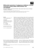
The influence of chemokine receptor expression on B
cell migration
To test whether low expression of CXCR3 on B cells is sufficient to respond to the inflammatory chemokine CXCL10,
PBMC from patients with RA were tested with the use of a
transmigration assay (Fig. 11). A cytometric analysis of the
migrated cells revealed that both fractions of memory B cells,
CXCR3hi and CXCR3lo B cells, migrated toward CXCL10
(Fig. 11). In contrast, in an analysis of migration towards the
chemokine CXCL12, only CXCR4+ B cells responded;
CXCR4- B cells did not (Fig. 11). The in vitro chemotaxis assay
suggested that CXCR3-low-expressing B cells are functionally reactive towards the chemokine CXCL10.
from healthy individuals and also from patients with OA. In
patients with RA and patients with SLE a fraction of B cells
showed decreased expression of CXCR5, CXCR4 and
CCR6, chemokine receptors that have been associated with
B cell homing into follicles [26,27]. In contrast, the expression
of CXCR3, a receptor reactive to inflammatory chemokines
[4,10], was increased. These changes in chemokine receptor
expression seem to be associated with chronic inflammation,
because they were not observed when B cells from patients
with OA were analyzed. Importantly, a comparison of B cells
from patients with RA and patients with SLE showed distinct
disease signatures. A negative correlation of CXCR5 and
CXCR3 expression in B cells was seen only in patients with
RA.
Discussion
The analysis of peripheral blood B cells from patients with RA
and patients with SLE showed significant differences in their
chemokine receptor expression when compared with B cells
R1008
CXCR5 was previously shown to be expressed on most
mature circulating B cells [8]. This receptor is the main chemokine receptor responsible for the controlled migration of B
Available online />
Figure 6
total B cells
naive B cells
(***)
(***)
100
75
75
50
50
25
CXCR3 hi B cells [%]
100
25
0
con
OA
RA
SLE
0
con
memory B cells
OA
RA
SLE
plasma cells
(**)
100
100
75
75
50
50
25
25
0
con
OA
RA
SLE
0
con
OA
RA
SLE
subpopulations
Chemokine receptor CXCR3hi expression on B cell subpopulations. The percentage of total B cells, naive B cells, memory B cells and plasma cells
expressing CXCR3hi are given. Box plots show the median values, 25th and 75th quartile and the range of values. Significant differences from controls are shown (**P < 0.005, ***P < 0.0005).
cells into secondary lymphoid organs [28]. The analysis of
blood samples from healthy individuals showed that after activation of B cells and their differentiation into plasma cells and
to some extent into memory cells, CXCR5 becomes downregulated (Fig. 3). In line with these results are data from an experiment in vitro in which stimulation with anti-CD40 antibodies
led to a downregulation of CXCR5 [29]. The finding of a significant decrease in the fraction of CXCR5+ B cells in patients
with RA and also in patients with SLE by our study may reflect
the chronic activation of B cells as reported for these groups
of patients [30].
Lower levels of chemokine receptor CXCR5 and higher levels
of CXCR3 on peripheral blood B cells may represent a generalized change in the profile and might be seen on all of the
activated B cells in the systemic compartment or these
changes in chemokine receptor expression might serve in
selective recruitment into the inflamed tissue. In line with this
interpretation we observed CXCR3 expression on synovial tissue B cells. However, comprehensive studies are under way
to further delineate the expression of chemokine receptors and
their function in migration into the effected tissue.
CXCR3 was described as a marker for malignant B cells, and
its expression on normal peripheral blood B cells has been
controversial [20,21]. Our results show that practically all B
cells express CXCR3, although with a low mean fluorescence
intensity. After differentiation, the level of CXCR3 expression
seems to be upregulated because the frequency of CXCR3hi
B cells increases from naive to memory B cells. Activation of
human B cells using a culture system in vitro showed that after
stimulation with cytokines the expression of CXCR3 is
R1009
Arthritis Research & Therapy
Vol 7 No 5
Henneken et al.
Figure 7
Figure 8
+
CXCR5 B cells [%]
100
75
50
25
0
r = –0.59
P = 0.007
0
10
20
30
40
50
CXCR3hi B cells [%]
Negative correlation in the frequency of CXCR3hi and CXCR5+ B cells
in patients with RA
in patients with RA. Correlation factor (Spearman's) and P value are
indicated (GraphPad software).
Figure 9
0.4
0.3
OD450
Modulation of CXCR3 and CXCR5 expression in patients A representpatients.
ative fluorescence-activated cell sorting analysis for chemokine receptor expression on peripheral B cells (CD19+) is shown. The
percentages of CXCR5+ and CXCR3hi B cells are given for healthy
controls (con) and patients with osteoarthritis (OA), rheumatoid arthritis
(RA) and systemic lupus erythematosus (SLE).
0.2
0.1
upregulated [31]. Similarly, it was shown for T lymphocytes
that after activation with interleukin-2 the expression of
CXCR3 is upregulated [32]. The observed increase in the frequency of CXCR3hi B cells may result from the chronic activation and differentiation of B cells in patients with RA.
A significant increase in the fraction of CXCR3hi B cells was
observed only when blood samples from patients with RA
were analyzed (Fig. 6). In sera of patients with SLE but not in
those from patients with RA, Narumi and colleagues [33]
described high titers of CXCL10. Because CXCR3 expression may be influenced by the level of CXCL10, sera from
healthy controls and from the different patient groups were
tested for the presence of this chemokine. However, we did
not find elevated titers of CXCL10 in our patients with SLE
(Fig. 9). These results exclude the possibility that a ligandinduced receptor internalization might underlie the lower frequency of CXCR3hi-expressing B cells in patients with SLE.
Using a transmigration assay we were able to show that low
levels of CXCR3 expression are sufficient to permit a
response to the migrational stimulus of the chemokine
CXCL10. However, these chemotaxis results in vitro are not
R1010
0
con
OA
RA
SLE
CXCL10 concentrations in sera. Sera of healthy controls and of
sera
patients with rheumatoid arthritis, systemic lupus erythematosus and
osteoarthritis were tested in a standard ELISA test. A D450 of 0.06 corresponds to 200 pg/ml CXCL10 (concentration after Ficoll centrifugation); for each of the groups analyzed median values are indicated. Con,
control; OA, osteoarthritis; RA, rheumatoid arthritis; SLE, systemic
lupus erythematosus.
necessarily predictive of their lymphocyte-recruiting activity in
vivo. The different levels of chemokine receptor CXCR3 on
peripheral blood B cells may still affect B cell migration. Elevated levels of the interferon-γ-inducible chemokines CXCL9
and CXCL10, both ligands for CXCR3, have been found in
chronically inflamed synovial tissue [10]. These chemokines,
which are normally involved in the chemotaxis of neutrophils, T
cells and mast cells, might also influence the migration of
CXCR3+ B cells. Further experiments will be required to show
whether the significant upregulation of CXCR3 on peripheral
Available online />
Figure 10
total B cells
naive B cells
(*)
100
75
50
25
0
/c
Fth
tre
un
er
ap
ds
at
co
oi
y
er
ap
Fth
SA
ID
N
SA
ID
N
hi
y
CXCR3
TN
/c
o
un
rti
c
tre
oi
at
ds
n
co
[%]
+
TN
0
or
tic
25
ed
50
CXCR5
n
75
ed
100
(*)
(*)
(*)
(**)
25
0
ap
s
at
er
th
F-
/c
o
ID
N
SA
TN
rti
tre
er
th
TN
F-
un
ap
ds
oi
N
SA
ID
/c
un
or
tre
tic
n
co
id
0
co
25
y
50
co
n
50
ed
75
y
75
(**)
100
at
ed
100
Effect of treatment on CXCR3 and CXCR5 expression. Data are shown for healthy individuals, untreated patients with rheumatoid arthritis and
expression
patients treated with corticoid and/or non-steroidal anti-inflammatory drugs or treatment with anti-tumor necrosis factor-α therapy. The percentage of
CXCR3hi and CXCR5+ B cells is shown for total B cells and the subpopulation of naive B cells. Box plots show the median values, 25th and 75th
quartile and the range of values. Significant differences from controls are shown (*P < 0.05, **P < 0.005). Con, control; RA, rheumatoid arthritis;
SLE, systemic lupus erythematosus.
blood B cells supports their accumulation in the inflamed synovial tissue.
for RF. A correlation of chemokine receptor expression and the
level of RF was therefore not seen.
Whereas B cells from healthy controls and patients with OA
showed little inter-individual variation in the expression of
chemokine receptors, individual patients with RA and patients
with SLE gave a rather heterogeneous picture (Fig. 7). For
each of the chemokine receptors analyzed, the fractions of
negative, low and highly positive B cells varied tremendously
and were seen on both B cells and memory cells.
An attempt to correlate the level of chemokine receptor
expression with age or sex of the patients, with disease duration or with disease activity failed. There was no clear-cut
correlation to be seen. Furthermore, from our data it seems
unlikely that the variability in chemokine receptor expression
results from the different treatment regimes of individual
patients with RA (Fig. 10). Individual variability of chemokine
receptor expression on B cells was as great in recently
diagnosed, yet untreated, patients with RA as in those receiving anti-TNF-α therapies, which suggests that TNF-α itself is
unlikely to be the cause of receptor modulation.
Little is known about the mechanisms controlling chemokine
receptor expression and what might cause the variability in
their expression level on peripheral blood B cells. One possibility might be that the modulation of chemokine receptor
expression is associated with rheumatoid factor (RF) antibody
titers. The majority of patients with RA analyzed were positive
RA and SLE are chronic inflammatory diseases, and both are
characterized by a continuous activation of B cells. Whereas
R1011
Arthritis Research & Therapy
Vol 7 No 5
Henneken et al.
CD27
Figure 11
CXCR3
CXCR4
control
input
migrated
Chemokine receptor expression and B cell migration Peripheral blood mononuclear cells were tested in a transmigration assay. A cytometric analymigration.
sis showed CXCR3 (upper row) and CXCR4 (lower row) expression on CD19+ B cells before analysis (control), after 90 min incubation in medium
without chemokines (input) and after migration towards the chemokines CXCL10 (upper row) or CXCL12 (lower row) respectively (migrated).
SLE is a more systemic inflammatory disease, in most patients
with RA the inflammation is localized primarily to the synovial
membrane. To what extent the described differences in chemokine receptor expression between B cells from patients with
RA and patients with SLE might influence the migrational pattern of B cells needs to be delineated by continuing studies,
potentially permitting new therapeutic avenues.
Conclusion
Here we show that chronic inflammation influences chemokine
receptor expression on peripheral blood B cells. Receptors for
homeostatic chemokines, like CXCL13 and CXCL12 showed
reduced levels of expression whereas CXCR3 a receptor for
inflammatory chemokines becomes unregulated. Differences
between RA and SLE patients suggest a disease specific regulation of chemokine receptor expression, which may influence
the migrational behavior of B cells.
Competing interests
The author(s) declare that they have no competing interests.
Authors' contributions
MH made acquisition of data and their interpretation, performed the statistical analysis and drafted the article. TD was
responsible for assessment of patients and revising the article
critically. G-RB was involved in the analysis and careful discussion of the data. CB coordinated the study, was involved in the
critical discussion of results and their interpretation and
helped to draft the article. All authors read and approved the
final manuscript.
R1012
Acknowledgements
The work was supported by the SFB 421 and by the Network of Competence, Rheumatology (BMBF grant C2.2 to CB). The DRFZ is supported by the Berlin Senate of Research and Education.
References
1.
Firestein GS: The immunopathogenesis of rheumatoid
arthritis. Curr Opin Rheumatol 1991, 3:398-406.
2. Szekanecz Z, Kim J, Koch AE: Chemokines and chemokine
receptors in rheumatoid arthritis. Semin Immunol 2003,
15:15-21.
3. Baggiolini M, Loetscher P: Chemokines in inflammation and
immunity. Immunol Today 2000, 21:418-420.
4. Zlotnik A, Yoshie O: Chemokines: a new classification system
and their role in immunity. Immunity 2000, 12:121-127.
5. Yoshie O, Imai T, Nomiyama H: Chemokines in immunity. Adv
Immunol 2001, 78:57-110.
6. Moser B, Loetscher P: Lymphocyte traffic control by
chemokines. Nat Immunol 2001, 2:123-128.
7. Buckley CD: Why do leucocytes accumulate within chronically
inflamed joints? Rheumatology (Oxford) 2003, 42:1433-1444.
8. Loetscher P, Moser B: Homing chemokines in rheumatoid
arthritis. Arthritis Res 2002, 4:233-236.
9. Katschke KJ Jr, Rottman JB, Ruth JH, Qin S, Wu L, LaRosa G,
Ponath P, Park CC, Pope RM, Koch AE: Differential expression
of chemokine receptors on peripheral blood, synovial fluid,
and synovial tissue monocytes/macrophages in rheumatoid
arthritis. Arthritis Rheum 2001, 44:1022-1032.
10. Patel DD, Zachariah JP, Whichard LP: CXCR3 and CCR5 ligands
in rheumatoid arthritis synovium. Clin Immunol 2001, 98:39-45.
11. Shi K, Hayashida K, Kaneko M, Hashimoto J, Tomita T, Lipsky PE,
Yoshikawa H, Ochi T: Lymphoid chemokine B cell-attracting
chemokine-1 (CXCL13) is expressed in germinal center of
ectopic lymphoid follicles within the synovium of chronic
arthritis patients. J Immunol 2001, 166:650-655.
12. Nanki T, Hayashida K, El-Gabalawy HS, Suson S, Shi K, Girschick
HJ, Yavuz S, Lipsky PE: Stromal cell-derived factor-1-CXC
chemokine receptor 4 interactions play a central role in CD4+
T cell accumulation in rheumatoid arthritis synovium. J
Immunol 2000, 165:6590-6598.
Available online />
13. Matsui T, Akahoshi T, Namai R, Hashimoto A, Kurihara Y, Rana M,
Nishimura A, Endo H, Kitasato H, Kawai S, et al.: Selective
recruitment of CCR6-expressing cells by increased production
of MIP-3 alpha in rheumatoid arthritis. Clin Exp Immunol 2001,
125:155-161.
14. Ruth JH, Rottman JB, Katschke KJ Jr, Qin S, Wu L, LaRosa G,
Ponath P, Pope RM, Koch AE: Selective lymphocyte chemokine
receptor expression in the rheumatoid joint. Arthritis Rheum
2001, 44:2750-2760.
15. Forster R, Mattis AE, Kremmer E, Wolf E, Brem G, Lipp M: A putative chemokine receptor, BLR1, directs B cell migration to
defined lymphoid organs and specific anatomic compartments of the spleen. Cell 1996, 87:1037-1047.
16. Hauser AE, Debes GF, Arce S, Cassese G, Hamann A, Radbruch
A, Manz RA: Chemotactic responsiveness toward ligands for
CXCR3 and CXCR4 is regulated on plasma blasts during the
time course of a memory immune response. J Immunol 2002,
169:1277-1282.
17. Kim HJ, Krenn V, Steinhauser G, Berek C: Plasma cell development in synovial germinal centers in patients with rheumatoid
and reactive arthritis. J Immunol 1999, 162:3053-3062.
18. Takemura S, Braun A, Crowson C, Kurtin PJ, Cofield RH, O'Fallon
WM, Goronzy JJ, Weyand CM: Lymphoid neogenesis in rheumatoid synovitis. J Immunol 2001, 167:1072-1080.
19. Finke D, Acha-Orbea H, Mattis A, Lipp M, Kraehenbuhl J:
CD4+CD3- cells induce Peyer's patch development: role of
α4β1 integrin activation by CXCR5. Immunity 2002,
17:363-373.
20. Jones D, Benjamin RJ, Shahsafaei A, Dorfman DM: The chemokine receptor CXCR3 is expressed in a subset of B-cell lymphomas and is a marker of B-cell chronic lymphocytic leukemia.
Blood 2000, 95:627-632.
21. Trentin L, Agostini C, Facco M, Piazza F, Perin A, Siviero M, Gurrieri C, Galvan S, Adami F, Zambello R, Semenzato G: The chemokine receptor CXCR3 is expressed on malignant B cells and
mediates chemotaxis. J Clin Invest 1999, 104:115-121.
22. Hutloff A, Buchner K, Reiter K, Baelde HJ, Odendahl M, Jacobi A,
Dorner T, Kroczek RA: Involvement of inducible costimulator in
the exaggerated memory B cell and plasma cell generation in
systemic lupus erythematosus. Arthritis Rheum 2004,
50:3211-3220.
23. Arnett FC, Edworthy SM, Bloch DA, McShane DJ, Fries JF, Cooper
NS, Healey LA, Kaplan SR, Liang MH, Luthra HS, et al.: The American Rheumatism Association 1987 revised criteria for the
classification of rheumatoid arthritis. Arthritis Rheum 1988,
31:315-324.
24. Debes GF, Hopken UE, Hamann A: In vivo differentiated
cytokine-producing CD4+ T cells express functional CCR7. J
Immunol 2002, 168:5441-5447.
25. Odendahl M, Jacobi A, Hansen A, Feist E, Hiepe F, Burmester GR,
Lipsky PE, Radbruch A, Dorner T: Disturbed peripheral B lymphocyte homeostasis in systemic lupus erythematosus. J
Immunol 2000, 165:5970-5979.
26. Bowman EP, Campbell JJ, Soler D, Dong Z, Manlongat N, Picarella
D, Hardy RR, Butcher EC: Developmental switches in chemokine response profiles during B cell differentiation and
maturation. J Exp Med 2000, 191:1303-1318.
27. Cyster JG, Ngo VN, Ekland EH, Gunn MD, Sedgwick JD, Ansel
KM: Chemokines and B-cell homing to follicles. Curr Top
Microbiol Immunol 1999, 246:87-92. discussion 93
28. Forster R, Emrich T, Kremmer E, Lipp M: Expression of the Gprotein – coupled receptor BLR1 defines mature, recirculating
B cells and a subset of T-helper memory cells. Blood 1994,
84:830-840.
29. Kim CH, Broxmeyer HE: Chemokines: signal lamps for trafficking of T and B cells for development and effector function. J
Leukoc Biol 1999, 65:6-15.
30. Lindenau S, Scholze S, Odendahl M, Dorner T, Radbruch A, Burmester GR, Berek C: Aberrant activation of B cells in patients
with rheumatoid arthritis. Ann NY Acad Sci 2003, 987:246-248.
31. Muehlinghaus G, Cigliano L, Huehn S, Peddinghaus A, Leyendeckers H, Hauser AE, Hiepe F, Radbruch A, Arce S, Manz RA:
Regulation of CXCR3 and CXCR4 expression during terminal
differentiation of memory B cells into plasma cells. Blood
2005, 105:3965-3971.
32. Sorensen TL, Roed H, Sellebjerg F: Chemokine receptor expression on B cells and effect of interferon-beta in multiple
sclerosis. J Neuroimmunol 2002, 122:125-131.
33. Narumi S, Takeuchi T, Kobayashi Y, Konishi K: Serum levels of
ifn-inducible PROTEIN-10 relating to the activity of systemic
lupus erythematosus. Cytokine 2000, 12:1561-1565.
R1013