Báo cáo y học: "Expression of CD147 on monocytes/macrophages in rheumatoid arthritis: its potential role in monocyte accumulation and matrix metalloproteinase production" ppt
Bạn đang xem bản rút gọn của tài liệu. Xem và tải ngay bản đầy đủ của tài liệu tại đây (1.29 MB, 11 trang )
Open Access
Available online />R1023
Vol 7 No 5
Research article
Expression of CD147 on monocytes/macrophages in rheumatoid
arthritis: its potential role in monocyte accumulation and matrix
metalloproteinase production
Ping Zhu
1
, Jin Ding
1
, Jun Zhou
2
, Wei-Jia Dong
1
, Chun-Mei Fan
1
and Zhi-Nan Chen
2
1
Department of Clinical Immunology, Xijing Hospital, Fourth Military Medical University, Xi'an, Shaanxi Province, China
2
Department of Cell Biology, Fourth Military Medical University, Xi'an, Shaanxi Province, China
Corresponding author: Zhi-Nan Chen,
Received: 5 Feb 2005 Revisions requested: 15 Mar 2005 Revisions received: 9 May 2005 Accepted: 31 May 2005 Published: 23 Jun 2005
Arthritis Research & Therapy 2005, 7:R1023-R1033 (DOI 10.1186/ar1778)
This article is online at: />© 2005 Zhu et al.; licensee BioMed Central Ltd.
This is an Open Access article distributed under the terms of the Creative Commons Attribution License ( />2.0), which permits unrestricted use, distribution, and reproduction in any medium, provided the original work is properly cited.
Abstract
Monocytes/macrophages play an important role in rheumatoid
arthritis (RA) pathogenesis. They can activate fibroblasts
through many molecules, including IL-1 and tumor necrosis
factor-alpha, but there have been very few reports on the role of
CD147 in RA. In our study, the results of flow cytometry reveal
that the mean fluorescence intensity (MFI) of CD147 expression
on CD14+ monocytes of peripheral blood from RA patients was
higher than that in normal control and ankylosing spondylitis
(AS) patients. The MFI of CD147 expression on the CD14+
monocytes in RA synovial fluid was higher than that in RA
peripheral blood. Immunohistochemical staining shows that
CD147 expression in RA synovium correlated with matrix
metalloproteinase (MMP)-1 expression. A double
immunofluorescent assay shows that CD147 was expressed on
CD68+ cells in RA synovium. The potential role of CD147 in
cyclophilin A (CyPA)-mediated cell migration was studied using
a chemotaxis assay in vitro and it was found that the addition of
anti-CD147 antibody or a CD147 antagonistic peptide
significantly decreased the chemotactic index of the
mononuclear cells. The role of CD147 in MMP production and
cell invasion in vitro were studied through the co-culture of
human CD14+ monocytes or monocytic line THP-1 cells and
human fibroblasts, as well as by gel zymography and an invasion
assay. Significantly elevated release and activation of MMP-9
and/or MMP-2 were seen in the co-culture of human
monocytes/THP-1 cells and fibroblasts compared with cultures
of the cells alone. An increased number of cells invading through
the filters in the invasion assays was also observed in the co-
cultured cells. The addition of CD147 antagonistic peptide had
some inhibitory effect, not only on MMP production but also on
cell invasion in the co-culture. Our study demonstrates that the
increased expression of CD147 on monocytes/macrophages in
RA may be responsible for elevated MMP secretion, cell
invasion and CyPA-mediated cell migration into the joints, all of
which may contribute to the cartilage and bone destruction of
RA. These findings, together with a better understanding of
CD147, CyPA and RA, will help in the development of innovative
therapeutic interventions for RA.
Introduction
Monocytes/macrophages are known to play an important role
in the pathogenesis of rheumatoid arthritis (RA). The number
of monocytes/macrophages infiltrating into the rheumatoid
synovium correlates with the extent of the inflammation in syn-
ovial tissues [1]. At the cartilage-pannus junction, macro-
phages, together with fibroblasts and endothelial cells, are
important sources of matrix metalloproteinases (MMPs), which
have been demonstrated to be involved in the process of car-
tilage and subchondral bone degradation [2,3]. The potential
of macrophages to degrade cartilage matrix components may
be modest, however, compared with that of synovial fibrob-
lasts, which are thought to be possibly one of the principle
cells involved in effecting the destructive response [4,5]. Thus,
rather than the primary effector of tissue destruction, macro-
phages may act as an amplifier of the pathogenetic cascade,
AP = antagonistic peptide; AS = ankylosing spondylitis; BSA = bovine serum albumin; CsA = cyclosporine A; CyPA = cyclophilin A; EMMPRIN =
extracellular matrix metalloproteinase inducer; ERK = extracellular signal-regulated kinase; IFN = interferon; IL = interleukin; LSCM = laser scanning
confocal microscope; MFI = mean fluorescence intensity; MMP = matrix metalloproteinase; OA = osteoarthritis; PBS = phosphate buffered saline;
PMA = phorbol myristate acetate; RA = rheumatoid arthritis; SP = streptavidin/peroxides; TIMP-1 = tissue inhibitors of metalloproteinases; TNF =
tumor necrosis factor.
Arthritis Research & Therapy Vol 7 No 5 Zhu et al.
R1024
especially via activation of fibroblasts by molecules such as IL-
1 and tumor necrosis factor (TNF)-alpha. Other molecules,
such as CD147, also participate in this process and may play
important roles in RA pathogenesis, but very few reports have
been presented on their precise functions.
CD147 (also known as extracellular MMP inducer
(EMMPRIN), basigin, tumor cell-derived collagenase stimula-
tory factor, human leukocyte activation-associated M6 antigen,
or HAb18G) is a highly glycosylated immunoglobulin super-
family transmembrane protein [6,7]. It was initially identified on
the surface of human cancer cells and has been proven to
stimulate the adjacent stromal cells to produce several MMPs,
including MMP-1, MMP-2, MMP-3, membrane type 1 MMP
(MT1-MMP) and MT2-MMP [8-10]. Cellular expression analy-
sis using the monoclonal antibodies from an international
workshop on HLA indicates that CD147 is broadly expressed
on haemopoietic and non-haemopoietic cell lines [11]. The
CD147 expressed by monocytes/macrophages may similarly
induce MMP production by fibroblasts and play an essential
role in articular cartilage lesion development in RA. The
expression of CD147 is upregulated in the rheumatoid arthritis
synovial membrane and correlates with MMP-1, MMP-2, and
MMP-3 upregulation [12,13]. There has been to date, how-
ever, no study reported on the expression of CD147 on mono-
cytes/macrophages of synovial fluid and macrophage-like
synoviocytes in RA.
The study reported here was designed to investigate the
expression of CD147 on monocytes/macrophages of periph-
eral blood, synovial fluid and synovium in RA and to explore the
possible functions of CD147 in the pathogenesis of RA. We
found that CD147 was highly expressed on the monocytes of
peripheral blood and synovial fluid in RA, and also that CD147
was expressed on CD68+ cells in RA synovium. Our in vitro
functional assays of a co-culture of human monocytes or THP-
1 cells and fibroblasts reveal a significantly elevated produc-
tion of MMP-9 and/or MMP-2 and a significant increase in the
number of cells invading through the Matrigel layer and filter
compared with those in the respective cultures of these cells
alone. CD147 antagonistic peptide had some inhibitory effect
on MMP production and cell invasion in the co-culture. In the
cyclophilin A (CyPA)-mediated cell migration assays, the addi-
tion of anti-CD147 antibody or CD147 antagonistic peptide
significantly decreased the chemotactic index of the peripheral
blood mononuclear cells.
Materials and methods
Patients
Samples of peripheral blood and synovial fluid were obtained
from 15 patients with active RA. Joint synovium specimens
were obtained from 12 patients with RA undergoing joint
replacement surgery either at an affiliated hospital of Beijing
University in Beijing or at Xijing Hospital in Xi'an. All the RA
patients met the 1987 revised diagnostic criteria of the Amer-
ican College of Rheumatology [14]. The mean age of the
patients was 56 years (range 28 to 74) and the mean disease
duration was 9 years. The 15 patients with active RA from
whom samples were obtained had received no treatment or
were treated only with nonsteroidal anti-inflammatory drugs.
Their mean erythrocyte sedimentation rate was 52 ± 28 mm/h
and the C-reactive protein was 30 ± 28 mg/l. Samples used
as control were obtained from another 15 patients with anky-
losing spondylitis (AS) who met the 1984 modified New York
criteria [15]. The mean age of the patients was 35 years (range
14 to 49) and the mean disease duration was 4 years. Joint
specimens were obtained as control from five patients with
osteoarthritis (OA) and three patients with AS. The normal
control samples of peripheral blood were taken from 15
healthy human donor volunteers, with no significant age or sex
differences compared to the RA patients. Ethics approval was
granted for this study and all the subjects provided their
informed consent.
Flow cytometry analysis
Mononuclear cells from heparinized synovial fluid of RA
patients were incubated with hyalidase (Sigma, Saint Louris,
Missouri, USA) at 37°C for 30 minutes before being isolated
using the Ficoll-Hypaque (Sigma) gradient centrifugation
method. Peripheral blood cells were activated for 2 h using 50
u/ml IFN-γ (Sigma, Saint Louis, Missouri, USA). The concen-
tration and incubation time were optimized by pre-experiment.
According to the manufacturer's instructions, the whole blood
cells or separated synovial fluid cells were labeled with FITC-
conjugated anti-CD147 monoclonal antibody (or FITC-conju-
gated Mouse IgG1 for the control) (BD Pharmingen, San
Diego, CA, USA) and PerCP-conjugated anti-CD14 mono-
clonal antibody (Becton-Dickinson, San Jose, CA, USA). The
red cells in whole blood were lysed with a lysis reagent (Bec-
ton-Dickinson). Cells were analyzed with FACS Calibur flow
cytometry (Becton-Dickinson). CD14+ cells were gated and
5000 events were measured. Data were processed using the
Cell Quest software (Becton-Dickinson).
Immunohistochemistry staining of synovium
Immunohistochemistry staining of the synoviums from 12 RA
patients and controls (5 OA, 3 AS) was performed using a
streptavidin/peroxides (SP) kit (Zymed, San Francisco, CA,
USA) according to the manufacturer's instructions. The mono-
clonal antibodies used were anti-CD147 mab (Becton-Dickin-
son), anti-MMP-1 mab (NeoMarkers, Fremont, California,
USA), and anti-TIMP-1 mab (NeoMarkers). Sections were
reacted in turn with biotin labeled goat-anti-mouse IgG, horse-
radish peroxidase (HRP) labeled streptavidin avidin, and
diaminobenzidine (DAB) (Zymed) before they were restained
with haematoxylin for visulization of nuclei. For negative con-
trols, primary antibodies were substituted by PBS instead of
CD147 or MMP antibodies. In the positive section the cell
membrane and/or cytoplasm were clear brown-yellowish in
color.
Available online />R1025
Laser scanning confocal microscope analysis of
synovium
After fixation, the frozen-sections of synovial tissue were incu-
bated first with rabbit anti-human CD147 polyclonal antibody
(Zymed) and mouse anti-human CD68 monoclonal antibody
(Serotec, Oxford, UK) and then with FITC-labeled goat anti-
mouse IgG (Sigma) and CY3 labeled goat anti-rabbit IgG
(Sigma). The sections were washed, mounted and then ana-
lyzed and photographed with an Olympus FV300 laser scan-
ning confocal microscope (LSCM; Olympus FV300, Tokyo,
Japan). Five hundred cells were counted in every section and
distinct red, green or yellow fluorescence were observed in
the membrane or cytoplasm of positive cells.
Chemotaxis assay
The mononuclear cells were obtained from heparinized venous
blood by the Ficoll-Hypaque (Sigma) gradient centrifugation
method. The chemotaxis of CyPA was assessed in a 48-well
modified Boyden chamber (Neuro Probe, Gaithersburg, Mary-
land, USA) with the two compartments separated by a polyvi-
nylpyrrolidone-free polycarbonate filter with a 5 mm pore size
(Neuro Probe). The mononuclear cells (1 × 10
6
cells/ml) in
serum-free RPMI-1640 supplemented with 2% BSA were
added to the compartment above the filter, and chemoattract-
ants and negative controls (serum-free RPMI-1640 supple-
mented with 2% BSA) diluted in the same medium were put
below the filters. The chambers were incubated at 37°C and
5% CO
2
for 90 minutes before the filters were recovered, fixed
and stained with Giemsa (Sigma, Saint Louis, Missouri, USA)
reagent. The number of cells appearing on the lower face of
the filter was recorded in four high-power fields for each well,
and each experimental condition was assayed in triplicate
wells. The number of the cells migrating to the bottom side of
the filter was counted and the chemotactic index was calcu-
lated as the number of cells migrating toward the test sample
divided by the number of cells migrating toward the negative
control medium. N-Formyl-Met-Leu-Phe (10
-7
M) was used as
a positive control. Cyclosporine A (CsA, 10
-6
M), anti-CD147
antibody (50 µg/ml) and antagonistic peptide 9 (AP9; 100
nM) were added separately to the upper cells to investigate
their effects. The anti-CD147 antibody and antagonistic pep-
tide we used were produced in our laboratory as described
previously [16-18].
Cell culture
The human CD14+ monocytes were isolated from peripheral
blood of the patients with RA or from controls using Dynal
magnetic human CD14 monocyte isolation kits (Dynal Bio-
tech, Oslo, Norway) according to the manufacturer's direc-
tions. The human monocytic THP-1 cells (American Type
Culture Collection, Manassas, VA, USA) were cultured in
RPMI 1640 medium supplemented with 10% fetal bovine
serum (Gibco, Los Angeles, CA, USA), 1% penicillin/strepto-
mycin and 2% L-glutamin at 37°C in a humidified atmosphere
of 5% CO
2
. For the induction of cell differentiation, cells (5 ×
10
5
to 10
6
per ml) were seeded in RPMI 1640 medium with
200 nM phorbol myristate acetate (PMA) for 24 h [19]. The
human skin fibroblast cells (a kind gift from the Department of
Dermatology, Xijing Hospital, Xi'an, China) were cultured in
DMEM medium supplemented with 10% fetal bovine serum
(Gibco), 1% penicillin/streptomycin and 2% L-glutamin at
37°C in a humidified atmosphere of 5% CO
2
. For the cell co-
culture, a fixed number of human CD14+ monocytes/THP-1
cells (1 × 10
4
) or fibroblast cells (1 × 10
4
) were cultured alone
or together in serum free DMEM medium supplemented with
1% penicillin/streptomycin and 2% L-glutamin. After a 24 h
culture, the supernatant was collected and used for gel
zymography.
Gel zymography
To assess MMP expression in the co-culture of human CD14+
monocytes/THP-1 and fibroblast, media were collected and
MMP activity was determined by SDS-PAGE zymography.
AP9 (200 µg/ml) was added 24 h in advance to the co-culture
of cells to study its effect. Media samples were centrifuged to
remove cellular debris, and the supernatant was collected and
stored at -20°C. Each sample suspension (30 µl) was mixed
with SDS sample buffer without reducing agent and loaded
onto a 10% polyacrylamide gel containing 0.1% gelatin. After
electrophoresis, gels were washed in 2.5% w/v Triton X-100
and incubated in low salt collagenase buffer containing 50
mmol/l Tris, 0.2 mol/l NaCl, 5 mmol/l anhydrous CaCl
2
and
0.02% w/v Brij detergent at 37°C for 30 minutes. The gels
were subsequently stained with 0.5% Comassie blue (G-250)
and were destained with buffer consisting of 20% methanol,
10% acetic acid and 70% distilled water for 30 minutes to vis-
ualize the zymogen bands. The zymography gels were
scanned and analyzed using US National Institutes of Health
Image 1.6 software.
Invasion assay
The cell invasion assay was performed using 24-well transwell
units (Costar, Cambridge, NY, USA) with an 8 µm pore size
polycarbonate filter coated with Matrigel (5 µg/ml in cold
medium; Becton-Dickinson) to form a continuous thin layer.
Prior to the addition of a cell suspension of fixed number (3 ×
10
5
), the dried layer of Matrigel matrix was rehydrated with
fetal bovine serum free medium (450 µl). AP9 (200 µg/ml) was
added in advance to the co-culture of human CD14+ mono-
cytes/THP-1 cells and fibroblasts to study its effect. The cells
were then cultured for 24 h at 37°C in a humidified atmos-
phere of 5% CO
2
. The cells remaining in the upper compart-
ment were completely removed with gentle swabbing. The
filter was fixed and stained with haematoxylin and eosin. The
cells invading the lower surface of the filter in five microscopic
fields of 150 × magnification were counted in each filter. Trip-
licate samples were acquired and the data were expressed as
the average cell number of 15 fields.
Arthritis Research & Therapy Vol 7 No 5 Zhu et al.
R1026
Statistical analysis
Results were expressed as the mean ± SD. The difference
between CD147 expression on monocytes in peripheral blood
before and after stimulation in the control and RA groups was
calculated with a LSD t-test. The difference between CD147
expression on monocytes in the peripheral blood before stim-
ulation in the control and RA groups was calculated with
ANOVA. The difference between CD147 expression on
monocytes from peripheral blood and synovial fluid in the RA
and AS groups was calculated with a Student's t-test. The dif-
ferences in MMP release and invading cell number in the co-
culture of human monocytes/THP-1 cells and fibroblasts in the
AP9 and control groups were calculated with Student's t-test.
The differences in the chemotactic index between treatment
groups and the CyPA control group were calculated with a
Dunnett t-test. Spearmen's rho correlation analysis was con-
ducted for the correlation study between CD147 expression
and MMP-1 expression, and between CD147 expression and
tissue inhibitor of metalloproteinases (TIMP)-1 expression.
SPSS software was used for the above analyses and a p-value
<0.05 was considered significant.
Results
Expression of CD147 on monocytes
Comparison between peripheral blood from RA patients
and control
The expression of CD147 on CD14+ monocytes was evalu-
ated by flow cytometry. Specifically, two parameters of flow
cytometry were used: the mean fluorescence intensity (MFI)
and the percentage of positive staining cells. The MFI of
CD147 expression on CD14+ monocytes before stimulation
was higher in the RA (96.37 ± 14.07) than in the normal con-
trol (58.40 ± 8.54) and AS (61.77 ± 15.59) groups, with no
significant difference between normal control and AS. It
remained almost unchanged after stimulation in the RA group
(92.27 ± 22.50) but increased significantly in the normal con-
trol group (130.76 ± 17.00, p < 0.05). The percentage of
CD147 positive staining cells in CD14+ cells was high in all
three groups (normal, 96.82 ± 3.36%; RA, 98.53 ± 2.09%;
AS 95.84 ± 3.44%) (Fig. 1a) and no marked change was seen
before and after stimulation.
Comparison between peripheral blood and synovial fluid
from RA patients and control
In the RA group, the MFI of CD147 expression on CD14+
cells of synovial fluid was 131.88 ± 21.04, higher than that in
peripheral blood (96.37 ± 14.07, p < 0.01). In the AS group,
the MFI of CD147 expression on CD14+ cells of synovial fluid
(154.76 ± 27.74) was also higher than that in peripheral blood
(61.77 ± 15.59, p < 0.01).
Expression of CD147, MMP-1 and TIMP-1 on synovium
from RA patients and control
The immunohistochemistry results show that the immunoreac-
tivity of CD147 and MMP-1 was more intense and more wide-
spread in RA synovium than in OA and AS synovium. CD147
was expressed predominantly on the macrophage-like cells
and fibroblast-like synovial cells in the lining and sublining lay-
ers (Fig. 2). MMP-1 was expressed predominantly on the
fibroblast-like synovial cells, macrophage cells and some vas-
cular endothelial cells. Both OA (data not shown) and RA syn-
ovium showed positive expression of TIMP-1 in the lining and
the sublining layers. The expression of CD147 correlated
significantly with that of MMP-1 (p < 0.05), but the expression
of CD147 and MMP-1 did not correlate with that of TIMP-1 (p
> 0.05) (Table 1). Tissue sections were double immunofluo-
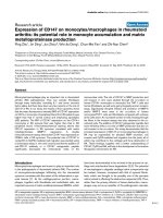
Figure 1
Flow cytometry analysis of CD147 expression on CD14+ cells of peripheral bloodFlow cytometry analysis of CD147 expression on CD14+ cells of peripheral blood. (a) CD14 (x-axis) and CD147 (y-axis) staining of leukocytes in
peripheral blood of RA patients. In CD14+ cells, the percentage of CD147 positive staining cells was 98.53 ± 2.09%. (b) Expression of CD147 on
CD14+ cells of peripheral blood in RA and normal control group. The black line shows the isotype control, the red and green lines shows anti-
CD147 antibody staining in the normal control and the RA group, respectively. The mean fluorescence intensity (MFI) of CD147 expression on
CD14+ cells was higher in the RA than in the normal control group.
Available online />R1027
rescent stained and then observed under a LSCM. CD68+/
CD147+ cells were observed in the lining layer and the sub-
lining layer of RA synovium (Fig. 3).
MMP secretion in co-culture of human monocytes/THP-
1 cells and fibroblasts
The gel zymography results show that MMP-9 and MMP-2
secretion increased significantly in the co-culture of isolated
human CD14+ monocytes and fibroblasts for both the RA and
normal control groups compared with the cultures of human
monocytes or fibroblasts alone (Fig. 4). In the RA group, the
CD14+ monocytes alone secreted more pro-MMP-9 and
MMP-9 than those in the normal control group.
To verify the above findings, we also used a PMA-induced cell
differentiation model of the human monocytic cell line THP-1
cells. THP-1 cells were used because of their high expression
of CD147 shown in previous reports [7] and our own experi-
ments (data not shown). The co-culture of undifferentiated
THP-1 cells and fibroblasts showed a significantly elevated
level of release and activation of MMP-2 and particularly MMP-
9 compared with the cultures of THP-1 cells or fibroblasts
alone (Fig. 5a). PMA-induced differentiated THP-1 cells also
secreted more pro-MMP-9 and MMP-9 compared with undif-
ferentiated THP-1 cells (Fig. 5b). Elevated secretion of pro-
MMP-2 and MMP-2 was observed in the co-culture of differen-
tiated THP-1 cells and fibroblasts (Fig. 5b).
Invasive ability of cells in co-culture of human
monocytes/THP-1 cells and fibroblasts
The functional cell invasion assay showed that the invasive
ability of human CD14+ monocytes in the RA group and nor-
mal control group were different. The number of human
CD14+ monocytes that invaded through the filter in the RA
group (858.3 ± 57.5) was higher than that in the normal con-
trol group (602.3 ± 126.7, p < 0.05). In the co-culture of
human monocytes and fibroblasts, the cell number in the RA
group (1235.7 ± 137.6) was also higher than that in the nor-
mal control group (918.3 ± 117.5, p < 0.05).
The pictures of filters show that the number of cells that
invaded through the filter in the co-culture of undifferentiated
THP-1 cells and fibroblasts was significantly increased com-
pared with the cultures of THP-1 cells or fibroblasts alone (p
< 0.05) (Fig. 6a). When the THP-1 cells were induced to dif-
ferentiate, the number of invading cells (865.7 ± 113.9) was
higher than that of undifferentiated THP-1 cells (478 ± 70.1, p
< 0.05), and the number of cells that invaded in the co-culture
of differentiated THP-1 cells and fibroblasts (1493.7 ± 417.5)
was also higher than that in the co-culture of undifferentiated
THP-1 cells and fibroblasts (1108.3 ± 73.4, p < 0.05).
Effect of AP9 on MMP release, activation and the
invasive ability of cells in co-culture of human
monocytes/THP-1 cells and fibroblasts
To identify the relationship between CD147 and the produc-
tion of MMPs, AP9 was added into the co-culture system of
human monocytes/THP-1 cells and fibroblasts and its effect
on MMP activity and the invasive ability of these cells was
observed. AP9 had some inhibitory effect on the secretion of
pro-MMP9 (control, 33.7 ± 18.5%; RA, 31.8 ± 16.2), MMP-9
(control, 41.9 ± 5.8%; RA, 43.8 ± 5.1%) and MMP-2 (control,
100%; RA, 63.9 ± 6.2%) in the co-culture of human mono-
cytes and fibroblasts in both the normal control and RA groups
(Fig. 4c).
A significantly reduced release (26.5 ± 11.7%) of MMP-9 in
the co-culture of undifferentiated THP-1 cells and fibroblasts
was observed in the AP9 group compared with that without
AP9 (p < 0.01; Fig. 5c). The pro-MMP-9, MMP-9 and MMP-2
secretion in the co-culture of differentiated THP-1 cells and
fibroblasts decreased significantly (27.5 ± 12.7%, 25.4 ±
12.4% and 25.5 ± 3.8%, respectively) in the AP9 group com-
pared with those without AP9 (p < 0.01; Fig. 5d).
In the cell invasion assays, the number of cells that invaded
through the Matrigel coated filter decreased following a 24 h
treatment with AP9 (200 µg/ml) in the co-culture cells (Fig. 6b,
c). AP9 inhibited cell invasion in the co-culture of human
monocytes and fibroblasts by 28.9 ± 5.9% in the normal con-
trol group and by 22.8 ± 3.8% in the RA group. AP9 inhibited
cell invasion by 55.1 ± 4.5% in the co-culture of
undifferentiated THP-1 cells and fibroblasts and by 44.1 ±
Table 1
Results of immunohistochemistry staining of the synovium of 12 rheumatoid arthritis patients
123456789101112
CD147+++++++++ ++++ ++++++
MMP-1+++ ++++ ++++ ++++++
TIMP-1++++ - ++++++ -
Positive intensity is indicated as: -, positive cell ratio <5%; +, ratio 5% to 50%; ++, ratio >50%. Spearmen's rho analysis showed that expression
of CD147 correlated significantly with matrix metalloproteinase-1 (MMP-1) expression (p < 0.05), but that the expression of CD147 and MMP-1
did not correlate with tissue inhibitors of metalloproteinases-1 (TIMP-1) expression (p > 0.05).
Arthritis Research & Therapy Vol 7 No 5 Zhu et al.
R1028
22.9% in the co-culture of differentiated THP-1 cells and
fibroblasts.
Chemoattraction of CyPA for peripheral mononuclear
cells in RA and its blockage by anti-CD147 antibody and
AP9
Based on the reports that CD147 is a high affinity receptor for
CyPA and responsible for a cyclophilin signaling cascade that
culminates in extracellular signal-regulated kinase (ERK) acti-
vation and chemotaxis [20,21], we examined the
chemoattraction of CyPA for peripheral mononuclear cells in
RA and the blockage effect of anti-CD147 antibody and AP9
on this. The optimum chemotaxis dose of CyPA was found to
be 100 ng/ml. The CyPA chemotactic index for peripheral
mononuclear cells in RA patients (350 ± 52% control) was
higher than that in the normal control group (252 ± 63% con-
trol, p < 0.05), indicating that CyPA had some significant
chemotactic effect on these cells (p < 0.05). The chemotactic
indexes decreased significantly when anti-CD147 antibody or
AP9 was added to the mononuclear cells in RA (120 ± 27%
and 150 ± 40% control, respectively; p < 0.01) (Fig. 7). The
blockage of CyPA chemoattraction for the mononuclear cells
by anti-CD147 antibody and AP9 was more obvious than that
caused by CsA (p < 0.05).
Discussion
Our results on the expression of CD147 on peripheral blood
monocytes in normal subjects are consistent with previous
studies demonstrating that CD147 is expressed on the sur-
face of activated monocytes [7,22-24]. The expression of
CD147 on normal inflammatory cells and many normal tissues
[25-27] is suggestive of its physiological role in some situa-
tions, perhaps in which increased MMP expression is involved
in tissue remodeling. In RA patients, however, we found that
the expression density of CD147 on monocytes from periph-
eral blood, as well as those from synovial fluid, was much
higher than normal. Following stimulation with IFN-γ, the
expression of CD147 on monocytes from normal subjects was
upregulated. But in vitro IFN-γ stimulation seemed to have little
effect on peripheral monocytes from RA patients, possibly
because of the already activated condition of RA monocytes
in vivo. These results indicate that the upregulation of CD147
Figure 2
Immunohistochemistry staining of synoviumImmunohistochemistry staining of synovium. The top four images show
the expression of CD147 (RA CD147), matrix metalloproteinase-1 (RA
MMP-1), tissue inhibitor of metalloproteinases-1 (RA TIMP-1) and blank
contrast (RA blank) in the lining and sublining layers in RA synovium (×
400). The bottom two images show the expression of CD147 in anky-
losing spondylitis (AS CD147) and osteoarthritis (OA CD147) syn-
ovium. CD147 was predominantly expressed by macrophage-like cells
and fibroblast-like synovial cells in the lining layer and partly by endothe-
lial cells in the rheumatoid arthritis (RA) synovium. The primary antibody
used for the blank contrast for streptavidin/peroxides staining was PBS
(× 400).
Figure 3
Laser scanning confocal microscope (× 400) image of the double immunofluorescent assayLaser scanning confocal microscope (× 400) image of the double
immunofluorescent assay. The expression of CD68+ (green) and
CD147+ (red) cells, and the co-expression of CD68+CD147+ cells
(yellow) in the lining and sublining layers of rheumatoid arthritis syn-
ovium is apparent.
Available online />R1029
expression on monocytes is possibly associated with the
pathogenesis of RA and CD147 may play an important role in
the cartilage and bone destruction of RA.
To confirm the expression of CD147 in synovium and to char-
acterize the CD147 expressing cells, immunohistochemical
staining and immunofluorescence were performed. The immu-
noreactivity of CD147 was more intense and more wide-
spread in RA synovium than in OA and AS synovium, and the
expression of CD147 correlated with MMP-1 expression.
These results are in part consistent with some previous reports
[12,13], but our immunohistochemical staining and immun-
ofluorescence results further confirmed that CD147 is
expressed in synovium not only on the fibroblast-like cells and
Figure 4
Matrix metalloproteinase (MMP) secretion in a co-culture of CD14+ monocytes and human fibroblast cells (HFC)Matrix metalloproteinase (MMP) secretion in a co-culture of CD14+ monocytes and human fibroblast cells (HFC). (a) Gel zymography detection of
MMP-2 and MMP-9 in human CD14+ monocytes from rheumatoid arthritis (RA) patients, in HFC and in a co-culture of the two. An elevated release
and activation of MMP-9 and MMP-2 was seen in the co-culture cells compared with those in the cultures of human monocytes or HFC alone. (b)
Gel zymography detection of MMP-2 and MMP-9 in human CD14+ monocytes of normal control monocytes, in HFC, and in a co-culture of the two.
MMP-2 and MMP-9 release was higher in the co-culture cells. (c) Effects of antagonistic peptide 9 (AP9) on pro-MMP-9, MMP-9 and MMP-2
release in a co-culture of human CD14+ monocytes and HFC. The zymography gels above were scanned and the gray density of strips was
counted. The release and activation of pro-MMP-9, MMP-9 and MMP-2 was significantly reduced when AP9 was added. Asterisks indicate a p-value
<0.01. HFC, Human fibroblast cells.
Arthritis Research & Therapy Vol 7 No 5 Zhu et al.
R1030
granulocytes, but also on the CD68+ macrophage-like cells,
which are believed to be peripheral monocyte-derived. As the
expression of CD147 is high in peripheral blood monocytes in
RA patients, it is highly possible that this will be maintained
and will stimulate MMP production after the monocytes infil-
trate into joints and differentiate into macrophage-like synovio-
cytes. The findings of Major et al. [28] that CD147 expressed
on differentiated monocytes and CD68-positive macrophages
in human atherosclerotic plaques also support our findings.
The upregulation of CD147 expression on monocytes/macro-
phages suggests that CD147 may be important in both the
autocrine and paracrine stimulation of MMP expression. It has
been shown that homophilic CD147-binding occurs in the
Figure 5
Matrix metalloproteinase (MMP) secretion in a co-culture of THP-1 cells and human fibroblast cells (HFC)Matrix metalloproteinase (MMP) secretion in a co-culture of THP-1 cells and human fibroblast cells (HFC). (a) Gel zymography detection of MMP-2
and MMP-9 in undifferentiated THP-1 cells, in HFC, and in a co-culture of the two. Elevated release and activation of MMP-2 and especially MMP-9
was seen in the co-culture cells compared to the cultures of THP-1 cells or HFC alone. (b) Gel zymography detection of MMP-2 and MMP-9 in phor-
bol myristate acetate (PMA)-induced differentiated THP-1 cells, in HFC, and in a co-culture of the two. Pro-MMP-2 and MMP-2 release was higher in
the co-cultured cells. PMA-induced differentiated THP-1 cells secreted more pro-MMP-9 and MMP-9 compared with undifferentiated THP-1 cells.
(c) Effects of CD147 antagonistic peptide 9 (AP9) on MMP-9 release in a co-culture of undifferentiated THP-1 cells and HFC. The zymography gels
above were scanned and the gray density of strips was counted. The release and activation of MMP-9 was significantly reduced when AP9 was
added. The asterisk indicates a p-value < 0.01. (d) Effects of AP9 on the release of pro-MMP-9, MMP-9 and MMP-2 in a co-culture of differentiated
THP-1 cells and HFC. Pro-MMP-9, MMP-9 and MMP-2 secretion all decreased significantly in the AP9 group. Asterisks indicate a p-value < 0.01.
HFC, Human fibroblast cells.
Available online />R1031
context of both heterotypic and homotypic cell-cell interac-
tions and that CD147 can itself be a receptor to induce MMP
production not only in primary fibroblast cells but also in tumor
cells [10]. On the basis of this, we presume that the increased
expression of CD147 on macrophages in RA synovium could
possibly induce MMP production through interaction with
surrounding fibroblast-like synoviocyts and also with other
macrophages. Our in vitro studies show that a co-culture of
human CD14+ monocytes/THP-1 cells and human fibroblasts
resulted in higher levels of MMP-2 and MMP-9 in culture
supernatants compared to human monocytes or fibroblasts
alone. This fibroblast triggered enhanced production could be
suppressed, however, by a peptide antagonistic to CD147/
HAb18G produced as described before [16-18]. (HAb18G is
abundantly expressed in human hepatoma tissues and on the
cell surface of several hepatoma cell lines with a highly meta-
static potential [18] and is a new member of CD147 family
[6,29].) The results of our cell invasion assay confirmed the
results of our gel zymography assay, which partly prove that
CD147 upregulation on macrophages may increase MMP
production through both autocrine and paracrine stimulation
and that macrophages may act as an amplifier of the pathoge-
netic cascade in RA via an increase in MMP production by
interacting macrophages and fibroblasts.
Bukrinsky and colleagues have reported that CD147 is a high
affinity receptor for CyPA and have demonstrated that it is
responsible for a cyclophilin signaling cascade that culminates
in ERK activation and chemotaxis [20,21]. It is also found that
CyPA, which belongs to the immunophilin family of peptidyl-
prolyl cis-trans isomerases [30-32], have chemotactic activity
towards eosinophils or neutrophils [33] and accumulate in
Figure 6
Effect of CD147 antagonistic peptide 9 (AP9) on the invasive ability of the co-culture cellsEffect of CD147 antagonistic peptide 9 (AP9) on the invasive ability of the co-culture cells. (a) The haematoxylin and eosin staining results of the
lower surface filters show that the cells had invaded through the filter and attached to the lower side of the filter (× 150). In the co-culture of undiffer-
entiated THP-1 cells and HFC, the number of cells was increased compared with the cultures of THP-1 cells or HFC alone. (b) Effect of AP9 on the
number of cells that invaded through the Matrigel coated filter in the co-culture of undifferentiated or differentiated THP-1 cells and HFC. The
number of cells that invaded through the filter was counted, showing that when AP9 was added the number of cells decreased. Asterisks indicate a
p-value <0.01. (c) Effect of AP9 on the number of cells that invaded through the Matrigel coated filter in a co-culture of human CD14+ monocytes
and HFC. The number of cells decreased when AP9 was added. Asterisks indicate a p-value < 0.05. HFC, Human fibroblast cells.
Arthritis Research & Therapy Vol 7 No 5 Zhu et al.
R1032
synovial fluids of patients with RA [34]. If CyPA is released into
the medium during inflammation, it is highly possible that it
could act as a ligand of CD147 to induce the accumulation of
inflammatory cells that highly express CD147 in the joints of
RA patients. The results of our in vitro chemotaxis assays con-
firm that CyPA has a chemotactic effect on peripheral blood
mononuclear cells. Moreover, we have also found that this
chemotaxis can be significantly suppressed by adding an anti-
CD147 monoclonal antibody or antagonists of CD147. This
suggests that CyPA does interact with CD147, although the
actual role of CyPA in CD147 function and in RA still needs to
be elucidated.
Conclusion
We conclude in this study that the increased expression of
CD147 on monocytes/macrophages in RA may be
responsible for elevated MMP secretion, cell invasion and the
CyPA-mediated cell migration into joints, all of which may con-
tribute to the cartilage and bone destruction of RA. These find-
ings, together with a better understanding of the relationship
between CD147, CyPA and RA and of the possible mecha-
nism and regulation of the effect of CD147 on MMP produc-
tion, will help in the development of innovative therapeutic
interventions for RA.
Competing interests
The authors declare that they have no competing interests.
Authors' contributions
PZ participated in the design of the study and drafted the man-
uscript. JD carried out the flow cytometry assay and the chem-
otaxis assay, performed the statistical analysis and helped to
draft the manuscript, and is one of the co-first authors. JZ per-
formed the invasion and gel zymography assays, and is one of
the co-first authors. WD participated in the immunohistochem-
istry staining and immunofluorescent assay, and is one of the
co-first authors. CF carried out the flow cytometry assays. ZC
participated in the design of the study and helped to draft the
manuscript, and is the corresponding author. All authors read
and approved the final manuscript.
Acknowledgements
This research was supported by grants from the National High Technol-
ogy Research and Development Program of China (863 Program)
(2001AA215061).
References
1. Tak PP, Smeets TJ, Daha MR, Kluin PM, Meijers KA, Brand R,
Meinders AE, Breedveld FC: Analysis of the synovial cell infil-
trate in early rheumatoid synovial tissue in relation to local dis-
ease activity. Arthritis Rheum 1997, 40:217-225.
2. Cunnane G, FitzGerald O, Hummel KM, Youssef PP, Gay RE, Gay
S, Bresnihan B: Synovial tissue protease gene expression and
joint erosions in early rheumatoid arthritis. Arthritis Rheum
2001, 44:1744-1753.
3. Konttinen YT, Ainola M, Valleala H, Ma J, Ida H, Mandelin J, Kinne
RW, Santavirta S, Sorsa T, Lopez-Otin C, Takagi M: Analysis of
16 different matrix metalloproteinases (MMP-1 to MMP-20) in
the synovial membrane: different profiles in trauma and rheu-
matoid arthritis. Ann Rheum Dis 1999, 58:691-697.
4. Pap T, Muller-Ladner U, Gay RE, Gay S: Fibroblast biology: role
of synovial fibroblasts in the pathogenesis of rheumatoid
arthritis. Arthritis Res 2000, 2:361-367.
5. Firestein GS: Invasive fibroblast-like synoviocytes in rheuma-
toid arthritis. Passive responders or transformed aggressors?
Arthritis Rheum 1996, 39:1781-1790.
6. Toole BP: Emmprin (CD147), a cell surface regulator of matrix
metalloproteinase production and function. Curr Top Dev Biol
2003, 54:371-389.
7. Kasinrerk W, Fiebiger E, Stefanova I, Baumruker T, Knapp W,
Stockinger H: Human leukocyte activation antigen M6, a mem-
ber of the Ig superfamily, is the species homologue of rat OX-
47, mouse basigin, and chicken HT7 molecule. J Immunol
1992, 149:847-854.
8. Suzuki S, Sato M, Senoo H, Ishikawa K: Direct cell-cell interac-
tion enhances pro-MMP-2 production and activation in co-cul-
ture of laryngeal cancer cells and fibroblasts: involvement of
EMMPRIN and MT1-MMP. Exp Cell Res 2004, 293:259-266.
9. Guo H, Zucker S, Gordon MK, Toole BP, Biswas C: Stimulation
of matrix metalloproteinase production by recombinant extra-
cellular matrix metalloproteinase inducer from transfected
Chinese hamster ovary cells. J Biol Chem 1997, 272:24-27.
10. Sun J, Hemler ME: Regulation of MMP-1 and MMP-2 production
through CD147/extracellular matrix metalloproteinase
inducer interactions. Cancer Res 2001, 61:2276-2281.
11. Stockinger H, Ebel T, Hansmann C, Koch C, Majdic O, Prager E,
Patel DD, Fox A, Horejsi V, Sagawa K, Shen D: Endothelial cell
antigens. In Leukocyte Typing VI. Part 8 Edited by: Kishimoto T,
Kikutani H, von dem Borne AEGK, Goyert SM, Mason DY, Miya-
saka M, Moretta L, Okumura K, Shaw S, Springer TA. New York:
Garland Publishing; 1997:760-763.
12. Konttinen YT, Li TF, Mandelin J, Liljestrom M, Sorsa T, Santavirta
S, Virtanen I: Increased expression of extracellular matrix met-
alloproteinase inducer in rheumatoid synovium. Arthritis
Rheum 2000, 43:275-280.
13. Tomita T, Nakase T, Kaneko M, Shi K, Takahi K, Ochi T, Yoshikawa
H: Expression of extracellular matrix metalloproteinase
inducer and enhancement of the production of matrix metallo-
proteinases in rheumatoid arthritis. Arthritis Rheum 2002,
46:373-378.
14. Arnett FC, Edworthy SM, Bloch DA, McShane DJ, Fries JF, Cooper
NS, Healey LA, Kaplan SR, Liang MH, Luthra HS: The American
Rheumatism Association 1987 revised criteria for the classifi-
cation of rheumatoid arthritis. Arthritis Rheum 1988,
31:315-324.
Figure 7
Effect of anti-CD147 antibody (Ab) and antagonistic peptide 9 (AP9) on the chemoattraction of cyclophilin A (CyPA)Effect of anti-CD147 antibody (Ab) and antagonistic peptide 9 (AP9)
on the chemoattraction of cyclophilin A (CyPA). The chemotactic
indexes decreased significantly when anti-CD147 antibody or AP9 was
added. CsA, cyclosporine A. C, CyPA. fmlp, n-Formyl-Met-Leu-Phe,
positive control.
Available online />R1033
15. Van der Linden S, Valkenburg HA, Cats A: Evaluation of diagnos-
tic criteria for ankylosing spondylitis. A proposal for modifica-
tion of the New York criteria. Arthritis Rheum 1984, 27:361-368.
16. Qian AR, Shang P, Luo ZQ, Huang BC, Chen ZN: Analyzing
HAb18G/CD147 antagonistic peptides using bioinformatics. J
Tumor Marker Oncol 2003, 18:29-36.
17. Jiang JL, Chan HC, Zhou Q, Yu MK, Yao XY, Lam SY, Zhu H, Ho
LS, Leung KM, Chen ZN: HAb18G/CD147-mediated calcium
mobilization and hepatoma metastasis require both C-termi-
nal and N-terminal domains. Cell Mol Life Sci 2004,
61:2083-2091.
18. Jiang JL, Zhou Q, Yu MK, Ho LS, Chen ZN, Chan HC: The involve-
ment of HAb18G/CD147 in regulation of store-operated cal-
cium entry and metastasis of human hepatoma cells. J Biol
Chem 2001, 276:46870-46877.
19. Tsuchiya S, Kobayashi Y, Goto Y, Okumura H, Nakae S, Konno T,
Tada K: Induction of maturation in culture human monocytic
leukemia cells by phorbol diester. Cancer Res 1982,
42:1530-1536.
20. Yurchenko V, Zybarth G, O'Connor M, Dai WW, Franchin G, Hao
T, Guo H, Hung HC, Toole B, Gallay P: Active-site residues of
cyclophilin A are crucial for its signaling activity via CD147. J
Biol Chem 2002, 277:22959-22965.
21. Pushkarsky T, Zybarth G, Dubrovsky L, Yurchenko V, Tang H, Guo
H, Toole B, Sherry B, Bukrinsky M: CD147 facilitates HIV-1 infec-
tion by interacting with virus-associated cyclophilin A. Proc
Natl Acad Sci USA 2001, 98:6360-6365.
22. Koch C, Staffler G, Huttinger R, Hilgert I, Prager E, Cerny J, Stein-
lein P, Majdic O, Horejsi V, Stockinger H: T cell activation-asso-
ciated epitopes of CD147 in regulation of the T cell response,
and their definition by antibody affinity and antigen density. Int
Immunol 1999, 11:777-786.
23. Nabeshima K, Suzumiya J, Nagano M, Ohshima K, Toole BP,
Tamura K, Iwasaki H, Kikuchi M: Emmprin, a cell surface inducer
of matrix metalloproteinases (MMPs), is expressed in T-cell
lymphomas. J Pathol 2004, 202:341-351.
24. Felzmann T, Gadd S, Majdic O, Maurer D, Petera P, Smolen J,
Knapp W: Analysis of function-associated receptor molecules
on peripheral blood and synovial fluid granulocytes from
patients with rheumatoid and reactive arthritis. J Clin immunol
1991, 11:205-212.
25. Marmorstein AD, Gan YC, Bonilha VL, Finnemann SC, Csaky KG,
Rodriguez-Boulan E: Apical polarity of N-CAM and EMMPRIN in
retinal pigment epithelium resulting from suppression of
basolateral signal recognition. J Cell Biol 1998, 142:697-710.
26. Noguchi Y, Sato T, Hirata M, Hara T, Ohama K, Ito A: Identifica-
tion and characterization of extracellular matrix metalloprotei-
nase inducer in human endometrium during the menstrual
cycle in vivo and in vitro. J Clin Endocrinol Metab 2003,
88:6063-6072.
27. DeCastro R, Zhang Y, Guo H, Kataoka H, Gordon MK, Toole B,
Biswas G: Human keratinocytes express EMMPRIN, an extra-
cellular matrix metalloproteinase inducer. J Invest Dermatol
1996, 106:1260-1265.
28. Major TC, Liang L, Lu XK, Rosebury W, Bocan TM: Extracellular
matrix metalloproteinase inducer (EMMPRIN) is induced upon
monocyte differentiation and is expressed in human
atheroma. Arterioscler Thromb Vasc Biol 2002, 22:1200-1207.
29. Gabison EE, Hoang-Xuan T, Mauviel A, Menashi S: EMMPRIN/
CD147, an MMP modulator in cancer, development and tissue
repair. Biochimie 2005, 87:361-368.
30. Fruman DA, Burakoff SJ, Bierer BE: Immunophilins in protein
folding and immunosuppression. FASEB J 1994, 8:391-400.
31. Liu J: FK506 and cyclosporin, molecular probes for studying
intracellular signal transduction. Immunol Today 1993,
14:290-295.
32. Sherry B, Yarlett N, Strupp A, Cerami A: Identification of cyclo-
philin as a proinflammatory secretory product of Lipopolysac-
charide-activated macrophages. Proc Natl Acad Sci USA 1992,
89:3511-3515.
33. Xu Q, Leiva MC, Fischkoff SA, Handschumacher RE, Lyttle CR:
Leukocyte chemotactic activity of cyclophilin. J Biol Chem
1992, 267:11968-11971.
34. Billich A, Winkler G, Aschauer H, Rot A, Peichl P: Presence of
cyclophilin A in synovial fluids of patients with rheumatoid
arthritis. J Exp Med 1997, 185:975-980.