Báo cáo y học: "Association between anti-nucleophosmin and anti-cardiolipin antibodies in (NZW × BXSB)F1 mice and human systemic lupus erythematosus" doc
Bạn đang xem bản rút gọn của tài liệu. Xem và tải ngay bản đầy đủ của tài liệu tại đây (649.2 KB, 10 trang )
Open Access
Available online />R1394
Vol 7 No 6
Research article
Association between anti-nucleophosmin and anti-cardiolipin
antibodies in (NZW × BXSB)F
1
mice and human systemic lupus
erythematosus
Aurelia Lartigue
1
, Laurent Drouot
1
, Fabienne Jouen
2
, Roland Charlionet
1
, François Tron
1,2
and
Danièle Gilbert
1,2
1
INSERM U519 and Institut Fédératif de Recherche Multidisciplinaire sur les Peptides, Faculté de Médecine et Pharmacie, 22 boulevard Gambetta,
76183 Rouen Cedex, France
2
Laboratoire d'Immunopathologie Clinique et Expérimentale, CHU de Rouen, 1 rue de Germont, 76000, Rouen cedex, France
Corresponding author: François Tron,
Received: 8 Jun 2005 Revisions requested: 18 Jul 2005 Revisions received: 11 Aug 2005 Accepted: 9 Sep 2005 Published: 13 Oct 2005
Arthritis Research & Therapy 2005, 7:R1394-R1403 (DOI 10.1186/ar1838)
This article is online at: />© 2005 Lartigue et al.; licensee BioMed Central Ltd.
This is an Open Access article distributed under the terms of the Creative Commons Attribution License ( />2.0), which permits unrestricted use, distribution, and reproduction in any medium, provided the original work is properly cited.
Abstract
We showed previously that nucleophosmin (NPM), a nucleolar
phosphoprotein, is recognized by sera from (NZW × BXSB)F
1
(WB) mice, a model of systemic lupus erythematosus (SLE) and
anti-phospholipid syndrome. In the present study we analysed
the prevalence and kinetics of anti-NPM autoantibodies in WB
mice by a solid-phase ELISA with recombinant human (rh) NPM
as the antigen and showed that most male WB mouse sera had
anti-NPM antibodies that were responsible for their indirect
immunofluorescence staining pattern on Hep-2 cells. Anti-NPM
antibodies were significantly associated with anti-cardiolipin
(aCL) antibodies. This antibody profile mirrored that observed in
certain human SLE sera because anti-NPM antibodies were
detected in 28% of the sera from patients with SLE and were
similarly associated with aCL antibodies. The demonstration
that rhNPM bound to cardiolipin (CL) in vitro and increased the
CL-binding activity of a WB-derived aCL monoclonal antibody
indicates that NPM can interact with CL to form SLE-related
immunogenic particles that might be responsible for the
concomitant production of anti-NPM and aCL antibodies.
Introduction
(NZW × BXSB)F
1
(WB) mice develop an autoimmune disease
whose histological and immunological manifestations resem-
ble those of human systemic lupus erythematosus (SLE) [1].
Male WB produce anti-nuclear antibodies (ANA), including
anti-deoxyribonucleic acid (DNA) autoantibodies and high lev-
els of anti-cardiolipin (aCL) antibodies that are thought to con-
tribute to the pathogenesis of myocardial infarction and
thrombocytopenia observed in these animals [2]. aCL antibod-
ies present in male WB mice require a plasma cofactor such
as β
2
-glycoprotein I (β
2
GPI), to bind to cardiolipin (CL) and
thus possess binding properties similar to those of aCL anti-
bodies observed in the serum of patients with SLE [3,4]. Male
WB mice are therefore considered an appropriate model for
the secondary anti-phospholipid syndrome associated with
SLE.
The precise nature of epitopes recognized by β
2
GPI-depend-
ent aCL antibodies remains a matter of debate. Some groups
consider that aCL antibodies do not recognize CL or β
2
GPI
alone but bind to either a CL-β
2
GPI complex [5] or cryptic
epitopes generated by their association [6]. Others think that
aCL antibodies bind to β
2
GPI in the absence of CL [7]. The
complexity of the interaction of anti-phospholipid antibodies
with their respective antigens is further illustrated by the dem-
onstration that β
2
GPI is not the unique cofactor involved in
their binding activity. Indeed, other phospholipid-binding pro-
teins have been described, such as prothrombin, protein C,
2D = two-dimensional; aCL = anti-cardiolipin; ANA = anti-nuclear antibodies; β
2
GPI = β
2
-glycoprotein I; BSA = bovine serum albumin; CL = cardi-
olipin; DTT = dithiothreitol; ELISA = enzyme-linked immunosorbent assay; FCS = fetal calf serum; mAb = monoclonal antibodies; MS = mass spec-
trometry; NPM = nucleophosmin; NTA = nitrilotriacetate; PBS = phosphate-buffered saline; PCR = polymerase chain reaction; rhNPM = recombinant
human nucleophosmin; RU = resonance units; SLE = systemic lupus erythematosus; WB = (NZW × BXSB)F
1
.
Arthritis Research & Therapy Vol 7 No 6 Lartigue et al.
R1395
protein S or annexin V, most of which participate in the coag-
ulation cascade [8,9].
We previously observed that WB mouse-derived monoclonal
antibodies (mAbs) selected for their capacities to react with
CL in the presence of FCS also reacted with nuclear antigens,
as shown by their nucleolar immunofluorescence-labelling pat-
tern on HEp-2 cells [10]. One of these mAbs, 4B7, reacted
with nucleophosmin (NPM; also known as B23), a nucleolar
protein involved in the assembly and transport of ribosomes
[11]. Subsequently, we showed by immunoscreening of a two-
dimensional (2D) PAGE-separated HL-60 cell protein map
with WB mouse sera and mass spectrometry (MS) that the
males of this strain mount an ordered autoimmune B-cell
response directed against various antigens, consistently
including NPM [12].
These observations led us to analyse further the prevalence
and kinetics of anti-NPM antibodies, their relationships with
aCL antibodies in WB mouse and human SLE sera and the
mechanisms that might account for the association between
these two antibody populations.
Materials and methods
Mice and sera
Female NZW and male BXSB mice were purchased, respec-
tively, from Bomholtgard Breeding and Research Center (Ry,
Denmark) and The Jackson Laboratory (Bar Harbor, ME,
USA), maintained in our animal facilities and crossbred to
obtain WB offspring. Control male CD1 mice were purchased
from Charles River Laboratories (Saint-Germain-sur-l'Arbresle,
France). All mice were housed in the same room and fed on
the same diet. Mice were bled every two months throughout
their lifetime. Sera were stored at -20°C until used. Sera from
(NZB × NZW)F
1
and MRL
lpr/lpr
mice were also tested. Mouse
studies were approved by the animal Ethical Committee of
Normandy (ceean number 1004-27).
Patients and sera
Serum samples were collected from 82 patients with SLE that
met the criteria of the American College of Rheumatology. The
serological profile of these patients was analysed for ANA by
indirect immunofluorescence using HEp-2 cells, anti-DNA
antibodies by the Farr assay and anti-β
2
GPI by ELISA (Varelisa
β
2
-glycoprotein I IgG antibody EIA kit; Pharmacia Diagnostics,
Freiburg, Germany). Serum samples were obtained from 103
healthy blood donors (agreement 31/10/2003). The study
was approved by the Ethical Committee of Haute Normandie
(number 99135HP).
Indirect immunofluorescence assay
The immunofluorescence pattern of sera from WB mice and
patients with SLE was determined on Hep-2 cells as
described previously [10]. Inhibition experiments were per-
formed by preincubating an anti-NPM mouse mAb (Invitrogen
immunodetection, San Francisco, CA, USA), mouse and
patient sera for 1 hour with 20 µg of recombinant NPM.
Generation of mAb 4B7
Splenocytes from a four-month-old male WB mouse were
fused with P3 × 63Ag8.653 myeloma cell line and the result-
ing hybridoma secreting mAb 4B7 was selected on the basis
of the capacity of its supernatant to react with CL in ELISA.
mAb 4B7 was purified as described elsewhere [10].
Production and purification of recombinant human NPM
in Escherichia coli
Total RNA was extracted from HL-60 cells by using TRIzol rea-
gent (Invitrogen, Eragny, France). The NPM cDNA was
obtained by transcription of oligo(dT) primer RNA with Molo-
ney murine leukaemia virus reverse transcriptase (Invitrogen).
An 879-base-pair DNA fragment was amplified from the cDNA
by PCR with the primers NPM-NdeI (5' -GGAATTCCATAT-
GGAAGATTCGATGGACATGGAC-3', sense) and NPM-
BamHI (5' -CCGGATCCTTACTTGTCATCGTCGTCCTTG-
TAGTCCGTACGAAGAGACTTCCTCCACTGCC-3', anti-
sense) designed to create a FLAG tag followed by a stop
codon and a BamHI site. The purified PCR product was
cloned into Nde-BamHI-digested pET-15b (Novagen, Madi-
son, WI, USA) downstream from the histidine tag. The result-
ing plasmid was transfected into E. coli BL21(DE3) pLysE
(Novagen, Darmstadt, Germany). Expression was induced by
incubation with isopropyl β-D-thiogalactoside (final concentra-
tion 1 mM) for 2.5 hours. E. coli pellets were suspended and
lysed in lysis buffer (50 mM NaH
2
PO
4
, 300 mM NaCl, 10 mM
imidazole, 1 mg/ml lysozyme and 1 mg/ml protease inhibitors,
pH 8). The suspension was sonicated at 40% intensity for 2.5
minutes (Vibra Cell; Bioblock Scientific, Illkirch, France) and
centrifuged at 7,000 g for 15 minutes at 4°C. The supernatant
was subjected to repeated aspiration and expulsion through a
fine needle to mechanically break DNA and then passed
through 0.22 µm filter. Batch purification was performed with
Ni
2+
-nitrilotriacetate (NTA)-Sepharose (Qiagen, Hilden, Ger-
many). The resin was washed with buffer (50 mM NaH
2
PO
4
, 1
M NaCl, 20 mM imidazole, 20% (v/v) ethanol, pH 8) until UV
absorbance at 280 nm became negative. The recombinant
human nucleophosmin (rhNPM) was eluted with 50 mM
NaH
2
PO
4
, 300 mM NaCl, 150 mM imidazole, pH 8. The
rhNPM-enriched fractions were dialysed against water and
then freeze-dried. This purification was performed for a sec-
ond time with the same protocol. The protein concentration
was determined with the Bradford protein assay kit (Bio-Rad
Laboratories Inc., Marnes-la-Coquette, France) and the yield
was about 2.5 mg per 150 ml of E. coli culture. Purity was con-
trolled by SDS-PAGE analysis with a 10% polyacrylamide gel
followed by western blotting with an anti-histidine-tag mAb
(Sigma-Aldrich Corp., St Louis, MO, USA). rhNPM was used
to detect anti-NPM antibodies, including those present in
mouse sera, because they were previously shown to bind
human NPM expressed by a human cell line [12] and there is
Available online />R1396
95% identity between human NPM and NO38, the murine
equivalent of human NPM.
ELISA for anti-NPM autoantibodies
High-binding plates (96-well, Microlon; Dutscher, Issy-les-
Moulineaux, France) were coated with 10 µg/ml purified
rhNPM in 0.05 M carbonate-bicarbonate buffer (pH 9.5) and
incubated overnight at 4°C. After being washed in PBS con-
taining 0.05% Tween, wells were blocked with PBS contain-
ing 5% (w/v) BSA for 2 hours at 23°C. Plates were washed
three times with 0.05% PBS-Tween, followed by the addition
to duplicate wells of mouse or human of sera, diluted 1:50 or
1:100, respectively, in diluting buffer (1% (w/v) BSA-PBS),
and then incubated for 2 hours at room temperature. After
being washed, biotin-conjugated goat anti-mouse IgG (Caltag
Laboratories, Hamburg, Germany) diluted 1:10,000 was
added, and incubated for 1 hour at room temperature. After
three washes, alkaline phosphatase-conjugated streptavidin
(1:10,000 dilution; Caltag Laboratories) was added and incu-
bated for 10 minutes at room temperature. After three washes,
plates were revealed with p-nitrophenyl phosphate (Sigma).
The absorbance at 405 nm (A
405
) was read. Horseradish per-
oxidase-conjugated goat anti-human IgG (Sigma) was added
and revealed with 3,3',5,5'-tetramethylbenzidine (Sigma). A
405
was read. Positive mouse sera were defined as those giving an
A
405
reading greater than the mean value plus 2 SD of sera
from 60 normal control CD1 male mice. Positive human sera
were defined as those giving an A
405
reading greater than the
mean value plus 3 SD of sera from 103 healthy controls.
ELISA for anti-CL antibodies
Each well of polystyrene microtitre plates (96-well; Luxlon,
Nemours, France) was coated with 50 µl of bovine heart CL
(10 µg/ml) in absolute alcohol. The plates were incubated
overnight at 4°C to allow the ethanol to evaporate. After block-
ing of non-specific binding sites by incubation with 10% FCS,
the plates were washed with PBS and incubated with sera
diluted in FCS. The plates were washed three times followed
by incubation with alkaline phosphatase-conjugated goat anti-
mouse IgG (Rockland, Gilbertsville, PA, USA) or with horse-
radish peroxidase-conjugated goat anti-human IgG. Reactivity
was determined as described above. Positive mouse sera
were defined as above, whereas positive human sera were
defined as those giving an A
405
reading of more than 20 immu-
noglobulin G phospholipid international units (GPLU).
NPM binding to CL
To determine whether NPM reacts with CL, CL-coated plates
were incubated with different rhNPM concentrations for 1
hour at room temperature. After blocking of non-specific bind-
ing sites with 0.5% gelatin, plates were washed three times
with PBS. Anti-histidine or anti-FLAG (Sigma-Aldrich) mAb
was added and incubated for 1 hour. After three washes, alka-
line phosphatase-conjugated goat anti-mouse IgG was added
and revealed with p-nitrophenyl phosphate.
Biacore analysis
The Biacore Biosensor system (Biacore, Uppsala, Sweden)
was used to study the interaction between rhNPM and CL.
Vesicles were prepared by drying 5 mg of CL under vacuum
and hydrating the lipid in 1 ml of PBS as described previously
[13]. The vesicles (400 µg/ml) were captured on the surface
of sensor chip L1, consisting of dextran modified with
lipophilic compounds. We successively injected into the sys-
tem 10 µl of 20 mM CHAPS, 150 µl of vesicles and 10 µl of
10 mM NaOH to stabilize the baseline, yielding about 4,000
to 5,000 resonance units (RU) of bound antigen. Then differ-
ent rhNPM concentrations (100 to 500 µg/ml) were injected
and the sensor chip was regenerated with 20 mM CHAPS.
Haemoglobin (Sigma) and human recombinant envoplakin
produced in our laboratory were used as controls. In a second
series of experiments, rhNPM was immobilized on the surface
of sensor chip NTA, allowing the binding of histidine-tagged
proteins. In brief, we injected 20 µl of 500 µM NiCl
2
and then
40 µl of 200 µg/ml NPM, which yielded about 1,500 RU of
antigen. The CL vesicles (400 µg/ml) were injected and the
sensor chip was regenerated with 350 mM EDTA.
Effect of rhNPM on aCL-binding activity
To examine the effect of rhNPM on aCL-binding activity, puri-
fied 4B7 mAb (0.8 mg/ml) was incubated on CL-coated plates
in the presence or absence of NPM, in an ELISA as described
above.
Preparation of HL-60 cell-protein extract for 2D PAGE
Human promyelocytic leukaemia cell line (American Type Cul-
ture Collection, Manassas, VA, USA) was grown at 37°C in a
humidified atmosphere (95% air, 5% CO
2
) in 50 ml of RPMI
1640 (Invitrogen, Eragny, France), supplemented with 10%
FCS Sigma), antibiotics (Invitrogen) and 1 mM sodium pyru-
vate (Sigma). Cells were washed three times with PBS and
isolated by centrifugation at 15,000 g for 5 minutes at room
temperature. HL-60 cells (2 × 10
8
) were suspended in 10%
(v/v) trichloroacetic acid, 0.12% (w/v) dithiothreitol (DTT) and
stored overnight at -20°C. Cells were centrifuged at 4°C for
30 minutes at 15,000 g. Cell pellets were suspended again in
0.12% (w/v) DTT and kept at -20°C for 1 hour before being
centrifuged. Dry pellets were suspended in lysis buffer (9 M
urea, 2% (w/v) CHAPS, 1% (w/v) DTT, 2% (v/v) protease
inhibitors (Sigma)) and then centrifuged at 4°C for 20 minutes
at 1,500 g. The protein content was determined with the Plu-
sOne 2D Quant kit (Amersham, Buckinghamshire, UK).
This lysate was subjected to 2D PAGE as previously
described [12]. The immunoreactive spots were detected with
human sera, diluted 1:100. After being washed, membranes
were incubated with alkaline phosphatase-conjugated goat
anti-human IgG (Amersham) and revealed with Nitro Blue
Tetrazolium salt and 5-bromo-4-chloroindol-3-yl phosphate
substrate (Roche, Meylan, France).
Arthritis Research & Therapy Vol 7 No 6 Lartigue et al.
R1397
Protein identification
The immunoreactive spots were excised from polyacrylamide
gels with Ettan Spot Picker (Amersham) and digested by
trypsin (proteomics grade; Sigma) with Ettan Digester (Amer-
sham). Samples were analysed by matrix-assisted laser des-
orption/ionization-time-of-flight MS to obtain peptide mass
information. Spectra obtained were compared with those reg-
istered in protein databases (SWISS-PROT and NCBInr).
Data were matched against the databases with the use of the
MS-Fit program (accessible through ProteinProspector).
Statistical analysis
Absorbances of sera obtained from male and female WB mice
of the same age were compared with the Mann-Whitney U-
test. The percentages of different antibodies in the sera of
patients with SLE were compared by using a χ
2
test. The rela-
tionship between the titres of different antibodies in mouse
sera was evaluated with Spearman's correlation test.
Results
Male WB mouse sera frequently react with NPM
To determine the prevalence of anti-NPM antibodies, sera col-
lected from normal CD1 (n = 54) and WB lupus-prone mice
at different times of life were analysed by solid-phase ELISA
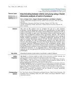
using rhNPM. As shown in Fig. 1a, anti-NPM antibodies were
present early in life in male WB mice and were detected in
more than 75% of sera from animals more than three months
old. Anti-NPM antibodies appeared later in female WB mice
(25% at 3 months), were less frequent (40% at 4 to 6 months)
and gave low A
405
values. The A
405
of male WB sera were sig-
nificantly higher than those of control mouse sera at each
period (P = 0.01) and tested dilution (Fig. 1b), whereas female
WB A
405
values differed significantly from normal mouse sera
only at four months (P = 0.03). All anti-NPM antibody-positive
mouse sera, tested by immunoblotting on 2D PAGE-sepa-
rated HL-60 cell-protein map, bound to the native human
NPM, thereby confirming their previously reported reactivity
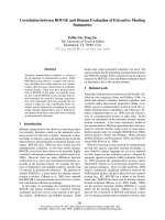
with this nucleolar autoantigen [12]. Anti-NPM positive mouse
sera gave a nucleolar staining pattern on Hep-2 cells by indi-
rect immunofluorescence analysis similar to that observed
with an anti-NPM mouse mAb (Fig. 2a,c). Preincubation of the
mAb and mouse sera with recombinant NPM abrogated the
nucleolar staining (Fig. 2b,d).
NPM-binding activity in other mouse strains
We then tested WB parental strains, female NZW and male
BXSB mice, but no reactivity against NPM was observed. Sim-
ilarly, offspring of NZB and NZW mice also did not develop
anti-NPM antibodies. In contrast, anti-NPM antibodies were
present in 60% of 20 sera from MRL
lpr/lpr
mice more than three
months old.
Anti-NPM and aCL antibodies are associated in male WB
mice
Because WB lupus-prone mice are also characterized by aCL
antibody production, we looked for an association between
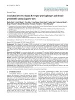
anti-NPM and aCL antibodies in their sera. Indeed, anti-NPM
and aCL antibodies were found to be associated in sera from
three-month-old male WB mice (χ
2
= 18.14; P < 0.0001), and
their A
405
values were positively correlated (r = 0.750; P <
0.0001; Fig. 3a). Similarly, A
405
values of anti-NPM and aCL
antibodies in sera from MRL
lpr/lpr
mice more than three months
old were positively correlated (r = 0.585; P < 0.01; Fig. 3b).
Figure 1
Nucleophosmin-binding activity of WB lupus-prone and control mouse seraNucleophosmin-binding activity of WB lupus-prone and control mouse
sera. (a) Sera from male (◆) and female (■) WB mice, and from male
CD1 (▲) mice, collected during months 1 to 2, 2 to 3, 3 to 4 and 4 to
6, were tested in solid-phase ELISA. The horizontal dotted line repre-
sents the cutoff value (mean A
405
of controls + 2 SD). Sera with A
405
<
0.522 were considered negative. (* P = 0.03, ** P = 0.01, *** P =
0.0001; Mann-Whitney test). (b) Different dilutions of male (◆) and
female (■) WB and male (▲) CD1 mouse sera collected at 3.5 months
were tested in ELISA.
Available online />R1398
Anti-NPM antibodies are present in patients with SLE
and are associated with aCL antibodies
The demonstration that anti-NPM antibodies are frequently
produced in male WB mice prompted us to search for anti-
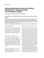
NPM antibodies in the sera of 82 patients with SLE. Indeed,
23 (28%) of these sera were positive for anti-NPM antibody
(Fig. 4) and the antibodies seemed to be more frequent
(although not significantly) in males than in females (5/9 ver-
sus 18/73; Table 1). These anti-NPM antibody-positive SLE
sera were analysed by immunoblotting on HL-60 cell protein
separated by 2D PAGE (Fig. 5a); most of them (80%) reacted
consistently with a spot of molecular mass (36 kDa) and pI
(4.5) that had the same coordinates as that recognized by
male WB mice and was characteristic of NPM (Fig. 5c). The
immunoreactive spots bound by human and mouse sera were
excised and analysed by matrix-assisted laser desorption/ioni-
zation-time-of-flight MS and corresponded to NPM (Fig. 5b, d).
Anti-NPM antibody-positive SLE sera yielded homogeneous
nuclear and nucleolar staining on Hep-2 cells by indirect
immunofluorescence analysis (Fig. 2e). The nucleolar staining
disappeared when sera were preincubated with recombinant
NPM (Fig. 2f).
aCL antibodies were detected in 32 (39%) of the sera from
patients with SLE. Pertinently, as in WB mice, the analysis of
the distribution of anti-NPM and aCL antibodies in these SLE
sera (Table 1) indicates that these autoantibodies were asso-
ciated (χ
2
= 9.2; P = 0.002). In contrast, ANA, anti-DNA and
anti-β
2
GPI antibodies rates did not differ significantly between
anti-NPM-positive and anti-NPM-negative patients. Interest-
ingly, of the 23 anti-NPM-positive sera, 18 (78%) did not react
with β
2
GPI; conversely, of the 12 anti-β
2
GPI-aCL-positive
Figure 2
Immunofluorescence staining pattern of anti-nucleophosmin-positive WB mouse and SLE sera on HEp-2 cellsImmunofluorescence staining pattern of anti-nucleophosmin-positive
WB mouse and SLE sera on HEp-2 cells. An anti-nucleophosmin (anti-
NPM) mouse mAb and anti-NPM antibody-positive mouse sera gave a
nucleolar pattern (a,c). Anti-NPM-antibody-positive systemic lupus ery-
thematosus (SLE) sera yielded homogeneous nuclear and nucleolar
staining (e). The nucleolar staining was abrogated when the mAb and
sera from mouse (b,d) or patient (f) were previously incubated with 10
µg of recombinant human nucleophosmin.
Figure 3
Correlation between anti-nucleophosmin and anti-cardiolipin antibodies in lupus miceCorrelation between anti-nucleophosmin and anti-cardiolipin antibodies
in lupus mice. A positive correlation between anti-nucleophosmin and
anti-cardiolipin antibody reactivities in 38 sera from male WB mice (a)
and 20 sera from MRL
lpr/lpr
mice (b) more than 3 months old.
Arthritis Research & Therapy Vol 7 No 6 Lartigue et al.
R1399
sera, only three were positive for anti-NPM, suggesting that
the presence of these two autoantibody populations is mutu-
ally exclusive.
NPM interacts in vitro with CL
The demonstration that anti-NPM and aCL antibodies were
associated in WB mice and certain patients with SLE was
reminiscent of the previously described association between
anti-β
2
GPI and aCL antibodies; it led us to ask whether NPM
could bind to CL. CL-coated plates were incubated with
increasing concentrations of rhNPM, which was revealed with
an anti-histidine or anti-FLAG mAb. Figure 6a shows that NPM
bound to CL-coated wells in a dose-dependent manner. This
binding was confirmed by Biacore analysis. CL vesicles were
captured on the surface of sensor chip L1 and NPM was
injected at a concentration of 400 µg/ml. Figure 7 shows that
CL vesicles bound to NPM (Fig. 7a, with a difference of 3,550
RU) but did not bind to irrelevant proteins (haemoglobin and
envoplakin; Fig. 7b). Similarly, NPM captured on an NTA sen-
sor chip bound CL vesicles, giving a response of 2,885 RU
(Fig. 7c).
NPM increases the CL binding of a murine mAb
The demonstration that NPM could bind to CL prompted us to
study the effect of rhNPM on the CL-binding activity of purified
4B7, an aCL mAb derived from a WB mouse whose serum
contained both aCL and anti-NPM antibodies [10]. Indeed, in
ELISA, 4B7 reactivity with CL increased markedly in the pres-
ence of NPM, which therefore acted as a cofactor (Fig. 6b).
Similarly, with the use of sensor chip L1 coated with CL vesi-
cles or CL vesicles plus NPM, the binding of 4B7 to the chip
was enhanced 1.5-fold (2,578 versus 3,989 RU; Fig. 8).
Discussion
NPM (B23) is an abundant nucleolar phosphoprotein with
multiple functions: the assembly and/or transport of ribosomes
[11], chaperone activities [14] and a regulatory role in cell pro-
liferation [15,16]. NPM was previously shown to be targeted
by antibodies produced in patients with either non-organ-spe-
cific autoimmune diseases [17-19] or cancer, namely hepato-
cellular and breast carcinoma [20,21].
Figure 4
Nucleophosmin-binding activity of sera from patients with SLENucleophosmin-binding activity of sera from patients with SLE. (a) Sera
from patients with systemic lupus erythematosus (SLE) (◆) or from
healthy controls (-) were analysed by ELISA using recombinant human
nucleophosmin. The dotted line represents the cutoff value (mean A
405
of controls + 3 SD). Sera giving an A
405
< 0.286 were considered neg-
ative. (b) Different dilutions of serum from an SLE patient (◆) and a
control (■) were tested in ELISA.
Table 1
Demographic and serological profiles of patients with SLE
Characteristic SLE patients Anti-NPM
antibodies
(n = 82) Positive (n = 23) Negative (n = 59)
Age, years
(mean ± SD)
38.3 ± 12.3 39.8 ± 14.9 37.8 ± 11.5
Females 73 (89.0) 18* 55
Males 9 (10.8) 5* 4
aCL 32 (39) 15 (65.2)** 17 (28.8)**
ANA-positive 66 (80.5) 23 (100) 43 (72.8)
Anti-DNA 46 (56) 13 (56.5) 33 (55.9)
Anti-β
2
GPI 15 (18.3) 5 (21.7) 10 (16.9)
Anti-β
2
GPI-CL+ 12 (14.6) 3 (13) 9 (15.2)
aCL, anti-cardiolipin; ANA, anti-nuclear antibodies; β
2
GPI, β
2
-
glycoprotein I; CL, cardiolipin; NPM, nucleophosmin; SLE, systemic
lupus erythematosus. Values are n (%) unless indicated otherwise.
* χ
2
= 2.4; P = 0.12. ** χ
2
= 9.2; P = 0.002.
Available online />R1400
By using a sensitive ELISA with rhNPM as the antigen, we
showed that anti-NPM antibodies are present in most male
WB lupus-prone mice and are therefore a constant feature of
the antibody response in these animals: anti-NPM positivity
appeared early in life, increased with age, and peaked at three
to four months, before death. In contrast to our previous obser-
vations obtained by immunoblot analysis of 2D gel-separated
NPM [12], this sensitive ELISA enabled us to detect these
antibodies in female WB mice too, although later, less fre-
quently and initially at lower A
405
values. Pertinently, anti-NPM
antibodies in male WB mice were consistently associated with
aCL, and both antibody populations appeared concomitantly,
as shown by sequential analyses of WB sera. Anti-NPM anti-
bodies could also be detected in MRL
lpr/lpr
mice and again
were significantly associated with aCL antibodies, which are
frequently produced by this lupus mouse strain. This antibody
pattern mirrored that observed in certain human SLE sera.
Indeed, anti-NPM antibodies were present in 28% of our 82
patients with SLE; they were more frequent in males and were
significantly associated with aCL antibodies. The association
of anti-NPM and aCL antibodies was first suggested by Li and
colleagues [17]. Indeed, in their analysis of 164 sera obtained
from patients with various autoimmune diseases selected by
the presence of anti-nucleolar antibodies, 6 had anti-NPM and
aCL antibodies and SLE. However, our results, showing that
anti-NPM antibodies are constantly detected in WB mice and
frequently observed in patients with SLE, demonstrate that
anti-NPM antibodies constitute a frequent and new marker in
mouse and human lupus, establish a clear relationship
between anti-NPM and aCL antibodies and finally define a
subset of patients with aCL antibodies.
The association of aCL and anti-NPM antibodies in WB lupus-
prone mice and patients with SLE might be explained by two
different mechanisms: first, cross-reactivity due to the expres-
sion of a shared epitope by CL and NPM, or second, the ability
of NPM to interact with CL to form an immunogenic complex
able to induce the two antibody populations and/or a unique
antibody population able to react with both NPM and CL (dual
reactivity), as reported for lupus-related antigen particles [22-
24]. The former hypothesis is not supported by the presence
of anti-NPM-positive/aCL-negative sera and, conversely, aCL-
Figure 5
Immunoblot analyses of sera positive for anti-nucleophosmin antibodyImmunoblot analyses of sera positive for anti-nucleophosmin antibody. (a,c) Anti-nucleophosmin antibody obtained respectively from a patient with
systemic lupus erythematosus (SLE) (a) and a male WB mouse (c) using a 2D PAGE-separated HL-60 cell protein map as the substrate. (b,d) Mass
spectra of the proteins bound by the SLE (b) and mouse (d) serum.
Arthritis Research & Therapy Vol 7 No 6 Lartigue et al.
R1401
positive/anti-NPM-negative sera and by the demonstration
that anti-NPM antibodies induced by immunization of normal
mice do not react with CL (data not shown); this conclusion
was also reached by Li and colleagues [17], who showed that
affinity-purified anti-NPM antibodies from SLE sera did not
bind to CL. We therefore tested the second hypothesis, which
proposed that lupus-related antigens are made of physically
linked epitopes, such as DNA-histones [22] or Sm-DNA
[23,24] and implies that NPM is able to bind to CL and behave
as an aCL antibody cofactor. Plasmon resonance analysis of
Figure 6
Binding of rhNPM to cardiolipin and effects of nucleophosmin on cardi-olipin-binding activity of 4B7 mAbBinding of rhNPM to cardiolipin and effects of nucleophosmin on cardi-
olipin-binding activity of 4B7 mAb. (a) Cardiolipin was coated on plas-
tic plates and incubated with various concentrations of recombinant
human nucleophosmin (rhNPM). (b) mAb 4B7 was incubated on cardi-
olipin-coated plates in the absence or presence of rhNPM. Bars repre-
sent the mean A
405
of duplicate experiments and error bars represent
SD.
Figure 7
Biacore analysis of recombinant human nucleophosmin (rhNPM)-cardi-olipin interactionBiacore analysis of recombinant human nucleophosmin (rhNPM)-cardi-
olipin interaction. (a,b) Cardiolipin vesicles were captured on the sur-
face of the sensor chip L1 and rhNPM (a), haemoglobin or envoplakin
(b) were injected. (c) rhNPM was immobilized on the surface of the
Ni
2+
-nitrilotriacetate sensor chip and cardiolipin vesicles were injected.
Available online />R1402
rhNPM-CL interaction clearly showed that NPM binds to CL in
vitro to form complexes. This binding might be attributed to the
functional N-terminal domain, which contains a high density of
hydrophobic residues involved in chaperone activity [25]. The
same technology enabled us to show that the reactivity of 4B7
mAb to NPM-CL complexes was markedly enhanced in com-
parison with its reactivity to either NPM or CL alone,
suggesting that 4B7 is representative of mAbs exhibiting a
dual specificity similar to that previously reported for certain
anti-histone murine mAbs, whose binding activity is increased
by DNA [22,26]. In human SLE sera, such autoantibodies
probably exist but their identification remains elusive because
of the polyclonal nature of sera, which may simultaneously
contain monoreactive anti-NPM and anti-CL antibodies.
These results could also lead us to consider that NPM acts as
an aCL cofactor, at least in this murine model of lupus. So far,
several cofactors of anti-phospholipid antibodies have been
described; most of them are soluble proteins involved in coag-
ulation [27,28]. We have showed here that a nuclear autoan-
tigen can behave as an aCL cofactor. This observation raises
important questions: is NPM the unique nuclear autoantigen
acting as an aCL cofactor? When and where does NPM inter-
act with CL to form an immunogenic complex able to initiate
antibody responses to both proteins? NPM is an abundant
nuclear protein [29] that can translocate to the cytoplasm
[30]. Recently, it was found to be localized in the cell
membrane and to be a component of the fructose lysine-spe-
cific receptor expressed by monocyte-like cell lines [31]. Thus,
NPM could well interact with anionic phospholipids at the cell
membrane to form a typical lupus-related immunogenic com-
plex, which could be released. Experiments are under way to
determine the cellular localization of NPM in different catego-
ries of cells and during various cellular processes, such as
apoptosis, which is thought to have a major role in the break-
age of B cell tolerance in SLE [32]. The answer to these ques-
tions will help to explain the high frequency of anti-
phospholipid antibodies in patients with SLE and to identify
other nuclear autoantigens able to behave as an aCL cofactor.
Another important objective is to precisely define the sensitiv-
ity and specificity of anti-NPM antibodies for lupus and to
determine whether their presence is correlated with disease
activity or clinical manifestations in patients with SLE. Such an
analysis is under way in a large series of patients with SLE and
various autoimmune diseases and will help to clarify the dis-
ease significance of this autoantibody population.
Conclusion
In this study we show that anti-NPM antibodies constitute a
frequent marker in WB and MRL
lpr/lpr
lupus-prone mice and in
human SLE, establish a clear relationship between anti-NPM
and aCL antibodies and define a subset of patients with aCL
antibodies. The demonstration that NPM binds to CL in vitro
and increases the CL-binding activity of a WB-derived aCL
mAb indicates that NPM can interact with CL to form SLE-
related immunogenic particles that might be responsible for
the concomitant production of anti-NPM and aCL antibodies.
Competing interests
The authors declare that they have no competing interests.
Authors' contributions
AL participated in the design of the study, performed the
immunoanalysis and wrote the manuscript. LD participated in
recombinant protein synthesis. FB provided sera from
patients. RC performed mass spectrometry analysis. FT partic-
ipated in the design of the study and wrote the manuscript. DG
performed Biacore analysis, participated in the design of the
study and wrote the manuscript. All authors read and
approved the final manuscript.
Acknowledgements
This study was supported by INSERM.
Figure 8
Biacore analysis of binding of 4B7 mAb to cardiolipin-nucleophosmin complexesBiacore analysis of binding of 4B7 mAb to cardiolipin-nucleophosmin
complexes. Cardiolipin vesicles were captured on the surface of sensor
chip L1. Binding of purified 4B7 mAb to cardiolipin vesicles alone (a) or
after the injection of recombinant human nucleophosmin (b).
Arthritis Research & Therapy Vol 7 No 6 Lartigue et al.
R1403
References
1. Hang LM, Izui S, Dixon FJ: (NZW × BXSB)F
1
hybrid. A model of
acute lupus and coronary vascular disease with myocardial
infarction. J Exp Med 1981, 154:216-221.
2. Koike T: Anticardiolipin antibodies in NZW × BXSB F
1
mice.
Lupus 1994, 3:241-246.
3. Hashimoto Y, Kawamura M, Ichikawa K, Suzuki T, Sumida T, Yosh-
ida S, Matsuura E, Ikehara S, Koike T: Anticardiolipin antibodies
in NZW × BXSB F
1
mice. A model of antiphospholipid
syndrome. J Immunol 1992, 149:1063-1068.
4. Gharavi AE, Cucurull E, Tang H, Silver RM, Branch DN: Effect of
antiphospholipid antibodies on beta(2) glycoprotein I-phos-
pholipid Interaction. Am J Reprod Immunol 1998, 39:310-315.
5. McNeil HP, Simpson RJ, Chesterman CN, Krilis SA: Anti-phos-
pholipid antibodies are directed against a complex antigen
that includes a lipid-binding inhibitor of coagulation: beta 2-
glycoprotein I (apolipoprotein H). Proc Natl Acad Sci USA
1990, 87:4120-4124.
6. Pengo Y, Biasiolo A, Fior MG: Autoimmune antiphospholipid
antibodies are directed against a cryptic epitope expressed
when beta 2-glycoprotein I is bound to a suitable surface.
Thromb Haemost 1995, 73:29-34.
7. Roubey RA, Eisenberg RA, Harper MF, Winfield JB: 'Anticardioli-
pin' autoantibodies recognize beta 2-glycoprotein I in the
absence of phospholipid. Importance of Ag density and biva-
lent binding. J Immunol 1995, 154:954-960.
8. Roubey RA: Immunology of the antiphospholipid antibody
syndrome. Arthritis Rheum 1996, 39:1444-1454.
9. Alarcon-Segovia D, Cabral AR: The concept and classification of
anti-phospholipid/cofactor syndromes. Lupus 1996,
5:364-367.
10. Thébault S, Gilbert D, Machour N, Marvin L, Lange C, Tron F, Char-
lionet R: Two-dimensional electrophoresis and mass spec-
trometry identification of proteins bound by a murine
monoclonal anti-cardiolipin antibody: a powerful technique to
characterize the cross-reactivity of a single autoantibody.
Electrophoresis 2000, 21:2531-2539.
11. Savkur RS, Olson MO: Preferential cleavage in pre-ribosomal
RNA by protein B23 endoribonuclease. Nucleic Acids Res
1998, 26:4508-4515.
12. Thébault S, Gilbert D, Hubert M, Drouot L, Machour N, Lange C,
Tron F: Orderly pattern of development of the autoantibody
response in (New Zealand White × BXSB)F
1
lupus mice: char-
acterization of target antigens and antigen spreading by two-
dimensional gel electrophoresis and mass spectrometry. J
Immunol 2002, 169:4046-4053.
13. Haruta K, Kobayashi S, Hirose S, Horiai A, Ohyanagi M, Tanaka M,
Kawano T, Shirai T, Takasaki Y, Hashimoto H: Monoclonal anti-
cardiolipin antibodies from New Zealand Black x New Zealand
White F
1
mice react to thrombomodulin. J Immunol 1998,
160:253-258.
14. Okuwaki M, Matsumoto K, Tsujimoto M, Nagata K: Function of
nucleophosmin/B23, a nucleolar acidic protein, as a histone
chaperone. FEBS Lett 2001, 506:272-276.
15. Feuerstein N, Chan PK, Mond JJ: Identification of numatrin, the
nuclear matrix protein associated with induction of mitogene-
sis, as the nucleolar protein B23: implication for the role of the
nucleolus in early transduction of mitogenic signals. J Biol
Chem 1988, 263:10608-10612.
16. Jiang PS, Yung BY: Down-regulation of nucleophosmin/B23
mRNA delays the entry of cells into mitosis. Biochem Biophys
Res Commun 1999, 257:865-870.
17. Li XZ, McNeilage LJ, Whittingham S: Autoantibodies to the
major nucleolar phosphoprotein B23 define a novel subset of
patients with anticardiolipin antibodies. Arthritis Rheum 1989,
32:1165-1169.
18. Pfeifle J, Anderer FA, Franke M: Characterisation of nucleolar
proteins as autoantigens using human autoimmune sera. Ann
Rheum Dis 1986, 45:978-986.
19. Ulanet DB, Wigley FM, Gelber AC, Rosen A: Autoantibodies
against B23, a nucleolar phosphoprotein, occur in sclero-
derma and are associated with pulmonary hypertension.
Arthritis Rheum 2003, 49:85-92.
20. Imai H, Ochs R, Kiyosawa K, Furuta S, Nakamura R, Tan EM:
Nucleolar antigens and autoantibodies in hepatocellular carci-
noma and other malignancies. Am J Pathol 1992,
140:859-870.
21. Brankin B, Skaar TC, Brotzman M, Trock B, Clarke R: Autoanti-
bodies to the nuclear phosphoprotein nucleophosmin in
breast cancer patients. Cancer Epidemiol Biomarkers Prev
1998, 7:1109-1115.
22. Losman MJ, Fasy TM, Novick KE, Monestier M: Monoclonal
autoantibodies to subnucleosomes from a MRL/Mp
-
+/+
mouse. Oligoclonality of the antibody response and recogni-
tion of a determinant composed of histones H2A, H2B and
DNA. J Immunol 1992, 148:1561-1569.
23. Retter MW, Eisenberg RA, Cohen PL, Clarke SH: Sm and DNA
binding by dual reactive B cells requires distinct VH, V kappa,
and VH CDR3 structures. J Immunol 1995, 155:2248-2257.
24. Retter MW, Cohen PL, Eisenberg RA, Clarke SH: Both Sm and
DNA are selecting antigens in the anti-Sm B cell response in
autoimmune MRL/lpr mice. J Immunol 1996, 156:1296-1306.
25. Hingorani K, Szebeni A, Olson MOJ: Mapping the functional
domains of nucleolar protein B23. J Biol Chem 2000,
275:24451-24457.
26. Losman MJ, Fasy TM, Novick KE, Monestier M: Relationships
among antinuclear antibodies from autoimmune MRL mice
reacting with histone H2A-H2B dimers and DNA. Int Immunol
1993, 5:513-523.
27. Galli M, Beretta G, Daldossi M, Bevers EM, Barbui T: Different
anticoagulant and immunological properties of anti-pro-
thrombin antibodies in patients with antiphospholipid
antibodies. Thromb Haemost 1997, 77:486-491.
28. Galli M, Comfurius P, Barbui T, Zwaal RF, Bevers EM: Anticoagu-
lant activity of beta 2-glycoprotein I is potentiated by a distinct
subgroup of anticardiolipin antibodies. Thromb Haemost 1992,
68:297-300.
29. Pasquali JL, Azerad G, Martin T, Muller S: The double reactivity
of a human monoclonal rheumatoid factor to IgG and histones
is related to distinct binding sites. Eur J Immunol 1988,
18:1127-1130.
30. Borer RA, Lehner CF, Eppenberger HM, Nigg EA: Major nucleolar
proteins shuttle between nucleus and cytoplasm. Cell 1989,
56:379-390.
31. Brandt R, Nawka M, Kellermann J, Salazar R, Becher D, Krantz S:
Nucleophosmin is a component of the fructoselysine-specific
receptor in cell membranes of Mono Mac 6 and U937 mono-
cyte-like cells. Biochim Biophys Acta 2004, 1670:132-136.
32. Casciola-Rosen L, Rosen A, Petri M, Schlissel M: Surface blebs
on apoptotic cells are sites of enhanced procoagulant activity:
implications for coagulation events and antigenic spread in
systemic lupus erythematosus. Proc Natl Acad Sci USA 1996,
93:1624-1629.