Báo cáo y học: "Between adaptive and innate immunity: TLR4-mediated perforin production by CD28null T-helper cells in ankylosing spondylitis" pptx
Bạn đang xem bản rút gọn của tài liệu. Xem và tải ngay bản đầy đủ của tài liệu tại đây (631.71 KB, 9 trang )
Open Access
Available online />R1412
Vol 7 No 6
Research article
Between adaptive and innate immunity: TLR4-mediated perforin
production by CD28
null
T-helper cells in ankylosing spondylitis
Bernd Raffeiner
1
*, Christian Dejaco
1
*, Christina Duftner
1
, Werner Kullich
2
, Christian Goldberger
1
,
Sandra C Vega
3
, Michael Keller
3
, Beatrix Grubeck-Loebenstein
3
and Michael Schirmer
1
1
Department of Internal Medicine, Innsbruck Medical University, Austria
2
Ludwig Boltzmann Institute for Rehabilitation of Internal Diseases, Saalfelden, Austria
3
Institute for Biomedical Aging Research, Austrian Academy of Science, Austria
* Contributed equally
Corresponding author: Michael Schirmer,
Received: 21 Apr 2005 Revisions requested: 14 Jun 2005 Revisions received: 26 Aug 2005 Accepted: 27 Sep 2005 Published: 18 Oct 2005
Arthritis Research & Therapy 2005, 7:R1412-R1420 (DOI 10.1186/ar1840)
This article is online at: />© 2005 Raffeiner et al.; licensee BioMed Central Ltd.
This is an Open Access article distributed under the terms of the Creative Commons Attribution License ( />2.0), which permits unrestricted use, distribution, and reproduction in any medium, provided the original work is properly cited.
Abstract
CD3
+
CD4
+
CD28
null
and CD3
+
CD8
+
CD28
null
T cells are
enriched in patients with immune-mediated diseases compared
with healthy controls. This study shows that CD4
+
CD28
null
T
cells express Toll-like receptors recognizing bacterial
lipopolysaccharides in ankylosing spondylitis, psoriatic arthritis
and rheumatoid arthritis. In ankylosing spondylitis, TLR4 (23.1 ±
21.9%) and, to a smaller extent, TLR2 (4.1 ± 5.8%) were
expressed on CD4
+
CD28
null
T cells, whereas expression was
negligible on CD4
+
CD28
+
and CD8
+
T cells. CD4
+
CD28
null
T
cells produced perforin upon stimulation with
lipopolysaccharide, and this effect was enhanced by autologous
serum or recombinant soluble CD14. Perforin production could
be prevented with blocking antibodies directed against CD14 or
TLR4. Incubation of peripheral blood mononuclear cells with
tumour necrosis factor alpha led to an upregulation of TLR4 and
TLR2 on CD4
+
CD28
null
T cells in vitro, and treatment of patients
with antibodies specifically directed against tumour necrosis
factor alpha resulted in decreased expression of TLR4 and
TLR2 on CD4
+
CD28
null
T cells in vivo. We describe here a new
pathway for direct activation of cytotoxic CD4
+
T cells by
components of infectious pathogens. This finding supports the
hypothesis that CD4
+
CD28
null
T cells represent an
immunological link between the innate immune system and the
adaptive immune system.
Introduction
Pattern recognition receptors (PRRs) are a family of receptors
of the innate immune system binding to conserved pathogen-
associated molecular patterns [1]. The most important PRRs
are the Toll-like receptors (TLRs), which allow monocytes,
neutrophils, dendritic cells, natural killer (NK) cells and B cells
to recognize bacterial components, viruses, fungi and host
material such as heat shock proteins [2-5]. Receptor engage-
ment leads to the translocation of NF-κB and to gene tran-
scription of proinflammatory cytokines. Lipopolysaccharide
(LPS) from Gram-negative bacteria is the main ligand for
TLR4. LPS binding to TLR4 is promoted by CD14, which can
be present either in a membrane-bound form (mCD14) or a
soluble form (sCD14) [6,7]. Whether LPS is a low-affinity lig-
and for TLR2 is still controversial [8].
The interactions between the innate and adaptive immune sys-
tems are crucial to promote proinflammatory reactions against
pathogens and to ensure maintenance of vital self-tolerance.
TLRs are expressed on both innate and adaptive immune cells
and are critically involved in this interplay. TLR-stimulated den-
dritic cells induce specific T cells to differentiate into memory
cells [9,10], and microbial induction of the TLR pathway on
dendritic cells also blocks the suppressive effects of regula-
tory T cells [11,12]. In addition, TLRs are themselves
expressed on T cells. In murine T cells, LPS signalling induces
AS = ankylosing spondylitis; ELISA = enzyme-linked immunosorbent assay; FCS = foetal calf serum; IFN-γ = interferon gamma; IL = interleukin; LPS
= lipopolysaccharide; mAb = monoclonal antibody; mCD14 = membrane-bound CD14; NF = nuclear factor; NK = natural killer; PBMC = peripheral
blood mononuclear cell; PBS = phosphate-buffered saline; PCR = polymerase chain reaction; PRR = pattern recognition receptor; PsA = psoriatic
arthritis; RA = rheumatoid arthritis; RT = reverse transcriptase; sCD14 = soluble CD14; TCR = T-cell receptor; TLR = Toll-like receptor; TNF-α =
tumour necrosis factor alpha.
Arthritis Research & Therapy Vol 7 No 6 Raffeiner et al.
R1413
production of INF-γ and T-helper-1 accentuated immune
responses [13,14]. After in vitro activation, mouse CD4
+
T
cells express TLR3 and TLR9, and treatment of these cells
with synthetic ligands for TLR3 and TLR9, viral dsRNA and
bacterial unmethylated DNA enhances their survival [15]. LPS
can directly activate regulatory T cells, and can thereby
increase their suppressive function [16]. Activated human T
cells express high levels of cell-surface TLR2 and produce ele-
vated levels of cytokines in response to bacterial lipopeptide,
a TLR2 ligand [17].
TLRs have also been shown to be important for the pathogen-
esis of immune-mediated diseases. In rheumatoid arthritis
(RA), for example, TLR2 expression occurs in inflamed synovial
tissue predominantly at sites of attachment and invasion into
the cartilage and bone [18]. The TLR2-mediated stimulation of
synovial fibroblasts with bacterial components promotes the
release of proinflammatory cytokines and leads to a higher
expression of TLR2.
In ankylosing spondylitis (AS), as in other immune-mediated
diseases, an unusual proinflammatory and cytotoxic CD4
+
T-
cell subgroup has been described, which lacks the co-stimu-
latory molecule CD28. These CD4
+
CD28
null
T cells depend
on alternative pathways for coactivation and can indeed obtain
such signals by NK receptors recognizing ubiquitous major
histocompatibility complex class I molecules [19-23]. Anoma-
lous expression of NKG2D on these T cells together with
upregulated MIC ligands in the inflamed synovial tissue of RA
have also been shown to provide co-stimulatory signals [24].
CD4
+
CD28
null
T cells have therefore been considered an
immunological link between the adaptive and the innate
defence system [25].
As CD4
+
CD28
null
T cells have NK cell features, and as NK
cells express TLRs on their surface [4], we investigated the
expression of TLRs as an alternative stimulatory pathway on
CD4
+
CD28
null
T cells.
Patients and methods
A total of 90 consecutive patients with spondyloarthropathies,
72 patients with RA and 64 age-matched healthy controls
were enrolled into the study. Spondyloarthropathy was
defined according to the European Spondyloarthropathy
Study Group criteria [26]. Out of the spondyloarthropathy-
defined patients, 65 patients had a diagnosis of AS according
to the modified New York criteria [27] and 25 patients had
psoriatic arthritis (PsA) as defined by the diagnostic criteria of
Moll and Wright [28]. RA was diagnosed according to the cri-
teria of the American College of Rheumatology [29]. Probands
did not show any history, clinical or laboratory sign for infec-
tions nor malignant diseases. Healthy controls had no history
of an immune-mediated disease. Heparinized blood samples
were drawn from peripheral veins after informed and written
consent according to the local ethics committee.
Patients' characteristics (including age, sex, the presence of
rheumatoid factor and anti-cyclic citrullinated peptide antibod-
ies, HLA-B27 status, axial involvement, erythrocyte sedimenta-
tion rate and C-reactive protein) are summarized in Table 1.
Nine of the AS patients were treated with antibodies directed
against tumour necrosis factor alpha (TNF-α) (infliximab =
Remicade
®
; Aesca, Vienna, Austria) at a dosage of 3 mg/kg
body weight.
Cell preparation and surface staining
Peripheral blood mononuclear cells (PBMCs) were isolated by
Ficoll density gradient centrifugation. For surface staining,
PBMCs were incubated, as appropriate, with fluorescein iso-
thiocyanate-conjugated anti-CD4, anti-CD8 or anti-CD28,
with phycoerythrin-conjugated anti-CD28 and with peridinin
chlorophyll protein-conjugated anti-CD3, anti-CD4 and anti-
CD8 monoclonal antibodies (Becton Dickinson, San Diego,
CA, USA). Specific mAbs directed against CD14 (fluorescein
isothiocyanate), TLR4 and TLR2 (phycoerythrin; eBioscience,
San Diego, CA, USA) were used to analyse expression of
PRRs. Cells were incubated for 30 minutes at 4°C with the
Table 1
Patients' characteristics
Controls
(n = 64)
Ankylosing spondylitis
(n = 65)
Psoriatic arthritis
(n = 25)
Rheumatoid arthritis
(n = 72)
Age (years) 49.2 ± 12.4 43.1 ± 10.9 52.2 ± 12 57.9 ± 11
Gender (% female) 50.8 28.4 45.8 75.6
Rheumatoid factor positivity (%) 0.0 n.d. n.d. 77.8
CCP-Ab positivity (%) 0.0 n.d. n.d. 80.5
HLA-B27 positivity (%) n.d. 78.6 31.3 n.d.
Axial involvement (% positive) 0.0 100.0 48.0 0.0
Erythrocyte sedimentation rate >15 mm/hour (% positive) 0.0 62.7 88.2 86.2
C-reactive protein >0.7 µg/dl (% positive) 0.0 26.1 41.7 46.8
CCP-Ab, antibodies directed against cyclic citrullinated peptides; n.d., not determined.
Available online />R1414
antibodies. After washing with PBS, cells were fixed in 4%
paraformaldehyde (cellfix; Becton Dickinson). Stained cells
were analysed on a FACS-Calibur analyser (Becton Dickin-
son). At least 100,000 events were counted for each acquisi-
tion. Data were analysed using WinMDI software (version 2.5,
Joseph Trotter; Scripps Research Institute, La Jolla, CA, USA).
Cells were considered positively stained when fluorescence
levels were higher than those of the corresponding isotype
controls.
For the isolation of CD4
+
CD28
+
and CD4
+
CD28
null
T cells,
the MACS
®
CD4
+
T-cell multisort kit and magnetic bead
labelled anti-phycoerythrin antibodies were applied according
to the manufacturer's instructions (Miltenyi Biotech, Amster-
dam, The Netherlands). To further increase the purity of T cells,
sorted fractions were incubated in 24-well plates for 2 hours
at 37°C to allow adherence of contaminating monocytes.
Purity of isolated fractions was determined by flow cytometry
as already described.
Reverse transcription-polymerase chain reaction
Total RNA was extracted from isolated cell fractions using 1 ml
Tri-Reagent (Sigma-Aldrich, St Louis, MO, USA), 200 µl chlo-
roform (Sigma-Aldrich), 0.5 ml isopropanol (Sigma-Aldrich)
and 1 µl glycogen (Roche, Basel, Switzerland) as previously
described [30]. RNA was reverse transcribed to cDNA apply-
ing the Reverse Transcription System (Promega, Madison, WI,
USA). PCR amplification was performed on 1 µg total RNA
and 10 pmol primers (TLR2: forward, 5'-GGCCAGCAAAT-
TACCTGTGTG-3'; reverse, 5'-CTGAGCCTCGTCCAT-
GGGCCACTCC-3'; TLR4: forward, 5'-
TGCAATGGATCAAGGACCAGAGGC-3'; reverse, 5'-
GTGCTGGGACACCACAACAATCACC-3'; and β
2
-
microglobulin: forward, 5'-CTCCGTGGCCTTAGCTGTG-3';
reverse, 5'-TTTGGAGTACGCTGGATAGCC-3') using the
HotStar Master Mix Kit (Qiagen Valencia, CA, USA). The PCR
reaction was performed according to a modified protocol [31]
on the PTC-100 Thermal Cycler (MJ Research, Waltham, MA,
USA) at 95°C for 3 minutes, at 95°C for 1 minute, 58°C for 2
minute and 72°C for 1 minute (35 cycles), and at 72°C for 2
minutes. Negative control samples were prepared by amplifi-
cation reactions in the absence of cDNA. PCR products were
separated on 1% agarose gel containing ethidium bromide
and were visualized by UV illumination.
LPS studies and intracellular cytokine staining
Isolated PBMCs were resuspended in RPMI 1640 containing
2 mM L-glutamine and were distributed on 24-well plates at a
density of 1 × 10
6
cells per well. LPS of Escherichia coli sero-
type 026:B6 (Sigma) was added at a concentration of 10 µg/
ml [16,17], sCD14 (R&D systems, Minneapolis, MN, USA) at
25 µg/ml and autologous serum at a concentration of 5% as
indicated.
To test inhibitory effects of blocking anti-CD14 or anti-TLR4
antibodies, cells were resuspended in RPMI 1640 with 5%
autologous serum and were incubated with 10 µg/ml blocking
anti-CD14 antibody (R&D Systems), 10 µg/ml anti-TLR4 anti-
body (Torrey Pines Biolabs, TX, USA) and 10 µg/ml isotype
control antibody (Becton Dickinson) for 1 hour before adding
LPS at a concentration of 10 µg/ml. Brefeldin A (Sigma) was
added to functional experiments at a concentration of 10 µg/
ml to avoid release of produced cytokines. After incubation for
16 hours at 37°C the cells were washed, stained with CD4-
peridinin chlorophyll protein mAbs and CD28-phycoerythrin
mAbs and were permeabilized with 0.05% Tween 20 to stain
intracellularly with fluorescein isothiocyanate-conjugated anti-
perforin mAbs or control immunoglobulin (Becton Dickinson).
Stimulation of short-term cell lines
Short-term cell lines were established after incubation of fresh
PBMCs on stimulating immobilized anti-CD3 mAbs (OKT3;
eBioscience) for 18 hours. Cells were then cultured in densi-
ties of 0.5 × 10
6
– 2 × 10
6
in RPMI 1640 containing 10%
FCS, 2 mM L-glutamine and 20 U/ml recombinant human IL-2
(Sigma). The medium was changed every 2–3 days. Cell lines
were used after 7 days for stimulation assays over 24 hours
with 20 ng/ml recombinant human TNF-α (Prepotech, London,
UK).
Enzyme-linked immunosorbent assay
Soluble levels of CD14 in blinded sera were tested in dupli-
cates with an ELISA kit (R&D systems) according to the man-
ufacturer's instructions.
Statistical analysis
Statistical analysis was performed using the SPSS program
(version 11.5; SPSS Inc., Chicago, IL, USA). The Kol-
mogorov–Smirnov test was used to test for normal distribu-
tion, and the Mann–Whitney U test and the Wilcoxon test
were used as appropriate. At least six assays were performed
for each experiment. P < 0.05 was considered significant and
P < 0.01 was highly significant. Data are shown as box plots
with the lines within the boxes representing the median, the
boxes representing the 25th–75th percentiles and the lines
outside the boxes including all values except mavericks.
Results
Comparison between the prevalences of circulating
CD4
+
CD28
null
and CD8
+
CD28
null
T cells in AS, PsA, RA and
healthy controls
Peripheral blood from patients with AS, PsA and RA showed
an increased prevalence of CD3
+
CD4
+
CD28
null
T cells and
CD3
+
CD8
+
CD28
null
T cells compared with age-matched
healthy controls. One representative example of a three-colour
flow cytometry analysis showing the prevalence of
CD3
+
CD4
+
CD28
null
and CD3
+
CD8
+
CD28
null
T cells is shown
in Figure 1a. The mean percentages of CD3
+
CD4
+
CD28
null
T
cells out of CD3
+
CD4
+
T cells were 5.1 ± 9.8%, 5.1 ± 6.8%,
Arthritis Research & Therapy Vol 7 No 6 Raffeiner et al.
R1415
4.6 ± 5.2% and 1.5 ± 4.5% for AS, PsA, RA and healthy con-
trols (each with P < 0.001), respectively. The mean percent-
ages of CD3
+
CD8
+
CD28
null
T cells out of CD3
+
CD8
+
T cells
were 38.4 ± 21.9% in AS, 44.4 ± 21.7% in PsA and 46 ±
26.2% in RA, compared with 22.3 ± 12.4% in the control
group (each with P < 0.001, Figure 1b).
Increased expression of pattern recognition receptors
on CD4
+
CD28
null
T cells
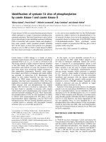
To test T cells for the expression of TLR2 and TLR4 mRNA,
monocyte-depleted CD4
+
CD28
null
and CD4
+
CD28
+
T cells
were isolated from patients as well as from healthy controls
(CD4
+
CD28
+
T cells). As shown in Figure 2a, purity was high
for both with the CD3
+
CD4
+
CD28
null
and CD3
+
CD4
+
CD28
+
fractions ranging between 94.2% and 99.7%. A representa-
tive example out of three independent RT-PCR experiments is
depicted in Figure 2b: CD4
+
CD28
null
T cells express TLR2
and TLR4 mRNA, whereas TLR4 transcripts were not
detected in CD4
+
CD28
+
T cells from patients and healthy
controls. In contrast, variable but significant levels of TLR2
mRNA were present in the CD3
+
CD4
+
CD28
+
population
even from healthy individuals.
To address PRR surface expression on T cells, PBMCs from
patients as well as from healthy controls were incubated with
mAbs against CD4, CD14, CD28, TLR4 and TLR2, and were
analysed by flow cytometry. As shown in Figure 3a, lym-
phocytes were gated on the forward and side scatter and spe-
cific gates were set to focus CD4
+
CD28
null
and CD4
+
CD28
+
T cells for the analysis of TLR expression.
Overall, all analysed PRRs (CD14, TLR4 and TLR2) were
detected on the surface of CD4
+
CD28
null
T cells but not on
CD28
+
T cells irrespective of the underlying diseases tested.
CD14 was expressed on 13.3 ± 20.4% of CD4
+
CD28
null
T
cells versus 0.7 ± 1% of CD4
+
CD28
+
T cells in AS, on 8.8 ±
15.7% of CD4
+
CD28
null
T cells versus 1.0 ± 2.3% of
CD4
+
CD28
+
T cells in PsA, and on 11.3 ± 17.2% of
CD4
+
CD28
null
T cells versus 0.6 ± 0.6% of CD4
+
CD28
+
T
cells in RA (each with P < 0.001). TLR-4, the main receptor for
LPS recognition, was significantly expressed on
CD4
+
CD28
null
T cells in AS (23.1 ± 21.9% versus 0.9 ±
1.2%), in PsA (12.4 ± 18.1% versus 0.4 ± 0.5%) and in RA
(23.1 ± 24.7% versus 0.6 ± 0.9%; each with P < 0.001).
TLR2 was more frequently expressed on CD4
+
CD28
null
T cells
than on CD28
+
cells (4.1 ± 5.8% versus 1.0 ± 1.1%, P <
0.001) in AS, but not in PsA or in RA (Figure 3b). Negligible
surface expression of PRRs without any difference between
CD28
null
and CD28
+
T cells were seen on CD8
+
T cells from
patients (data not shown). CD4
+
CD28
+
, CD8
+
CD28
+
and
CD8
+
CD28
null
T cells from healthy controls showed no signif-
icant expression of PRRs. CD4
+
CD28
null
T cells from healthy
controls expressed PRR to some extent but, as the prevalence
of this subpopulation is low, no significant data could be
acquired (data not shown).
LPS-mediated perforin production of CD4
+
CD28
null
T
cells depends on CD14 and TLR4
For functional testing of TLR-mediated lymphocytic stimula-
tion, fresh PBMCs were incubated for 16 hours with LPS and
Figure 1
Prevalence of circulating CD3
+
CD4
+
CD28
null
and CD3
+
CD8
+
CD28
null
cells in ankylosing spondylitis, psoriatic arthritis and rheumatoid arthritisPrevalence of circulating CD3
+
CD4
+
CD28
null
and CD3
+
CD8
+
CD28
null
cells in ankylosing spondylitis, psoriatic arthritis and rheumatoid arthri-
tis. (a) Representative example showing the prevalence of
CD3
+
CD4
+
CD28
null
T cells (percentage out of C3
+
CD4
+
) and
CD3
+
CD8
+
CD28
null
T cells (percentage out of C3
+
CD8
+
). The histo-
gram shows staining for CD3 (filled grey curve) and isotype control
antibody (black line). Dot plots are then gated on CD3
+
cells. (b) Box
plots summarize the prevalence of CD3
+
CD4
+
CD28
null
and
CD3
+
CD8
+
CD28
null
T cells in ankylosing spondylitis (AS), psoriatic
arthritis (PsA) and rheumatoid arthritis (RA) patients. The Mann-Whit-
ney U test was used to determine the statistical differences between
patients and the age-matched healthy control group (CO). ***P <
0.001.
Figure 2
Messenger RNA expression of TLR2 and TLR4 in CD3
+
CD4
+
CD28
null
and CD3
+
CD4
+
CD28
+
T cellsMessenger RNA expression of TLR2 and TLR4 in CD3
+
CD4
+
CD28
null
and CD3
+
CD4
+
CD28
+
T cells. (a) Fluorescence-activated cell sorting
analysis shows the purity of CD3
+
CD4
+
CD28
null
and
CD3
+
CD4
+
CD28
+
T cells. (b) mRNA expression of TLR2, TLR4 and
β2-microglobulin (β2m, housekeeping gene) in CD3
+
CD4
+
CD28
null
T
cells (CD28
-
) and in CD3
+
CD4
+
CD28
+
T cells (CD28
+
). Peripheral
blood mononuclear cells were used as positive control (pos co), and a
negative control (neg co) was performed in the absence of cDNA. A
representative example out of three independent experiments is given.
Available online />R1416
the T cells were analysed for their intracellular production of
perforin. As shown in Figure 4a,b, perforin was produced upon
LPS stimulation by CD4
+
CD28
null
T cells (13 ± 10.7% per-
forin
+
cells), but perforin expression was negligible in
CD4
+
CD28
+
T cells (0.4% ± 0.3% perforin
+
cells, P = 0.009).
Combining recombinant sCD14 with LPS doubled the per-
centage of perforin
+
CD4
+
CD28
null
T cells (24 ± 15.3% per-
forin
+
cells) compared with LPS stimulation alone (P =
0.0001). A comparable additional effect was seen when cul-
tures stimulated with LPS were supplemented with 5% autol-
ogous serum (26.9 ± 16.6% versus 13 ± 10.7% perforin
+
cells, P = 0.001) but not with FCS (data not shown).
CD4
+
CD28
+
T cells did not produce perforin after co-incuba-
tion with LPS, even after addition of sCD14 or autologous
serum (Figure 4a and data not shown).
These findings implicated the possible occurrence of natural
sCD14 in sera from AS patients. Blinded samples from
patients with AS and from healthy controls were therefore ana-
lysed using enzyme-linked immunoassays. As shown in Figure
5a, the levels of sCD14 were higher in AS patients than in
healthy controls (1,653.6 ± 463 pg/ml versus 1,170 ± 259
pg/ml, P = 0.008). To investigate whether sCD14 from autol-
ogous serum was crucial for a stronger response of
CD4
+
CD28
null
T cells to LPS, cells were incubated with a
blocking antibody directed against CD14 prior to the addition
of LPS. This antibody is capable of binding both sCD14 and
mCD14. As expected, LPS-induced perforin production of
CD4
+
CD28
null
T cells was reversed by blocking sCD14 and
mCD14 (24.0 ± 16.3% versus 3.1 ± 2.5% perforin
+
cells, P =
0.006) but not by isotype control antibody (Figure 5b).
Blocking assays with antibodies directed against TLR4 were
then performed to specifically address the role of TLR4 in
LPS-mediated perforin production of CD4
+
CD28
null
T cells.
As shown in Figure 5c, preincubation with anti-TLR4 antibody
inhibited activation of CD4
+
CD28
null
T cells by LPS (3.4 ±
2.3% with anti-TLR4 antibody versus 23.8 ± 14.7% perforin
+
cells with isotype control antibody, P = 0.002).
Figure 3
Surface expression of CD14, TLR4 and TLR2 on CD4
+
CD28
+
and CD28
null
cells in ankylosing spondylitis, psoriatic arthritis and rheuma-toid arthritisSurface expression of CD14, TLR4 and TLR2 on CD4
+
CD28
+
and
CD28
null
cells in ankylosing spondylitis, psoriatic arthritis and rheuma-
toid arthritis. (a) Representative dot plots and histograms show TLR4
expression (filled red curve, black line represents isotype control) on
CD4
+
CD28
+
and CD4
+
CD28
null
T cells. Gates were set on lym-
phocytes (forward scatter and sideward scatter) as well as on CD28
+
and CD28
null
cells expressing high levels of CD4. (b) Box plots summa-
rize the expression of CD14, TLR4 and TLR2 on CD4
+
CD28
+
and
CD28
null
T cells in patients as indicated. The Wilcoxon test was used to
determine the statistical differences between the groups. ***P < 0.001.
SSC, side scatter; FSC, forward scatter; AS, ankylosing spondylitis;
PsA, psoriatic arthritis; RA, rheumatoid arthritis.
Figure 4
Effects of LPS, sCD14 and autologous serum on perforin production by CD4
+
T cellsEffects of LPS, sCD14 and autologous serum on perforin production
by CD4
+
T cells. Fresh peripheral blood mononuclear cells of patients
with ankylosing spondylitis were incubated with medium (as a negative
control), soluble CD14 (sCD14) or 10 µg/ml lipopolysaccharide (LPS)
alone or with LPS in combination with 25 µg/ml sCD14 and 5% autolo-
gous serum for 16 hours. After staining with fluorescence-marked mon-
oclonal antibodies directed against perforin, CD4 and CD28, cells
were counted by flow cytometry. (a) Histograms show perforin produc-
tion by CD4
+
CD28
+
(upper row) and CD4
+
CD28
null
T cells (lower row)
in response to medium, sCD14, LPS, LPS + sCD14 and LPS + serum
as indicated (red curves). Black lines represent isotype control staining.
Values indicate the mean fluorescence intensity. Gates for
CD4
+
CD28
null
and CD4
+
CD28
+
T cells were set as shown in Figure
3a. (b) Box blots show percentages of perforin-producing
CD4
+
CD28
null
T cells from seven independent experiments. Differ-
ences were tested for significance using the Wilcoxon test. ***P ≤
0.001.
Arthritis Research & Therapy Vol 7 No 6 Raffeiner et al.
R1417
Effects of TNF-α in vitro and therapeutic blockade of
TNF-α in vivo on PRR expression of CD4
+
CD28
null
T cells
In vitro assays were performed to test the effect of TNF-α on
the expression of PRRs. Incubation of PBMCs with TNF-α for
24 hours increased the expression of TLR4 and TLR2, but not
of CD14 on CD4
+
CD28
null
T cells (from 9.2 ± 25.8% to 26.6
± 27.7% for TLR4, P < 0.001 and from 1.1 ± 3.1% to 2.4 ±
4.1% for TLR2, P = 0.008; Figure 6a). Expression of CD14,
TLR4 and TLR2 were neither induced on CD4
+
CD28
+
T cells
nor on CD8
+
T cells after incubation with TNF-α (data not
shown).
To examine the effects of TNF-α blocking treatment on the
expression of PRRs on CD4
+
CD28
null
T cells in vivo, periph-
eral CD4
+
CD28
null
T cells from AS patients were tested
before and after successful treatment with TNF-α-specific chi-
meric antibodies. As shown in Figure 6b, CD4
+
CD28
null
T
cells from patients during active AS disease showed higher
levels of PRRs than after successful treatment with the TNF-α
blocking agent. CD14 was reduced from 10.6 ± 16.6%
before treatment to 2.5 ± 2% after treatment (P = 0.011),
TLR4 was reduced from 46.9 ± 32.7% to 11.7 ± 12.5% (P =
0.008) and TLR2 was reduced from 11.7 ± 19.4% to 1.8 ±
2.9% (P = 0.012).
Discussion
The present study shows an increased expression of PRRs on
human circulating CD4
+
T cells lacking the CD28 co-stimula-
tory molecule. TLR4 can be considered an alternative
signalling pathway for cytotoxic CD4
+
CD28
null
T cells, but nei-
ther for their CD28
+
counterparts nor for CD8
+
T cells. The
concomitant expression of T-cell receptor (TCR) and PRRs on
the cell surface further supports the role of CD4
+
CD28
null
T
cells as an immunological link between the adaptive and the
innate defence system, and is in accordance with earlier
descriptions of co-existing NK receptors and TCR on these
cells [25]. In all chronic immune diseases tested (AS, PsA and
RA) more CD4
+
CD28
null
T cells expressed TLR4 than TLR2,
thus stressing the superior role of TLR4 over TLR2. Indeed, a
significant surface expression of TLR2 on CD4
+
CD28
null
T
cells has only been found in patients with AS, but not in
patients with PsA and RA (Figure 1b), which is consistent with
an earlier histological study in RA that did not find TLR2 on
CD3
+
T cells in the synovial tissue [32].
To assure a high purity of T-cell populations was a critical part
in this study. T cells were therefore not only purified by
MACS
®
technology for the RT-PCR assays, but were also
monocyte depleted. A high purity of CD3
+
CD4
+
CD28
null
cells
Figure 5
CD14 and TLR4-mediated effectsCD14 and TLR4-mediated effects. (a) ELISA assays were performed to
analyse levels of soluble CD14 (sCD14) in sera from patients with
ankylosing spondylitis (AS) (n = 50) and healthy controls (CO) (n =
23). The Mann-Whitney test was used to determine the statistical differ-
ences between the group of patients and the control group. **P < 0.01.
A blocking antibody (Ab) directed against (b) CD14 and (c) TLR4 or an
isotype control were added to peripheral blood mononuclear cells from
patients with AS maintained in 5% autologous serum. After 1 hour,
lipopolysaccharide (LPS) stimulation at a concentration of 10 µg/ml for
16 hours was started. Box blots show percentages of perforin-produc-
ing CD4
+
CD28
null
T cells from seven independent experiments. Differ-
ences were tested for significance using the Wilcoxon test. **P < 0.01.
Figure 6
Effects of TNF-α on expression of pattern recognition receptors in vitro and in vivoEffects of TNF-α on expression of pattern recognition receptors in vitro
and in vivo. (a) Peripheral blood mononuclear cells were stimulated
with 20 ng/ml tumour necrosis factor-α (TNF-α) for 24 hours, and
CD4
+
CD28
null
T cells were analysed for expression of CD14, TLR4 and
TLR2. Box plots summarize data from seven independent experiments.
Medians were compared using the Wilcoxon test. ***P < 0.001, **P <
0.01. (b) CD4
+
CD28
null
T cells in patients with active ankylosing
spondylitis treated with infliximab at a dosage of 3 mg/kg body weight
were tested for the expression of pattern recognition receptors (PRRs)
before and 3 weeks after injection (n = 8). The expression of CD14,
TLR4 and TLR2 was detected by flow cytometry. The Wilcoxon test
was used to determine differences in expression of PRRs before (pre)
and under successful TNF-α blocking treatment (post). **P < 0.01, *P
< 0.05.
Available online />R1418
ranging from 94.2% up to 99.1% was thus obtained, as
shown in Figure 2a. In a separate approach, surface expres-
sion of TLRs was studied by fluorescence-activated cell sort-
ing analysis with gates carefully set to focus on the lymphocyte
population on the forward scatter and the side scatter. An
additional gate was then set on the population expressing high
levels of CD4, ensuring monocytes that express lower levels of
CD4 were excluded [33]. Detection of mRNA and surface
expression of TLRs were therefore used as two independent
techniques to ensure the presence of TLRs in the examined
cell populations.
From the functional perspective, TLR4 is the key receptor for
Gram-negative bacteria. In our in vitro model the effect of bac-
terial exposure on the cytotoxic function of CD4
+
CD28
null
T
cells was simulated by addition of LPS from an E. coli strain.
TLR4 binds LPS and thus provides activating signals to the
CD4
+
CD28
null
T cells, which can be reversed with TLR4
blocking antibodies (Figure 5c). This mechanism clearly
depends on CD14, which allows signal transmission [34].
CD14 in AS may either occur on cell membranes of
CD4
+
CD28
null
T cells (Figure 3b) or as a soluble molecule in
the serum (Figure 5a). As the percentages of CD4
+
CD28
null
T
cells expressing mCD14 were much lower than the
percentages of cells expressing TLR4, we added either
recombinant CD14 or autologous sera for stimulation assays
(Figure 4a,b). Indeed, addition of CD14 nearly doubled the
percentage of perforin
+
CD4
+
CD28
null
T cells upon LPS stim-
ulation, whereas the addition of anti-CD14 antibody com-
pletely abolished the effect of LPS (Figure 5b). LPS binding
protein, another TLR-related molecule, is also known to sup-
port binding of LPS to TLR4. Serum levels of LPS binding pro-
tein correlate with inflammation in RA and reactive arthritis
[35], but have not so far been studied in AS. In our experi-
ments both the addition of recombinant sCD14 and autolo-
gous serum had a comparable additional effect on LPS-
induced perforin production of CD4
+
CD28
null
T cells, which
indicates serum LPS binding protein not to be indispensable
in AS. Taking these facts together, activated CD4
+
CD28
null
T
cells produce perforin upon LPS-mediated activation in a
CD14-dependent and TLR4-dependent manner.
As we used PBMCs for functional assays, we cannot exclude
that LPS also activated antigen-presenting cells within the
PBMCs. However, direct LPS-mediated effects on TLR4
+
T
cells appear more relevant: TLR
-
T cells were not activated in
the presence of LPS, and the percentage of per-
forin
+
CD4
+
CD28
null
T cells correlated well with the prevalence
of TLR4-expressing CD4
+
CD28
null
T cells (data not shown).
Antigen-presenting cells would not need addition of CD14 for
activation by LPS anyway, as CD14 is widely expressed on
antigen-presenting cells.
Direct TLR-mediated activation of human T cells has been pre-
viously shown for activated CD8
+
T cells and CD4
+
CD45RO
+
memory T cells from healthy individuals with high surface lev-
els of TLR2, but not TLR4 [17]. Although a number of
CD4
+
CD28
+
T cells express the memory marker CD45RO
(data not shown), we did not detect TLR2 on these cells. A
possible explanation for the discrepancy with our results may
be that the mAbs used recognize different epitopes or variants
of TLRs. We showed that expression of mRNA for TLR2 was
present in both CD4
+
CD28
+
and CD4
+
CD28
null
T cells to a
varying extent (Figure 2b). In contrast, we found TLR4 exclu-
sively in CD4
+
CD28
null
T cells on both the mRNA and the pro-
tein level. The activation of TLR4 on CD4
+
CD28
null
T cells was
independent of TCR-mediated stimulation for perforin produc-
tion, and TLR4 signalling did not lead to an additive effect on
concomitant cross-linking of TCR (data not shown). The high
affinity of TLR4 to LPS without the obligatory need of the TCR
signal may therefore have an influence on the susceptibility of
CD4
+
CD28
null
T cells from AS patients to Gram-negative bac-
terial components.
As TNF-α directly influences CD28 gene transcription and
may facilitate the emergence of CD4
+
CD28
null
T cells in
chronic inflammatory syndromes [36], we also studied the
effects of TNF-α on PRRs in vitro. In line with its effects on
monocytic TLRs on the mRNA level [37], TNF-α also resulted
in an increased protein expression of TLR4 and TLR2 on
CD4
+
CD28
null
T cells (Figure 6a). Accordingly, expression of
TLRs on CD4
+
CD28
null
T cells from patients with active AS
disease before treatment (Figure 6b) were higher than those
of unselected AS patients (examined for Figure 3b). The
expression of CD14, TLR4 and TLR2 was then reduced on
fresh CD4
+
CD28
null
T cells from AS patients treated with TNF-
α blocking agents, further indicating the important role of TNF-
α for the upregulation of surface expression of these PRRs
also on CD4
+
CD28
null
T cells (Figure 6b).
Conclusion
The finding of PRRs on cytotoxic CD4
+
CD28
null
T cells of
patients with AS, PsA or RA represents a new pathophysiolog-
ical link between the innate and the adaptive immune system.
In vitro activation of CD4
+
CD28
null
T cells by LPS is mediated
by TLR4 and depends on CD14. Additional work has to be
carried out to explain the downstream mechanisms of action
and the clinical implications of these findings.
Competing interests
The 'Verein zur Förderung der Hämatologie, Onkologie und
Immunologie' (Innsbruck, Austria) which sponsors the labora-
tory, had been supported to a minor extent by Aesca, Austria.
The authors declare that they have no competing interests.
Authors' contributions
BR, C Dejaco, C Duftner and CG carried out the cell culture
work, WK carried out the ELISAs. CG also helped to coordi-
nate the study. SCV and MK performed RT-PCR. BR, C
Dejaco, C Duftner and MS designed the study, performed the
Arthritis Research & Therapy Vol 7 No 6 Raffeiner et al.
R1419
statistical analysis and drafted the manuscript. BGL critically
provided important discussion on the data. All authors read
and approved the final manuscript.
Acknowledgements
This work was supported by the Innsbruck Medical University, the
'Verein zur Förderung der Hämatologie, Onkologie und Immunologie'
(Innsbruck, Austria) and by the 'Verein zur Förderung der Ausbildung
und wissenschaftlichen Tätigkeit von Südtirolern an der Universität Inns-
bruck' (Innsbruck, Austria) (to C Dejaco).
References
1. Janeway CA Jr: Approaching the asymptope? Evolution and
revolution in immunology. Cold Spring Harb Symp Quant Biol
1989, 54:1-13.
2. Iwasaki A, Medzhitov R: Toll-like receptor control of the adap-
tive immune responses. Nat Immunol 2004, 5:987-995.
3. Faure E, Equils O, Sieling PA, Thomas L, Zhang FX, Kirschning CJ,
Polentarutti N, Muzio M, Arditi M: Bacterial lipopolysaccharide
activates NF-κB through Toll-like receptor 4 (TLR-4) in cul-
tured human dermal endothelial cells. Differential expression
of TLR-4 and TLR-2 in endothelial cells. J Biol Chem 2000,
275:11058-11063.
4. Chalifour A, Jeannin P, Gauchat JF, Blaecke A, Malissard M,
N'Guyen T, Thieblemont N, Delneste Y: Direct bacterial protein
PAMP recognition by human NK cells involves TLRs and trig-
gers α-defensin production. Blood 2004, 104:1778-1783.
5. Ohashi K, Burkart V, Flohé S, Kolb H: Cutting edge: heat shock
protein 60 is a putative endogenous ligand of the Toll-like
receptor-4 complex. J Immunol 2000, 164:558-561.
6. Wright SD, Ramos RA, Tobias PS, Ulevitch RJ, Mathison JC:
CD14, a receptor for complexes of lipopolysaccharide (LPS)
and LPS binding protein. Science 1990, 249:1431-1433.
7. Poltorak A, HE X, Smirnova I, Liu MY, van Huffel C, Du X, Birdwell
D, Alejos E, Silva M, Galanos C, et al.: Defective LPS signalling
in C3H/HeJ and C57BL/10ScCr mice: mutations in Tlr4 gene.
Science 1998, 282:2085-2088.
8. Lien E, Sellati TJ, Yoshimura A, Flo TH, Rawadi G, Finberg RW,
Carroll JD, Espevik T, Ingalls RR, Radolf JD, Golenbock DT: Toll-
like receptor 2 functions as a pattern recognition receptor for
diverse bacterial products. J Biol Chem 1999,
274:33419-33425.
9. Maxwell JR, Rossi RJ, McSorley SJ, Vella AT: T cell clonal condi-
tioning: a phase occurring early after antigen presentation but
before clonal expansion is impacted by Toll-like receptor
stimulation. J Immunol 2004, 172:248-259.
10. Medzhitov R, Janeway CA Jnr: Innate immunity. N Engl J Med
2000, 343:338-344.
11. Pasare C, Medzhitov R: Toll-dependent control mechanisms of
CD4 T cell activation. Immunity 2004, 21:733-741.
12. Pasare C, Medzhitov R: Toll-pathway-dependent blockade of
CD4
+
CD25
+
T cell-mediated suppression by dendritic cells.
Science 2003, 299:1033-1036.
13. Matsuguchi T, Takagi K, Musikacharoen T, Yoshikai Y: Gene
expressions of lipopolysaccharide receptors, toll-like recep-
tors 2 and 4, are differently regulated in mouse T lymphocytes.
Blood 2000, 95:1378-1385.
14. Sobek V, Birkner N, Falk I, Wurch A, Kirschning CJ, Wagner H,
Wallich R, Lamers MC, Simon MM: Direct Toll-like receptor 2
mediated co-stimulation of T cells in the mouse system as a
basis for chronic inflammatory joint disease. Arthritis Res Ther
2004, 6:R433-R446.
15. Gelman AE, Zhang J, Choi Y, Turka LA: Toll-like receptor ligands
directly promote activated CD4+ T cell survival. J Immunol
2004, 172:6065-6073.
16. Caramalho I, Lopes-Carvalho T, Ostler D, Zelenay S, Haury M,
Demengeot J: Regulatory T cells selectively express Toll-like
receptors and are activated by lipopolysaccharide. J Exp Med
2003, 197:403-411.
17. Komai-Koma M, Jones L, Ogg GS, Xu D, Liew FY: TLR2 is
expressed on activated T cells as a costimulatory receptor.
Proc Natl Acad Sci USA 2004, 101:3029-3034.
18. Seibl R, Birchler T, Loeliger S, Hossle JP, Gay RE, Saurenmann T,
Michel BA, Seger RA, Gay S, Lauener RP: Expression and regu-
lation of Toll-like receptor 2 in rheumatoid arthritis synovium.
Am J Pathol 2003, 162:1221-1227.
19. Schmidt D, Goronzy JJ, Weyand CM: CD4+ CD7- CD28- T cells
are expanded in rheumatoid arthritis and are characterized by
autoreactivity. J Clin Invest 1996, 97:2027-2037.
20. Lamprecht P, Moosig F, Csernok E, Seitzer U, Schnabel A, Mueller
A, Gross WL: CD28 negative T cells are enriched in granuloma-
tous lesions of the respiratory tract in Wegener's
granulomatosis. Thorax 2001, 56:751-757.
21. Markovic-Plese S, Cortese I, Wandinger KP, McFarland HF, Martin
R: CD4+CD28- costimulation-independent T cells in multiple
sclerosis. J Clin Invest 2001, 108:1185-1194.
22. Duftner C, Goldberger C, Falkenbach A, Würzner R, Falkensam-
mer B, Pfeiffer KP, Maerker-Hermann E, Schirmer M: Prevalence,
clinical relevance and characterization of circulating cytotoxic
CD4
+
CD28
-
T cell in ankylosing spondylitis. Arthritis Res Ther
2003, 5:R292-R300.
23. Namekawa T, Snyder MR, Yen JH, Goehring BE, Leibson PJ, Wey-
and CM, Goronzy JJ: Killer cell activating receptors function as
costimulatory molecules on CD4
+
CD28
null
T cells clonally
expanded in rheumatoid arthritis. J Immunol 2000,
165:1138-1145.
24. Groh V, Bruhl A, El-Gabalawy H, Nelson JL, Spies T: Stimulation
of T cell autoreactivity by anomalous expression of NKG2D
and its MIC ligands in rheumatoid arthritis. Proc Natl Acad Sci
USA 2003, 100:9452-9457.
25. Warrington KJ, Takemura S, Goronzy JJ, Weyand CM: CD4+,
CD28- T cells in rheumatoid arthritis patients combine fea-
tures of the innate and adaptive immune systems. Arthritis
Rheum 2001, 44:13-20.
26. Dougados M, van der Linden S, Juhlin R, Huitfeldt B, Amor B, Calin
A, Cats A, Dijkmans B, Olivieri I, Pasero G, et al.: The European
Spondylarthropathy Study Group preliminary criteria for the
classification of spondylarthropathy. Arthritis Rheum 1991,
34:1218-27.
27. Goie The HS, Steven MM, van der Linden SM, Cats A: Evaluation
of diagnostic criteria for ankylosing spondylitis: a comparison
of the Rome, New York and modified New York criteria in
patients with a positive clinical history screening test for anky-
losing spondylitis. Br J Rheumatol 1985, 24:242-249.
28. Wright V, Moll JMH: Psoriatic arthritis. In Seronegative Polyar-
thritis Edited by: Wright V, Moll JMH. Amsterdam: North Holland
Publishing Company; 1976:169-223.
29. Arnett FC, Edworthy SM, Bloch DA, McShane DJ, Fries JF, Cooper
NS, Healey LA, Kaplan SR, Liang MH, Luthra HS, et al.: The Amer-
ican Rheumatism Association 1987 revised criteria for the
classification of rheumatoid arthritis. Arthritis Rheum 1988,
31:315-324.
30. Herndler-Brandstetter D, Schwaiger S, Veel E, Fehrer C, Cioca
DP, Almanzar G, Keller M, Pfister G, Parson W, Wurzner R, et al.:
CD25-expressing CD8+ T cells are potent memory cells in old
age. J Immunol 2005, 175:1566-1574.
31. Schaefer TM, Desouza K, Fahey JV, Beagley KW, Wira CR: Toll-
like receptor (TLR) expression and TLR-mediated cytokine/
chemokine production by human uterine epithelial cells.
Immunology 2004, 112:428-436.
32. Iwahashi M, Yamamura M, Aita T, Okamoto A, Ueno A, Ogawa N,
Akashi S, Miyake K, Godowski PJ, Makino H: Expression of Toll-
like receptor 2 on CD16+ blood monocytes and synovial tissue
macrophages in rheumatoid arthritis. Arthritis Rheum 2004,
50:1457-1467.
33. Filion LG, Izaguirre CA, Garber GE, Huebsh L, Aye MT: Detection
of surface and cytoplasmic CD4 on blood monocytes from
normal and HIV-1 infected individuals. J Immunol Methods
1990, 135:59-69.
34. Pugin J, Schürer-Maly C-C, Leturcq D, Moriarty A, Ulevitch RJ,
Tobias PS: Lipopolysaccharide activation of human endothelial
and epithelial cells is mediated by lipopolysaccharide-binding
protein and soluble CD14. Proc Natl Acad Sci USA 1993,
90:2744-2748.
35. Heumann D, Bas S, Gallay P, Le Roy D, Barras C, Mensi N,
Glauser MP, Vischer T: Lipopolysaccharide binding protein as a
marker of inflammation in synovial fluid of patients with arthri-
tis: correlation with interleukin 6 and C-reactive protein. J
Rheumatol 1995, 22:1224-1229.
Available online />R1420
36. Bryl E, Vallejo AN, Weyand CM, Goronzy JJ: Down-regulation of
CD28 expression by TNF-α. J Immunol 2001, 167:3231-3238.
37. Muzio M, Bosisio D, Polentarutti N, D'amico G, Stoppacciaro A,
Mancinelli R, van't Veer C, Penton-Rol G, Ruco LP, Allavena P,
Mantovani A: Differential expression and regulation of toll-like
receptors (TLR) in human leukocytes: selective expression of
TLR3 in dendritic cells. J Immunol 2000, 164:5998-6004.