Báo cáo y học: "Magnetic resonance imaging changes of sacroiliac joints in patients with recent-onset inflammatory back pain: inter-reader reliability and prevalence of abnormalities" potx
Bạn đang xem bản rút gọn của tài liệu. Xem và tải ngay bản đầy đủ của tài liệu tại đây (278.22 KB, 6 trang )
Open Access
Available online />Page 1 of 6
(page number not for citation purposes)
Vol 8 No 1
Research article
Magnetic resonance imaging changes of sacroiliac joints in
patients with recent-onset inflammatory back pain: inter-reader
reliability and prevalence of abnormalities
Liesbeth Heuft-Dorenbosch
1
, René Weijers
2
, Robert Landewé
1,3
, Sjef van der Linden
1,3
and
Désirée van der Heijde
1,3
1
Department of Internal Medicine, Division of Rheumatology, University Hospital Maastricht, The Netherlands
2
Department of Radiology, University Hospital Maastricht, The Netherlands
3
CAPHRI Research Institute, University Maastricht, The Netherlands
Corresponding author: Désirée van der Heijde,
Received: 6 Jul 2005 Revisions requested: 23 Aug 2005 Revisions received: 28 Oct 2005 Accepted: 2 Nov 2005 Published: 1 Dec 2005
Arthritis Research & Therapy 2006, 8:R11 (doi:10.1186/ar1859)
This article is online at: />© 2005 Heuft-Dorenbosch et al, licensee BioMed Central Ltd.
This is an open access article distributed under the terms of the Creative Commons Attribution License ( />),
which permits unrestricted use, distribution, and reproduction in any medium, provided the original work is cited.
Abstract
To study the inter-reader reliability of detecting abnormalities of
sacroiliac (SI) joints in patients with recent-onset inflammatory
back pain by magnetic resonance imaging (MRI), and to study
the prevalence of inflammation and structural changes at various
sites of the SI joints.
Sixty-eight patients with inflammatory back pain (at least four of
the five following criteria: symptom onset before age 40,
insidious onset, morning stiffness, duration >3 months,
improvement with exercise — or three out of five of these plus
night pain) were included (38% male; mean age, 34.9 years
[standard deviation 10.3]; 46% HLA-B27-positive; mean
symptom duration, 18 months), with symptom duration <2
years. A MRI scan of the SI joints was made in the coronal plane
with the following sequences: T1-weighted spin echo, short-tau
inversion recovery, T2-weighted fast-spin echo with fat
saturation, and T1-spin echo with fat saturation after the
administration of gadolinium. Both SI joints were scored for
inflammation (separately for subchondral bone and bone
marrow, joint space, joint capsule, ligaments) as well as for
structural changes (erosions, sclerosis, ankylosis), by two
observers independently. Agreement between the two readers
was analysed by concordance and discordance rates and by
kappa statistics.
Inflammation was present in 32 SI joints of 22 patients, most
frequently located in bone marrow and/or subchondral bone (29
joints in 21 patients). Readers agreed on the presence of
inflammation in 85% of the cases in the right SI joint and in 78%
of the cases in the left SI joint. Structural changes on MRI were
present in 11 patients. Ten of these 11 patients also showed
signs of inflammation.
Agreement on the presence or absence of inflammation and
structural changes of SI joints by MRI was acceptable, and was
sufficiently high to be useful in ascertaining inflammatory and
structural changes due to sacroiliitis. About one-third of patients
with recent-onset inflammatory back pain show inflammation,
and about one-sixth show structural changes in at least one SI
joint.
Introduction
Ankylosing spondylitis (AS) is a chronic rheumatic condition,
characterized by inflammation of the axial skeleton, particularly
the sacroiliac (SI) joints. Patients fulfil classification criteria for
AS if characteristic radiological changes of the SI joint are
present, together with defined clinical symptoms and findings
[1]. AS belongs to the group of seronegative spondyloar-
thritides (SpA). The European Spondylarthropathy Study
Group has developed classification criteria for SpA [2].
Sacroiliitis is a characteristic feature of AS and is frequently
found in patients with SpA, although it is not obligatory.
Patients with sacroiliitis experience chronic low back pain with
an inflammatory pattern that often begins in young adulthood.
AS = ankylosing spondylitis; IBD = inflammatory bowel disease; MRI = magnetic resonance imaging; SE = spin echo; SI = sacroiliac; SpA = spond-
yloarthritis; STIR = short-tau inversion recovery.
Arthritis Research & Therapy Vol 8 No 1 Heuft-Dorenbosch et al.
Page 2 of 6
(page number not for citation purposes)
Because chronic low back pain is common in the population,
sacroiliitis is often not considered as a cause of back pain.
Besides, early sacroiliitis is often not visible on conventional
radiographs, or is difficult to interpret, which may lead to a long
delay in establishing a diagnosis. Frequently, a mean duration
of more than eight years between the start of symptoms and
the diagnosis of AS is reported [3,4]. Such a delay is increas-
ingly unwarranted because of the availability of effective treat-
ment. Magnetic resonance imaging (MRI) is an imaging
modality that may shorten the delay between the start of symp-
toms and a classifying diagnosis of AS or SpA since MRI can
detect inflammation early [5-7]. Algorithms for diagnostic pur-
poses have recently been proposed in which MRI of the SI
joints was attributed a prominent place [8]. In order to judge
whether MRI is helpful in making an early diagnosis, the psy-
chometric properties of assessing inflammation and structural
changes by MRI should appropriately be tested in patients
with very early disease and not only in those with advanced
AS. The aim of this study was to evaluate whether MRI could
reliably assess inflammation and structural damage of SI joints
in patients with short-term inflammatory back pain.
Methods
Patients
Patients with inflammatory low back pain present for two years
at most, and without a confirmed rheumatologic diagnosis,
were eligible for this study. Inflammatory back pain was
defined according to the Calin criteria [9]. Inflammatory back
pain by these criteria is defined if at least four of the five follow-
ing characteristics are present: insidious onset, onset before
the age of 40 years, persistence for at least three months,
association with morning stiffness, and improvement with exer-
cise. Patients also could be included if three out of five of
these criteria were present plus night pain. Preferably, but this
is not obligatory, patients should have at least one feature of
SpA according to the European Spondylarthropathy Study
Group criteria: presence of a family member with AS, and
presence or history of psoriasis, inflammatory bowel disease
(IBD) or uveitis.
The study was approved by the institutional review board and
all patients gave written informed consent.
Magnetic resonance imaging
A MRI examination of the SI joints was performed using a 1.5
Tesla Philips Gyro scan ACS-NT (Philps, Best, The Nether-
lands). Patients were scanned in a supine position using a
Synergy-spine coil as the surface coil. We chose a coronal
oblique scan plane parallel to the length of the sacrum and two
slabs: one transversal slab was positioned cranially to the
region of interest, to diminish flow artefacts; and one was posi-
tioned frontally through the bowel and anterior abdominal wall,
to diminish motion artefacts of breathing and bowel move-
ments. The following sequences were used: T1-weighted spin
echo (SE), short-tau inversion recovery (STIR), T2-weighted
fast SE with fat saturation, and T1-weighted SE with fat sup-
pression after the intravenous administration of contrast
medium (gadolinium diethylenetriaminepentate, 0.1 mmol/kg
body weight).
Different relevant MRI findings with regard to sacroiliitis were
identified from the literature; a differentiation was made
between inflammatory changes and structural changes and
the different localization of these changes. Pathological
changes of interest were defined as inflammation and struc-
tural changes including erosions, sclerosis and ankylosis.
Regions of interest were the subchondral region, the bone
marrow, the joint capsule, the joint space and the retro-auricu-
lar ligaments.
Firstly, in different sessions, MRI scans were reviewed and
scored together by two observers (LHD and RW) and discrep-
ancies in scoring were extensively discussed. After these train-
ing sessions, inter-reader reliability was assessed for a small
subset of MRI scans. As the reliability appeared sufficiently
high, each MRI was thereafter independently scored by these
two observers, who were blind for the patient identity and for
clinical, laboratory and radiological data. Findings were graded
as 0 (absent), 1 (minimal), 2 (moderate) and 3 (extensive).
Inflammation was scored per SI joint in the subchondral region
(the region adjacent to the cortical lamella, extending 0.5 cm
into the bone marrow cavity), the bone marrow, the joint cap-
sule (the transition of the joint space to para-articular soft tis-
sue), the joint space (defined as the space between the
cortical lamellae) and the retro-auricular ligaments. Inflamma-
tion was defined as a low signal intensity on T1, with enhance-
ment after gadolinium administration, and/or high signal
intensity on STIR and/or T2 fast SE. Inflammation in ligaments
was defined as areas of low signal intensity running through
high signal intensity tissue on T1, which reflects interosseous
ligaments crossing juxta-articular fatty tissue.
Structural changes were scored per SI joint, and included ero-
sions (an irregularly delineated joint space on T1), sclerosis
(low signal intensity on T1, STIR and T2 fast SE, without
enhancement after gadolinium administration) and ankylosis
(the disappearance of the joint space in all sequences).
Inflammation and sclerosis were scored on the iliac and sacral
side of both SI joints separately. Erosions and ankylosis were
scored for the entire left and right SI joints. Active inflammation
was defined as inflammation in at least one of the joint regions
(subchondral bone, bone marrow, ligaments, joint capsule,
joint space) and the presence of structural damage as ero-
sions, sclerosis and/or ankylosis per SI joint.
Analysis
Agreement between both MRI readers with respect to inflam-
mation (per site) and chronic changes (sclerosis, erosions and
ankylosis) was analysed by cross-tabulation, by concordance
Available online />Page 3 of 6
(page number not for citation purposes)
and discordance rates, and by kappa statistics (unweighted
Cohen's kappa).
Results
Patients
Of the 70 patients that were selected for the study, two
patients were excluded (one because of claustrophobia and
one because of withdrawal of consent); therefore, complete
data for 68 patients were available for analysis. The character-
istics of the patients are presented in Table 1. One-half of the
patients were HLA-B27-positive. One-third of the patients
reported a history of either psoriasis, IBD or uveitis. Two addi-
tional patients reported a history of both uveitis and IBD, and
one patient reported psoriasis and IBD. Of the 25 patients
(37%) with a family history of AS, four patients had a medical
history of uveitis and two patients a history of IBD. Fifteen
patients (22%) did not have any of the additional SpA features.
Of these 15 patients, seven were HLA-B27-positive.
Agreement on MRI findings
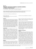
Figure 1 shows the frequency and localization of inflammation
(Figure 1a–d) and chronic changes (sclerosis [Figure 1e], ero-
sions [Figure 1f] and ankylosis [Figure 1g]) per SI joint per
observer. Inflammation of the subchondral region and the
bone marrow was the most frequently observed finding. It can
be seen that both readers use all grades, but pathological find-
ings were mostly scored as grade 2, representing moderate
involvement. For the further analyses all positive findings are
grouped together irrespective of the grade applied.
Table 2 presents data on the inter-observer agreement with
respect to the inflammatory and structural findings. Readers
agreed on the presence of inflammation at any site in 85% of
the cases in the right SI joint, and in 78% of the cases in the
left SI joint. Kappa values reflecting the agreement for detect-
ing inflammation were reasonable (right SI joint, 0.68; left SI
joint, 0.51). Table 2 also provides insight into the prevalence
of the various findings in this population with early inflamma-
tory back pain. Based on the concordance and discordance
rates, agreement is very similar for all assessed sites. How-
ever, due to the low prevalence of inflammation at several of
the locations and structural changes overall, Cohen's kappa
values are influenced negatively. The lowest kappa value and
also the lowest concordance rate is found for inflammation of
the joint capsule of the left SI joint, which was present in only
three patients.
Table 3 summarizes the concordant findings of both readers
with respect to inflammation and structural changes on a
patient level. Twenty-two out of 68 patients showed any sign
of inflammation; 10 patients in both SI joints and 12 patients
unilaterally. Only one patient had inflammation excluding the
bone marrow and subchondral bone. Ten of these 22 patients
with inflammation in one or both joints also had structural
changes. Only one patient had structural changes without
inflammation. Similarly to inflammation, about one half of the
patients with structural changes showed these changes bilat-
erally (six out of 11).
Discussion
One of the important aims of this study was to establish
whether inflammation and structural changes on MRI could
reliably be assessed. In order to allow a detailed judgement,
and to trace redundancies, we decided to score inflammation
Table 1
Baseline characteristics of 68 patients with chronic inflammatory low back pain
Characteristic All patients (N = 68)
Sex (% male) 38
Mean age (years) (SD) 34.9 (10.3)
Median symptom duration (months) (interquartile range) 18.0 [12.0–24.0]
Criteria for inflammatory low back pain
Three criteria present (%) 56
Four criteria present (%) 41
Five criteria present (%) 3
Night pain present (%) 96
a
HLA-B27 present (%) 46
History of inflammatory bowel disease present (%) 15
History of uveitis present (%) 15
History of psoriasis present (%) 24
Family history of ankylosing spondylitis present (%) 37
a
Forty-five of the 47 patients in whom night pain was explored were reported confirmatory.
Arthritis Research & Therapy Vol 8 No 1 Heuft-Dorenbosch et al.
Page 4 of 6
(page number not for citation purposes)
and structural changes per site and per type of lesion. It can
be concluded that the agreement between both readers about
the presence or absence of pathological findings on MRI was
reasonable, especially for inflammation at sites were it was
most prevalent. With agreement levels mostly around 85% for
the presence of inflammation overall and at different locations,
it seems sufficiently high to justify a conclusion of inflammation
made by one observer in clinical practice. Expectedly with ref-
erence to the population under study, the prevalence of
chronic changes on MRI was low. Because of this low preva-
lence of structural changes, the reliability of scoring these
changes is more difficult to assess. This similarly applies to the
assessment of inflammation in the joint space, capsule and lig-
aments. Notwithstanding this limitation, the overall agreement
for the different sites of the joint was comparable, with a
possible exception for inflammation in the joint capsule. An
explanation may be that the delineation of the joint capsule is
poorly defined, which may give rise to misinterpretations.
Figure 1
Schematic presentation of the number of pathological findings by the two observersSchematic presentation of the number of pathological findings by the two observers. All abnormalities are graded for extensiveness as 0 (absent), 1
(minimal), 2 (moderate) and 3 (extensive) in the right and left sacroiliac (SI) joints in 68 patients. For inflammation in subchondral bone and bone mar-
row, and for sclerosis, the data are presented for each site of the joint. For the other abnormalities, the grading for the entire joint per observer is
presented.
Available online />Page 5 of 6
(page number not for citation purposes)
Another important finding in this study was that it is probably
sufficient to look for bone marrow oedema and/or subchondral
inflammation. The contribution of other sites of the joint to
make a diagnosis of inflammation was only marginal. We found
only one patient in whom inflammation was restricted to joint
capsule and ligaments.
A few studies reported agreement with respect to lesions
found on MRI examination of the SI joints, but none of the
studies was performed in patients with recent onset inflamma-
tory back pain. Bigot and colleagues proposed 11 criteria
referring to both the synovial and the fibrous part of the SI joint
that point to sacroiliitis, and showed a good intra-observer and
inter-observer reliability (a kappa value of 0.89 for detecting
bone marrow oedema) [10]. However, this was a study in 22
SpA patients with established disease, in which grade 2
radiological sacroiliitis according to the New York criteria was
present in 80% of the SI joints. Puhakka and colleagues have
proposed a scoring system for MRI abnormalities of the SI
joints, which distinguished inflammatory activity as well as joint
damage [11]. Inter-observer reliability in this study, which
included 41 patients with SpA, of whom 20 patients had
Table 2
Agreement between two observers with respect to magnetic resonance imaging (MRI) characteristics per site per sacroiliac joint in
68 patients with inflammatory low back pain
MRI characteristic per site Sacroiliac joint Present (n)Absent (n) Concordance rate Discordance rate Cohen's kappa value
Inflammation
At any site Right 18 40 0.85 0.15 0.68
Left 14 39 0.78 0.22 0.51
Bone marrow ± subchondral bone Right 17 43 0.88 0.12 0.73
Left 12 47 0.87 0.13 0.65
Joint capsule Right 5 54 0.87 0.13 0.46
Left 3 47 0.74 0.26 0.12
Ligaments Right 4 57 0.90 0.10 0.49
Left 3 57 0.88 0.12 0.38
Joint space Right 6 55 0.90 0.10 0.58
Left 8 54 0.91 0.09 0.68
Chronic changes Right 6 49 0.81 0.19 0.37
Left 11 49 0.88 0.12 0.66
Table 3
Number of patients with abnormalities (inflammation, chronic changes or both) based on concordant observations by both readers
Abnormality Number of patients with involvement of sacroiliac joints
Only the left sacroiliac
joint
Only the right
sacroiliac joint
Both sacroiliac joints One or two sacroiliac
joints
Inflammation 4 8 10 22
Structural changes (ankylosis, sclerosis,
erosions)
50611
Inflammation as well as structural changes 5 3 2 10
Inflammation
Bone marrow or subchondral bone 4 9 8 21
Joint capsule 0 2 4 6
Ligaments 0 1 3 4
Joint space 4 2 4 10
Arthritis Research & Therapy Vol 8 No 1 Heuft-Dorenbosch et al.
Page 6 of 6
(page number not for citation purposes)
grade 2 sacroiliitis or more of at least one SI joint on radiogra-
phy, was importantly lower (a kappa value of 0.47 for bone
marrow enhancement, and of 0.67 for joint space enhance-
ment) as compared with our study. Finally, Docherty and col-
leagues found a kappa value of 0.63 for inter-observer
agreement with respect to inflammation on MRI in a study of
20 patients with established or suspected sacroiliitis on radio-
graphs, but contrast administration was not performed [12].
Our results are largely in accordance with the published liter-
ature, although comparability is limited due to the differences
in study population, with the prevalence of the abnormalities
largely influencing kappa values.
In this cohort of inflammatory back pain of less than two years
duration, inflammation in the SI joints on MRI could be
detected in about one third of the patients (22/68). Moreover,
one sixth of patients already showed signs of structural
changes on the MRI scan (11/68 patients). Although the
number of patients with structural changes is low, this finding
indicates that MRI might be a useful tool in the assessment of
patients with early inflammatory back pain. It is the amount of
variation in the outcome of interest (inflammation and/or struc-
tural changes) rather than the number of patients under study
that is important in judging whether the sample size is suffi-
cient to test reliability. As long as all kinds of abnormalities are
covered, it is possible to test reliability even in situations like
this, with only 11 patients showing abnormalities. Undoubt-
edly, however, the likelihood of covering all kinds of abnormal-
ities will increase by increasing patient number. The real value
of MRI will therefore be ascertained in future, by following the
patients longitudinally and obtaining more data, which will
occur in this cohort. The development and choice of an appro-
priate scoring system for sacroiliitis on MRI to be used in clin-
ical studies and trials will be subject of interest in an ongoing
ASAS-OMERACT working group [13].
Conclusion
MRI can reliably detect inflammation and structural changes in
SI joints in patients with early inflammatory back pain. Assess-
ing bone marrow and/or subchondral bone enhancement suf-
fices to detect inflammation. Inflammation in the joint space,
the joint capsule and the ligaments hardly contributes to this
detection, because it is associated with inflammation in the
bone marrow and/or subchondral bone. About one-third of
patients with recent-onset inflammatory back pain show
inflammation, and about one-sixth of patients show structural
changes in at least one SI joint, indicating that MRI might be a
useful tool to diagnose sacroiliitis in patients with inflammatory
back pain.
Competing interests
The authors declare that they have no competing interests.
Authors' contributions
DvdH designed the study and performed statistical analysis.
LHD collected data, read the MRI scans, and performed sta-
tistical analysis. RW read the MRI scans. RL performed statis-
tical analysis. All authors interpreted the results, and wrote and
commented on the manuscript.
Acknowledgements
This study was partly supported by a grant from the Dutch Arthritis
Association.
References
1. van der Linden SM, Valkenburg HA, de Jongh BM, Cats A: The
risk of developing ankylosing spondylitis in HLA-B27 positive
individuals. A comparison of relatives of spondylitis patients
with the general population. Arthritis Rheum 1984, 27:241-249.
2. Dougados M, van der Linden S, Juhlin R, Huitfeldt B, Amor B, Calin
A, Cats A, Dijkmans B, Olivieri I, Pasero G, The European Spond-
ylarthropathy Study Group, et al.: The European Spondylar-
thropathy Study Group preliminary criteria for the
classification of spondylarthropathy. Arthritis Rheum 1991,
34:1218-1227.
3. Feldtkeller E, Bruckel J, Khan MA: Scientific contributions of
ankylosing spondylitis patient advocacy groups. Curr Opin
Rheumatol 2000, 12:239-247.
4. Spoorenberg A, de Vlam K, van der Heijde D, de Klerk E, Douga-
dos M, Mielants H, van der Tempel , Boers M, van der Linden Sj:
Radiological scoring methods in ankylosing spondylitis: relia-
bility and sensitivity to change over one year. J Rheumatol
1999, 26:997-1002.
5. Braun J, Bollow M, Eggens U, Konig H, Distler A, Sieper J: Use of
dynamic magnetic resonance imaging with fast imaging in the
detection of early and advanced sacroiliitis in spondylarthrop-
athy patients. Arthritis Rheum 1994, 37:1039-1045.
6. Bollow M, Loreck D, Banzer D, Brandt H, Zerbes K, Kourik W, Mel-
lorowicz H, Backhaus M, Schmidt W, Bohl-Buhler M, et al.: Diag-
nostic imaging of inflammation in the axial skeleton. Zeitschr
Rheumatol 1999, 58:61-70.
7. Braun J, van der Heijde D: Imaging and scoring in ankylosing
spondylitis. Best Pract Res Clin Rheumatol 2002, 16:573-604.
8. Rudwaleit M, van der Heijde D, Khan MA, Braun J, Sieper J: How
to diagnose axial spondyloarthritis early. Ann Rheum Dis
2004, 63:535-543.
9. Calin A, Porta J, Fries JF, Schurman DJ: Clinical history as a
screening test for ankylosing spondylitis. JAMA 1977,
237:2613-2614.
10. Bigot J, Loeuille D, Chary-Valckenaere I, Pourel J, Cao MM, Blum
A: Determination of the best diagnostic criteria of sacroiliitis
with MRI. J Radiol 1999, 80:1649-1657.
11. Puhakka KB, Jurik AG, Egund N, Schiottz-Christensen B, Sten-
gaard-Pedersen K, van Overeem Hansen G, Christiansen J: Imag-
ing of sacroiliitis in early seronegative spondylarthropathy.
Assessment of abnormalities by MR in comparison with radi-
ography and CT. Acta Radiol 2003, 44:218-229.
12. Docherty P, Mitchell MJ, MacMillan L, Mosher D, Barnes DC, Hanly
JG: Magnetic resonance imaging in the detection of sacroiliitis.
J Rheumatol 1992, 19:393-401.
13. Landewé R, Hermann K-G, van der Heijde D, Baraliakos X, Jurik A-
G, Lambert R, Ostergaard M, Rudwaleit M, Salonen D, Braun J, the
ASAS/OMERACT MRI in AS Working Group: Scoring sacro-iliac
joints by magnetic resonance imaging. A multiple-reader reli-
ability experiment. J Rheumatol 2005, 32:2050-2055.