Báo cáo y học: "Therapeutic impact of the ethyl acetate extract of Tripterygium wilfordii Hook F on nephritis in NZB/W F1 mice" pot
Bạn đang xem bản rút gọn của tài liệu. Xem và tải ngay bản đầy đủ của tài liệu tại đây (1.24 MB, 11 trang )
Open Access
Available online />Page 1 of 11
(page number not for citation purposes)
Vol 8 No 1
Research article
Therapeutic impact of the ethyl acetate extract of Tripterygium
wilfordii Hook F on nephritis in NZB/W F1 mice
Xuelian Tao
1
, Fred Fan
1
, Victoria Hoffmann
2
, Nancy S Longo
1
and Peter E Lipsky
1
1
Autoimmunity Branch, National Institute of Arthritis and Musculoskeletal and Skin Diseases, National Institutes of Health, Rockville Pike, Bethesda,
MD 20892, USA
2
Office of Research Services Division of Veterinary Resources, National Institutes of Health, Rockville Pike, Bethesda, MD 20892, USA
Corresponding author: Peter E Lipsky,
Received: 1 Nov 2005 Revisions requested: 24 Nov 2005 Revisions received: 6 Dec 2005 Accepted: 6 Dec 2005 Published: 3 Jan 2006
Arthritis Research & Therapy 2006, 8:R24 (doi:10.1186/ar1879)
This article is online at: />© 2006 Tao et al.; licensee BioMed Central Ltd.
This is an open access article distributed under the terms of the Creative Commons Attribution License ( />),
which permits unrestricted use, distribution, and reproduction in any medium, provided the original work is properly cited.
Abstract
This study was designed to examine the potential use of the
ethyl acetate (EA) extract of Tripterygium wilfordii Hook F
(TwHF), a Chinese herbal medicine, in the treatment of systemic
lupus erythematosus. A total of 48 28-week-old female NZB/W
F1 mice were randomly divided into three groups and orally
administered vehicle or the EA extract of TwHF at 18.25 mg/kg
(EA
low
) or 36.5 mg/kg (EA
high
) for 14 weeks. Proteinuria and
serum anti-double-stranded (ds)DNA antibody titers were
assayed before and after treatment. At the end of treatment, all
animals were sacrificed and pathological changes in the kidneys
were examined by observers blinded to the treatment regimens.
Immunohistological studies were carried out on kidneys and
spleens. At 28 weeks of age, proteinuria (>30 mg/dl) and anti-
dsDNA antibodies were found in all mice in the three groups.
Fourteen, sixteen and fifteen mice in the vehicle, EA
low
and EA
high
groups, respectively, completed at least four weeks of
treatment. At the end of treatment, the mean proteinuria of the
EA
low
and EA
high
groups was significantly less than that of the
vehicle group and no different from proteinuria at the onset of
treatment. Histological evidence of glomerulonephritis,
glomerular deposition of IgG and complement 3 and cellular
infiltration in the interstitium and perivascular regions were
significantly less severe in the EA extract treated mice than in
vehicle treated mice. Treatment with the EA extract significantly
inhibited the progression of kidney disease in NZB/W F1 mice,
though had no significant effect on the levels of anti-dsDNA
antibody.
Introduction
The Chinese anti-rheumatic remedy Tripterygium wilfordii
Hook F (TwHF) has been reported to be effective in the treat-
ment of a variety of autoimmune diseases, including rheuma-
toid arthritis (RA), systemic lupus erythematosus (SLE), and
psoriasis [1,2]. The therapeutic benefit of TwHF preparations
in patients with a variety of kidney diseases, including IgA
nephropathy and Henoch-Schonlein purpura nephritis, has
also been described [3-6]. Moreover, in several uncontrolled
trials, improvement in clinical manifestations and laboratory
abnormalities was observed in as many as 94% of SLE
patients treated with a variety of TwHF preparations [7-10].
Different preparations of TwHF have been tested for their ther-
apeutic effect in the MRL-lpr/lpr murine model of lupus. The
TwHF preparations employed in these studies were crude
extracts and their composition was not known. Therefore, it is
difficult to assess the pharmacological impact of the material
or to standardize the extract for further development. Gu and
colleagues [11] found that a water extract of TwHF amelio-
rated glomerulonephritis and prolonged survival in MRL-lpr/lpr
mice, but only when therapy was begun before disease onset.
Consistent with this, Zhang and colleagues [12] reported
improvement in survival, proteinuria, arthritis and lymphaden-
opathy in MRL-lpr/lpr mice treated with another TwHF prepa-
ration. However, no improvement in renal histology was noted.
Although both of these studies suggest benefit in this model
of murine lupus when treatment was begun before disease
onset, the full extent of potential benefit was not established.
Importantly, no evidence was provided that the extract of
BSA = bovine serum albumin; BUN = blood urea nitrogen; C3 = complement 3; ds = double stranded; EA = ethyl acetate; FITC = fluorescein iso-
thiocyanate; HPLC = high-performance liquid chromatography; IL = interleukin; LD50 = lethal dose 50%; RA = rheumatoid arthritis; SLE = systemic
lupus erythematosus; TwHF = Tripterygium wilfordii Hook F.
Arthritis Research & Therapy Vol 8 No 1 Tao et al.
Page 2 of 11
(page number not for citation purposes)
TwHF was beneficial as treatment after onset of autoimmune
disease.
NZB/W F1 mice spontaneously develop autoantibodies
against double-stranded (ds)DNA; these antibodies form
immune complexes with dsDNA. Deposition of the immune
complex in the kidney induces activation of the complement
system, which consequently results in chronic glomerulone-
phritis, vasculitis and cellular infiltration in the interstitium of the
kidney [13,14]. This animal model has been commonly used
for screening of drugs for treatment of human SLE because of
its similarities to human SLE in clinical, immunopathological,
and genetic features [15-18]. Specifically, the high incidence
of SLE-like disease, characterized by gender selectivity,
chronic immune complex nephritis and high titers of anti-
dsDNA antibody, makes it possible to evaluate efficacy of
treatment easily in the NZB/W F1 mice. This animal model,
however, had not yet been employed to assess the impact of
the TwHF preparations.
An ethyl acetate (EA) extract of TwHF has been prepared and
used for the first time in the United States in a controlled, dou-
ble-blinded clinical trial of patients with RA [19,20]. Results
from the trial showed significant therapeutic benefit and good
tolerance in treated RA patients. The EA extract of TwHF has
been studied in detail for its content of active components,
namely the diterpenoids, triptolide and tripdiolide, as well as its
in vitro and in vivo anti-inflammatory and immunosuppressive
impact and toxicity [21-25]. Importantly, the EA extract of
TwHF can be standardized by quantitatively assessing its con-
tent of active components, as well as with regard to efficacy
and adverse events. To estimate the potential therapeutic
effect of this standardized extract on patients with SLE, exper-
iments with NZB/W F1 mice were undertaken. NZB/W F1
mice with established nephritis were treated orally with vehicle
only or the EA extract for a total of 14 weeks beginning at 28
weeks of life. Kidney disease significantly worsened in more
than 90% of the mice treated with vehicle. In contrast, kidney
disease was improved or controlled in NZB/W F1 mice
treated with either a low or a high dose of the EA extract, sug-
gesting an important therapeutic effect of this standardized
extract in this animal model of lupus.
Materials and methods
The ethyl acetate extract
The EA extract was prepared as described and analyzed for its
content of triptolide and tripdiolide, which are responsible for
up to 90% of the bioactivity of the EA extract [21]. In addition,
the EA extract was assessed for the amount that caused death
of 50% of treated C57BL/6j mice (LD50) as described.
Briefly, plant material for extraction was assessed by HPLC for
its content of diterpenoids, after which selected peeled roots
of TwHF were ground and extracted with EA. The EA extract
was concentrated to dryness and ground to a fine powder.
The final preparation contained 0.77 µg/mg of triptolide and
0.44 µg/mg of tripdiolide.
Animals and treatment regimens
Eight week old female NZB/WF1/J mice were purchased from
Jackson Laboratory (Bar Harbor, ME, USA) and maintained in
a conventional animal housing facility throughout the study. At
28 weeks of age, the animals were randomly divided into three
treatment groups (vehicle, EA
low
and EA
high
) and orally admin-
istered vehicle (2% dimethyl sulfoxide/ 5% Tween-20 in
water), the EA extract of TwHF at 18.25 mg/kg (equivalent to
1/20 of the LD50) or the EA extract of TwHF at 36.5 mg/kg
(equivalent to 1/10 of the LD50), respectively. This equated to
28.1 µg/kg of triptolide and 16 µg/kg of tripdiolide. The EA
extract was dissolved in vehicle solution to obtain an appropri-
ate concentration for oral administration (about 0.4 ml per ani-
mal per dose). Based on the information obtained from a
previous study demonstrating that the half life of triptolide was
6.2 h after oral administration, treatment was given daily, 5
days a week from Monday to Friday for a total of 14 weeks.
Body weight was monitored weekly. If an animal lost more than
15% of body weight, treatment was terminated and the animal
euthanized. Otherwise, mice were sacrificed after 14 weeks of
treatment. Blood, kidneys and spleen were collected from all
mice, including those dying before completion of treatment
and those completing the treatment protocol.
The study proposal (A-002-03-03) has been approved and all
procedures monitored by the Animal Care and Use Committee
(ACUC) in the Intramural Research Program of National Insti-
tutes of Health.
Urine collection and proteinuria assay
Urine from individual mice was collected biweekly from the age
of 24 weeks using metabolic cages. Proteinuria was tested by
dipstrip (Chemstrip, Roche (Indianapolis, IN 46256 USA) and
semiquantified as 0, ± (0 to 30 mg/dl), + (30 to 100 mg/dl),
++ (100 to 500 mg/dl) and +++ (>500 mg/dl). Proteinuria
was also analyzed by spectrophotometer using a bicin-
choninic acid based BCA protein assay kit (Pierce, Rockford,
IL, USA) and standardized with BSA. In the entire study
course, proteinuria was measured first with the urinary analysis
strips (Chemstick) followed by the spectrophotometer. It was
found that results obtained from the Chemstick correlated with
those by spectrophotometer. Since proteinuria was quantified
more accurately, data generated using the spectrophotometer
were used to plot the figures.
Assay for blood urea nitrogen
Serum blood urea nitrogen (BUN) was determined by the NIH
Diagnostic and Research Service Branch, the laboratory of the
Veterinary Resources Program.
Available online />Page 3 of 11
(page number not for citation purposes)
ELISA for anti-dsDNA antibodies [26]
EIA/RIA plates (96-well; Corning Inc., Corning, NY, 14831,
USA) were coated with 50 µl of 10 µg/ml type XV calf thymus
DNA (Sigma, St Louis, MO 63103, USA) overnight at 4°C.
After washing, the plates were blocked with 10% BSA/PBS.
The plates were incubated with diluted serum (first dilution of
1:20 followed by serial 3-fold dilutions) for 2 h at room temper-
ature. After washing with 0.5% Tween-20/PBS, the plates
were incubated with alkaline phosphatase conjugated goat
anti-mouse IgG (Fab) for 1 h followed by the alkaline phos-
phatase substrate p-nitrophenyl phosphate. The reaction was
stopped by addition of 25 µl of 5N NaOH solution. The plates
were assayed with a microplate spectrophotometer (Bio-Tek
Instrument Inc., Winooski, VT 05404-0998, USA) at 405 nm.
Sera from 10 C57BL/6j mice were employed as normal
controls.
Pathological study of kidneys
Kidneys and spleens were harvested from the mice after spon-
taneous death or euthanasia. One-half of the kidney from each
mouse was immersed in TBS tissue freezing medium (Triangle
Biomedical Sciences, Durham, NC, 27705, USA) and snap
frozen in methanol-dry ice. The other half of the freshly har-
vested kidney was fixed in buffered 10% formalin (Fisher Sci-
entific, Pittsburgh, PA 15275-9952, USA) and embedded in
paraffin blocks (Sugipath, Richmond, IL 60071, USA). Seven
micron thick sections were cut and stained with hematoxylin
and eosin (Histoserv, Germantown, MD 20874-1202, USA).
Sections were graded semi-quantitatively by two veterinary
pathologists for glomerular, interstitial and vascular lesions.
The grading scheme, a modification of the method reported by
Chan and colleagues [27], is shown in Table 1.
Flow cytometry analysis of spleen cells
Mononuclear cells separated from spleens of the NZB/W F1
mice were stained for B cells, T cells and dendritic cells using
Allophycocyanin (APC) conjugated rat anti-mouse CD19, flu-
orescein isothiocyanate (FITC) conjugated rat anti-mouse
CD3 and Phycoerythrin (PE) conjugated rat anti-mouse
CD11c antibodies, respectively (BD Biosciences, Pharmin-
gen, San Diego, CA 92121-8995, USA After staining, the
cells were analyzed by flow cytometry using a FACSCalibur
(Becton Dickson, San Jose, CA, USA) along with CellQuest
software (BD Biosciences, San Jose, CA 95131, USA) for
data acquisition and analysis.
Staining of IgG and complement 3 in kidneys
For determination of IgG deposition, kidney cryosections were
stained using FITC conjugated goat anti-mouse IgG antibody
(Cappel, MP Biomedical Inc., Aurora, OH 44202, USA). For
determination of complement 3 (C3) deposition, kidney cryo-
sections were stained with horseradish peroxidase conju-
gated goat anti-mouse C3 antibody (MP Biomedical Inc.)
followed by color development with the VIP substrate kit (Vec-
tor Laboratories, Burlingame, CA 94010, USA). The slides
were counter-stained with hematoxylin (Vector Laboratories).
Fluorescent staining of lymphoid cells in spleens and
kidneys
Frozen sections were used for examination of lymphoid cell
subsets in spleen and cell infiltration in kidneys. For determina-
tion of CD3
+
T cells, the frozen sections were first blocked
with 10% goat serum and stained with rat anti-mouse CD3
+
antibody (BD Pharmingen) followed by succinimidyl ester-
labeled goat anti-rat IgG antibody (Alexa Fluor 568, Molecular
Probes, Eugene, OR 9402-2209, USA). For determination of
CD11c
+
cells, the cryosection was blocked with 10% mouse
serum followed by sequential staining with hamster anti-
mouse CD11c (BD Pharmingen) and FITC conjugated mouse
anti-hamster IgG (BD Pharmingen). For identification of B
cells, cryosections were directly stained with FITC conjugated
rat anti-mouse IgD (BD Pharmingen).
Images were captured using a Hamamatsu camera system
connected to a microscope (Leica DMRB) along with the
IPLab Scientific Image Processing software (Scanalytic Inc.,
Rockville, MD 20850, USA). Two color photos were devel-
oped using the software Adobe Photoshop version 7.
Table 1
Grading of kidney lesions
Score Glomerulonephritis Interstitial nephritis Vessels
1+ Focal, mild or early proliferative 1–3 small foci (5–10 cells) of MNC * MNC around renal pelvis/blood vessels
2+ Multifocal proliferative with increased matrix
and inflammatory cells
Mild MNC infiltrates around individual
tubules; isolated atrophied tubules
MNC infiltrates around main arteries; small
foci 10–20 cells of MNC around
interlobular arteries
3+ Diffuse proliferative More extensive infiltrates with large foci of
tubular atrophy
Individual foci of MNC around small arterial
branches more extensive
4+ Extensive sclerosis/ crescents: proteinuria Extensive MNC infiltrates between tubules;
extensive tubule atrophy/necrosis
MNC infiltrates extend into surrounding
parenchyma/most vessels affected/
vasculitis
*MNC = mononuclear cells.
Arthritis Research & Therapy Vol 8 No 1 Tao et al.
Page 4 of 11
(page number not for citation purposes)
Statistical analysis
An intention-to-treat analysis was carried out, including all
NZB/W F1 mice that completed at least four weeks of the
treatment. All statistical tests were two sided. Comparison of
the mean values of individual variables measured at each time
to the corresponding base-line values for mice of the same
group was carried out using the Student's t test. The Kruskal-
Wallis test was employed to compare each variable between
groups before and after treatment. For proteinuria data, a last-
observation-carried-forward approach was used for the mice
that died before the end of study.
Results
Outcome
Initially, 16 mice were included in each group. One mouse
from each group had severe proteinuria at the beginning of
treatment and was dead within four weeks of starting the treat-
ment. One animal of the vehicle group unexpectedly died of
acute pulmonary edema caused by impropriate gavage. Data
from these four mice were excluded from analysis of treatment
efficacy. One animal from each group completed more than
four weeks of treatment, but died before the end of the treat-
ment course after development of severe proteinuria and ele-
vated BUN, suggesting disease related death. These mice
were included in the analysis. Two mice from the EA
low
group
were euthanized at the eighth and ninth week of the EA treat-
ment because of weight loss reaching 15% of baseline.
Despite that, there was no significant difference in the mean
body weight between the three groups either before or after
treatment (42.3 g, 41.6 g and 40.8 g before starting treatment
and 40.4 g, 39.5 g and 40.2 g after treatment for vehicle, EA
low
and EA
high
groups, respectively). These mice were also
included in the analysis.
Therapeutic effect of the EA extract on proteinuria and
renal function
All mice at age 28 weeks exhibited significant proteinuria (30
mg/dl or higher) before starting the treatment course. At this
time, the mean proteinuria for the vehicle, EA
low
and EA
high
groups was 100.93 mg/dl, 121.98 mg/dl and 95.91 mg/dl,
respectively (Fig 1a). Notably, during the treatment course,
proteinuria increased in the mice treated with vehicle, with a
mean proteinuria of 300 mg/dl at the age of 36 weeks. Of 14
animals in the vehicle group, 13 (93%) developed severe pro-
teinuria (>500 mg/dl) before the end of the study (age of 42
weeks). Only one mouse in the vehicle group continued to
maintain proteinuria below 100 mg/dl at the end of study. In
contrast, 12 mice (80%) from the EA
high
group had 30 to 100
mg/dl of proteinuria at the start of treatment. In 9 out of the 15
animals, proteinuria was improved or maintained at the same
low level (30 to 100 mg/dl) throughout the study. Proteinuria
worsened in only four animals. In two of the four mice, worsen-
ing of proteinuria was noted two and four weeks after starting
treatment. Two mice developed severe proteinuria before
starting treatment that was unchanged during the treatment
course. Similar to the EA
high
group, proteinuria was improved
or maintained at a low level throughout the 14 week treatment
in 7 out of 15 mice in the EA
low
group. In four animals from the
EA
low
group, proteinuria was maintained at a low level and
increased only at the very end of the treatment course. Four
mice from the EA
low
group had severe proteinuria in the begin-
ning of treatment without improvement. Figure 1b shows the
comparison of proteinuria between groups before and after
treatment. There was no significant difference in mean pro-
teinuria between the three groups at the beginning of treat-
ment. At the end of the treatment course, however, mean
proteinuria was 496 mg/dl, 204 mg/dl and 190 mg/dl for the
vehicle, EA
low
and EA
high
groups, respectively (p < 0.005, vehi-
cle versus EA
low
; p < 0.0001, vehicle versus EA
high
).
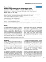
BUN was also determined and was increased and correlated
with the severity of proteinuria in the three groups. As shown
in Figure 2, 10 out of 12 tested animals from the vehicle group
had increased BUN ranging from 32 mg/dl to 159 mg/dl (nor-
mal range <27 mg/dl). In contrast, in 9 out of 12 mice from the
EA
low
group, BUN was within normal range and none of the
mice in the EA
high
group exhibited elevated BUN. The mean
BUN was 86.75, 29.73 and 19.09 mg/dl for the vehicle, EA
low
and EA
high
groups, respectively (p < 0.001).
Effect of the EA extract treatment on kidney pathology
At the end of the study, kidneys obtained from 14 animals from
the vehicle group and 15 from each of the EA treated groups
were examined for histological changes. All kidneys from the
vehicle group had glomerular, interstitial and vascular lesions
(Figure 3). Glomeruli were the most severely affected. Glomer-
ular lesions ranged from proliferative glomerulonephritis with
influx of mononuclear cells and rare neutrophils to diffuse
glomerular sclerosis with crescent formation, fibrinoid necro-
sis and proteinuria. Interstitial disease consisted of infiltration
of mononuclear cells around tubules with tubular atrophy, dila-
tion and thickened tubular basement membranes. Occasional
tubules had necrotic tubular epithelium. Perivascular disease
consisted of mononuclear cell infiltrates in a follicular pattern
around large arteries and interlobular arteries. In the most
severely affected mice, the mononuclear cells around blood
vessels were confluent with interstitial inflammation. After 14
weeks of treatment, most mice treated with either the low or
high dose of the EA extract had less severe kidney disease
with significantly diminished glomerular, interstitial and
perivascular lesions.
Figure 3d shows the comparison of the renal lesion scores
between the three groups. Glomerular, interstitial and perivas-
cular lesions were significantly less severe in the mice treated
with the EA extract than those treated with vehicle. Most
significant was the differences in glomerular lesions between
the groups; p < 0.01, EA
low
versus vehicle; p < 0.001, EA
high
versus vehicle.
Available online />Page 5 of 11
(page number not for citation purposes)
Treatment with the EA extract limits lymphoid cell
infiltration and immune complex deposition in kidneys
IgG and C3 staining was observed only in glomeruli and the
intensity of IgG and C3 was correlated with the severity of pro-
teinuria (Fig. 4a,b,e,f). Remarkable deposition of both IgG and
C3 was observed in the kidneys from all mice of the vehicle
group. In contrast, significantly less or no glomerular deposi-
tion of either IgG or C3 was noted in the mice treated with
either the low or high dose of the EA extract, similar to the nor-
mal control mice (Figure 4i,j).
Immunofluorescent staining was carried out to determine the
impact of treatment on mononuclear cells infiltrating the kid-
neys. Both the extent and the density of T cells, B cells and
macrophages/dendritic cells infiltrating the kidneys were
related to the severity of proteinuria. Significant numbers of
CD3
+
T cells, CD11c
+
myeloid cells, and IgD
+
B cells were
found in the area around vessels that extended into the inter-
stitium in the kidneys from the mice of the vehicle group (Fig-
ure 4c,d,g,h). Similar to that noted with normal C57BL/6j
mice, there was almost no cellular infiltration in the kidneys
from either the EA
low
or the EA
high
group.
Changes in serum levels of anti-dsDNA antibody
Autoantibody against dsDNA was examined before, 7 weeks
and 14 weeks after starting treatment. Compared to normal
C57BL/6j mice, sera from the NZB/WF1 mice of the three
groups contained higher titers of anti-dsDNA antibody at 28
weeks of age. The relative titers of anti-dsDNA at this time for
C57BL/6j mice ranged from 0.1 to 0.3, with an average of
0.23. All NZB/W F1 mice employed in the study exhibited
elevated titers of this autoantibody, with averages of 1.0, 1.3
and 1.6 for the vehicle, EA
low
and EA
high
groups, respectively
(Figure 5a). The levels of the anti-dsDNA autoantibody
increased during the treatment course for all of the three
groups. The increase of anti-dsDNA antibody at the end of
treatment compared to that before treatment was statistically
significant for the vehicle group (p < 0.001) and both the EA
low
(p < 0.001) and EA
high
(p < 0.01) groups. Moreover, there was
no significant difference in the mean titers of this autoantibody
between groups, either before or after treatment.
Since the initial titer of anti-DNA antibody was higher in the
mice treated with the high dose of the EA extract, the fold
increases in this antibody upon completion of the treatment
course were compared between the three groups. The mean
fold increases of anti-dsDNA antibody were 2.29, 1.81 and
0.81 for vehicle, EA
low
and EA
high
groups, respectively (Figure
Figure 1
Impact of treatment with Tripterygium wilfordii Hook F on proteinuria in NZB/W F1 miceImpact of treatment with Tripterygium wilfordii Hook F on proteinuria in
NZB/W F1 mice. Proteinuria was determined by spectrophotometry as
described in Materials and methods. Data are the means ± standard
error of the mean of each group. Numbers in parentheses indicate the
animals examined. (a) Changes in proteinuria in NZB/WF1 mice during
the treatment course. Statistical analysis was done for proteinuria
measured before and at each time point after treatment for each group.
Proteinuria increased significantly in the mice of the vehicle group (*p <
0.05; ***p < 0.001; ****p < 0.0001; before treatment verses after treat-
ment). (b) Proteinuria was compared between groups before and after
treatment. No significant difference was determined before treatment
between the three groups. After treatment, proteinuria was significantly
higher in the vehicle treated mice than in mice treated with the ethyl
acetate (EA) extract (**p < 0.0026, EA
low
versus vehicle; ****p <
0.0001, EA
high
versus vehicle).
Figure 2
Renal function (blood urea nitrogen (BUN)) of the NZB/WF1 mice after treatment with vehicle or the ethyl acetate (EA) extract of Tripterygium wilfordii Hook FRenal function (blood urea nitrogen (BUN)) of the NZB/WF1 mice after
treatment with vehicle or the ethyl acetate (EA) extract of Tripterygium
wilfordii Hook F. Serum was collected from the animals for assay of
BUN before euthanasia. Dotted lines show the normal range for BUN
(17 to 28 mg/dl). Numbers in parentheses indicate the animals exam-
ined. The horizontal bars indicate the median values of each group. **p
< 0.001, vehicle versus EA
low
; ***p < 0.01, vehicle versus EA
high
.
Arthritis Research & Therapy Vol 8 No 1 Tao et al.
Page 6 of 11
(page number not for citation purposes)
5b). Compared to the vehicle group, mice treated with the high
dose of the EA extract had less increase in serum anti-dsDNA
antibody, although this did not reach statistical significance (p
= 0.07).
Effect of treatment with the EA extract on spleen
lymphoid cells
Splenomegaly was observed in most NZB/WF1 mice of the
vehicle group. Mice treated with the EA extract had signifi-
cantly smaller spleens than those treated with vehicle. To
examine the effect of the EA extract in greater detail, spleen
mononuclear cells were analyzed by flow cytometry. There
was no significant difference in the percentage of CD3
+
T cells
over the total cells between the NZB/W F1 mice of the three
groups (Figure 6a). The ratio of CD11c
+
myeloid cells to the
total cells, however, was significantly lower in the NZB/W F1
mice treated with the higher dose of the EA extract than in
those treated with vehicle (p < 0.01).
To confirm the information obtained from flow cytometry, fluo-
rescent staining of spleen cryosections was carried out. Very
large follicles with abundant IgD
+
B cells, CD3
+
T cells as well
as CD11c
+
myeloid cells forming a greatly increased white
pulp were observed in the spleens from the mice of the vehicle
group (Fig. 6b). In contrast, spleens of mice treated with the
EA extract exhibited smaller follicles with significantly less
numbers of IgD
+
B cells. In addition, CD11c
+
myeloid cells
were only infrequently observed in the spleens of the mice
treated with the EA extract, consistent with the results from
flow cytometry. The densities of follicles and CD3
+
T cells in
the spleens of the EA treated mice were not significantly
different from those in the vehicle treated mice. The profile of
cells observed in the spleens of the EA extract treated mice
was very similar to that of the normal C57BL/6j mice.
Figure 3
Changes in renal pathology as a result of treatment with the ethyl acetate (EA) extract of Tripterygium wilfordii Hook FChanges in renal pathology as a result of treatment with the ethyl acetate (EA) extract of Tripterygium wilfordii Hook F. (a) Kidney from the vehicle
group showed glomerulonephritis, tubular dilation and atrophy, and heavy cell infiltration in perivascular and interstitial region. Kidney sections from
the mice of the (b) EA
low
and (c) EA
high
groups showed normal glomeruli and tubules. Slight cell infiltration in perivascular regions was seen in (b).
Results shown are representative of 14 animals of each group. Arrows indicate representative glomeruli. (d) Glomerular, interstitial and perivascular
disease was scored from (0) to (4+) as described in Materials and methods. Numbers of mice examined are indicated in parentheses. *p < 0.05, **p
< 0.01, ***p < 0.001, EA
low
or EA
high
versus vehicle. Original magnification × 400.
Figure 4
Immunohistochemical analysis of the kidney of NZB/WF1 mice after treatment with (a-d) vehicle or (e-h) the ethyl acetate (EA) extract of Tripterygium wilfordii Hook FImmunohistochemical analysis of the kidney of NZB/WF1 mice after
treatment with (a-d) vehicle or (e-h) the ethyl acetate (EA) extract of
Tripterygium wilfordii Hook F. Kidney was examined for deposition of
(a,e,i) IgG, (b,f,j) complement 3 (C3) and infiltration of (c,g,k) CD3
+
cells (in red) and CD11c
+
cells (in green) and (d,h,l) IGD
+
cells. Results
shown are representative of five mice per group. Arrows indicate repre-
sentative glomeruli.
Available online />Page 7 of 11
(page number not for citation purposes)
Discussion
In this study, we selected NZB/W F1 mice to examine the
potential therapeutic role of the EA extract of TwHF in SLE.
The study was designed to start therapy of the NZB/W F1
mice at the age of 28 weeks because all mice at this time had
positive anti-dsDNA antibody titers and more than 90% of the
mice had detectable proteinuria, indicating that autoimmune
nephritis was established before the start of treatment in these
mice. Results of the study show that 93% of mice treated with
vehicle had progressive glomerulonephritis, documented by
severe proteinuria, elevated BUN and histological
abnormalities at the end of the study. In contrast, proteinuria
and pathological changes in the kidneys were significantly
improved or maintained at mild levels of disease in most mice
treated with the EA extract, suggesting that the EA extract
exerted a therapeutic effect on established lupus nephritis in
NZB/W F1 mice. This result is important because therapy of
human SLE involves treatment of established disease. Thera-
peutic trials in NZB/W F1 mice frequently involve initiation of
therapy before disease onset [16,26,28]. Only a few agents
have demonstrated benefit after the onset of disease [15-17].
The finding that the EA extract of TwHF is effective as a ther-
apy for established NZB/W F1 murine lupus suggests that its
efficacy may be more easily translated to treatment of human
lupus.
It is important to note that the current results were obtained
with a standardized extract of TwHF. TwHF has been widely
used for several decades in China in the treatment of a variety
of autoimmune disorders, including RA and, to a lesser extent,
SLE. However, the extracts employed in these studies were
manufactured from wild TwHF plants collected from varying
locations. As the composition of these preparations was
uncertain, the treatment dose of the extract was determined
according to the weight of the raw plant materials and the
basis for the clinical effects could not be determined. Since
there was no quantitative standard for these TwHF prepara-
tions, reproducible clinical efficacy and toxicity could not be
ensured. The EA extract of TwHF employed in the present
study was prepared from plant material characterized by
HPLC and liquid chromatography-mass spectroscopy and by
a standardized manufacturing protocol with quantitative con-
trol of the major active components, triptolide and tripdiolide,
and their ratios [21]. The EA extract of TwHF was also quanti-
tatively evaluated and standardized for its in vitro bioactivities
and in vivo toxicity before being applied to the current studies
[24]. It is important to note that the extract employed in the cur-
rent study has already been tested in phase I and phase II clin-
ical trials in patients with RA and has been found to be both
safe and effective [19,20].
TwHF has been tested for its effect on the MLR-lpr/lpr lupus
model [11,12]; however, the preparations used in these two
studies were poorly standardized. In addition, treatment was
started before the development of autoimmune disease [11].
Notably, the results of the two studies differed in that in one
[12] no effect on renal histology was noted whereas improve-
ment was noted in the other [11]. This could be related to the
use of different extracts of TwHF, both of which were poorly
characterized. In the current study, a well characterized extract
was employed in a therapeutic approach in animals with estab-
lished disease; histological changes were well correlated with
clinical improvement of the kidney disease and the extract
showed considerable efficacy. Previously, treatment of MRL-
lpr/lpr mice with an extract of TwHF after onset of disease
resulted in improvement in proteinuria but not renal histopa-
thology [11]. Whether this related to differences in the
potency of the extract employed or differences in the murine
models is unknown.
The mechanism by which the EA extract reduced autoimmune
nephritis in the NZB/W F1 mice could relate to the immuno-
Figure 5
Serum anti-double-stranded (ds)DNA antibody in the NZB/WF1 mice before and after treatment with vehicle or the ethyl acetate (EA) extract of Tripterygium wilfordii Hook FSerum anti-double-stranded (ds)DNA antibody in the NZB/WF1 mice
before and after treatment with vehicle or the ethyl acetate (EA) extract
of Tripterygium wilfordii Hook F. (a) Serum was collected before, 7 and
14 weeks after starting treatment. Anti-dsDNA antibody was deter-
mined by ELISA as described in Materials and methods. The mean of
relative titers of anti-dsDNA antibody of serum obtained from normal
C57BL/6j mice was 0.23 (0.1 to 0.3). Numbers in parentheses indicate
the mice evaluated. Data are the mean ± standard error of the mean of
the optical density (OD) readings on 1:20 diluted serum of each group.
**p < 0.01, ***p < 0.001 at time 0 versus at 7 weeks or 14 weeks of
treatment for the same group. (b) The same data were analyzed as the
change in anti-dsDNA antibody titers from the baseline at 28 weeks of
age. Original magnification × 400.
Arthritis Research & Therapy Vol 8 No 1 Tao et al.
Page 8 of 11
(page number not for citation purposes)
suppressive effect of its active components. The immunosup-
pressive effects of the extract of TwHF have been documented
in both in vitro and in vivo studies. In vitro, the extract of TwHF
inhibited proliferation and IL-2 and interferon-γ production by
T cells in response to antigen and mitogen stimulation [23].
Inhibition of IL-2 production reflected an inhibition of IL-2 gene
transcription [25]. In agreement with the results of in vitro
studies, IL-2 production by spleen cells of mice treated with
the EA extract was less than that from control animals [29].
TwHF also suppressed humoral responses. In vitro, the extract
of TwHF inhibited proliferation and production of antibody by
purified human B cells in response to stimulation with polyclo-
nal B cell activators, indicating that triptolide and tripdiolide
directly affected the function of B cells as potently as that of T
cells [23]. In vivo treatment with TwHF or triptolide has also
been reported to inhibit antibodies against sheep red blood
cells (SRBC) in mice [30]. In addition, we have found that
treatment of normal mice with the EA extract of TwHF blocked
the induction of the primary antibody responses to immuniza-
tion with trinitrophenyl conjugated keyhole limpet hemocyanin
(TNP-KLH). However, delaying treatment until 30 days after
immunization when antibody titers were already elevated
resulted in no inhibitory effect (unpublished data). These
results suggest that the active components of the EA extract
of TwHF inhibited the initiation of antibody responses but not
ongoing antibody production by established plasma cells.
Notably, treatment of RA patients with an extract of TwHF sig-
nificantly reduced the production of IgM and IgM-rheumatoid
factor by non-stimulated or pokeweed mitogen-stimulated
peripheral blood mononuclear cells isolated from treated
patients [31]. These findings are all consistent with the possi-
bility that the EA extract of TwHF reduced autoimmune nephri-
tis in NZB/W mice and normalized splenic architecture by a
direct immunosuppressive action.
Some of the effects of the extract could also have related to an
anti-inflammatory effect of its active components. A direct anti-
inflammatory effect has been demonstrated by the finding that
the EA extracts or purified components of TwHF inhibited in
vitro production of many inflammatory mediators, including
Figure 6
Changes in splenic mononuclear cells as a result of treatment with the ethyl acetate (EA) extract of Tripterygium wilfordii Hook F (TwHF)Changes in splenic mononuclear cells as a result of treatment with the ethyl acetate (EA) extract of Tripterygium wilfordii Hook F (TwHF). (a) Flow
cytometric analysis of spleen mononuclear cells from NZB/WF1 mice after treatment with vehicle or the EA extract of TwHF. Horizontal bars indicate
the median value for each group. Numbers of mice analyzed are indicated in parentheses. *p < 0.05, EA
high
versus vehicle. (b) Immunohistochemical
analysis of spleen from NZB/WF1 mice after treatment with vehicle (A,C) or the EA extract of TwHF (B,D). CD3
+
cells (red) and CD11c
+
cells
(green) are shown in (A,B,C) and IgD
+
cells shown in (D,E,F). Results shown are representative of five mice per group. Original magnification × 400.
Available online />Page 9 of 11
(page number not for citation purposes)
prostaglandin E
2
(PGE
2
)and nitric oxide, and inhibited tran-
scription of relevant molecules, such as cyclooxygenase 2 and
inducible nitric oxide synthase [29,32,33]. Treatment of ani-
mals with extracts of TwHF also significantly suppressed pro-
duction of IL-6, tumor necrosis factor, PGE
2
and nitric oxide by
cultured spleen mononuclear cells from these animals [29,34].
These findings suggest that some of the benefit of the EA
extract of TwHF could relate to the anti-inflammatory effects of
its components. The combination of the anti-inflammatory and
immunosuppressive actions of the active components of
TwHF could explain its effectiveness in the treatment of
nephritis in the NZB/W F1 model of lupus.
The current study shows remarkable elimination of glomerular
deposition of IgG and C3 but no apparent effect on circulating
levels of anti-dsDNA antibodies in the EA treated mice. It is
notable that many other reagents that have been reported to
be effective in lupus nephritis in NZB/W F1 mice have not nec-
essarily had an effect on anti-dsDNA titers, depending on the
timing of therapy. For example, treatment of NZB/W F1 mice
with cyclophosphamide, prednisone or azathioprine has been
reported to reduce serum anti-dsDNA antibody when drug
administration was started before appearance of proteinuria
and anti-nuclear antibodies [16]. Similarly, NZB/W F1 mice
treated with cyclosporine from the age of 7 months showed
decreased serum levels of anti-dsDNA. However, anti-dsDNA
antibody levels were unaffected in mice treated from the age
of 8 months [35].
The mechanism for the decrease of kidney deposition of IgG
and C3 in the EA treated NZB/W F1 mice is uncertain, since
there was no significant reduction of serum anti-dsDNA anti-
body titers in these mice. It is possible that pathogenic anti-
dsDNA antibodies may have been produced locally within the
kidney and not be reflective of serum anti-dsDNA levels. In this
regard, plasma cells have been noted in the kidney of NZB/W
F1 mice [36]. Treatment with the EA extract of TwHF could
have suppressed the production of these locally secreted
autoantibodies but not those derived from long lived plasma
cells in the bone marrow. This is consistent with the findings
that EA extract treatment of NZB/W F1 mice completely elim-
inated the cellular infiltrate in the kidney. Second, it is possible
that glomerular deposition was accounted for by autoantibod-
ies other than anti-dsDNA, explaining the dichotomy between
serum anti-dsDNA and glomerular deposition of IgG. In this
regard, a variety of autoantibodies have been eluted from
murine lupus kidneys [37] and proliferative nephritis has been
noted in the absence of anti-dsDNA antibody in some murine
models of SLE [38-40]. Finally, the EA extract may have
changed the micro-environment of the basement membrane of
the glomeruli and thereby altered IgG deposition.
Besides the effects on glomerular deposition of IgG and C3,
treatment with the EA extract of TwHF also had a major impact
on the hypercellularity within the kidney and spleen. Although
the effect in the kidney could be secondary to the decrease in
immune complex deposition, it is possible that the impact on
hypercellularity in both kidney and spleen are related to direct
immunosuppressive activities of the active components of
TwHF. The effects were most striking on CD11c+ myeloid
cells and IgD+ B cells. The active components of TwHF have
been shown to have several immunosuppressive effects,
including inhibition of the transcription of cytokine genes and
genes encoding other molecules involved in the production of
inflammatory mediators. The effect of the active components
of TwHF can be explained by their capacity to inhibit the activ-
ity of transcription factors, such as NF-κB, the activator protein
1 (AP-1), the nuclear factor of activated T cells (NFAT) and the
Octamer transcription factor 1 (Oct-1) [41-43]. Whether the
active components of the EA extract of TwHF are exerting a
direct effect on lymphoid and myeloid cells in the NZB/W F1
mouse or an indirect effect by inhibiting the production of
cytokines and/or inflammatory mediators is currently unknown.
Regardless, it is important to note that therapy with the EA
extract of TwHF resulted in the loss of splenomegaly with a
normalization of splenic architecture. There was no evidence,
however, of pathological immunodepletion in either the NZB/
W F1 mice or in humans. Rather, therapy appeared to restore
immunological homeostasis. Regardless of the precise mech-
anism, it is clear that treatment with the EA extract of TwHF
stabilized renal function and improved renal pathology in NZB/
W F1 mice.
Conclusion
The EA extract of TwHF effectively controlled and reduced
autoimmune nephritis in NZB/W F1 mice and, therefore, may
provide a novel therapeutic approach in SLE patients.
Competing interests
The authors declare that they have no competing interests.
Authors' contributions
XT participated in the design of the study, performed the
experimental assays and wrote the manuscript. FF participated
in urine analysis and immunohistochemical assays. VH per-
formed pathological studies and wrote the manuscript. NL par-
ticipated in pathological studies and was involved in drafting
the manuscript. PL participated in the design of the studies
and was involved in drafting the manuscript and approval of
the version to be published.
Acknowledgements
We would like to thank Dr Chun Gao (National Eye Institute) for techni-
cal support for immunohistochemical studies and Dr Kristina Zale for
technical support for acquisition and analysis of imaging data.
References
1. Lipsky PE, Tao XL: A potential new treatment for rheumatoid
arthritis: Thunder God Vine. Semin Arthritis Rheum 1997,
26:713-723.
Arthritis Research & Therapy Vol 8 No 1 Tao et al.
Page 10 of 11
(page number not for citation purposes)
2. Tao XL, Lipsky PE: The Chinese anti inflammatory and immuno-
suppressive herbal remedy Tripterygium wilfordii Hook F.
Rheum Dis Clin North America 2000, 26:29-50.
3. Wang QW, Li LS, Zhang JH: Clinical studies of treatment of idi-
opathic IgA nephropathy with Tripterygium wilfordii Hook f.
Jiang Su Yi Yao 1991, 1:7-9.
4. Li H, Cheng QR, Dong DC: Observation on clinical effect on
treatment of IgA nephropathy with Tripterygium wilfordii Hook.
Shanghai Yi Xue 1993, 16:223-224.
5. Peng SY, Guo SJ, Dong SR: Treatment of Henoch-Shoenlein
purpura with total glycosides of Tripterygium wilfordii. Jiang
Su Yi Yao 1983, 9:38-40.
6. Pan YR: Treatment of purpura nephritis with Tripterygium
wilfordii Hook F. Acta Acad Med Sinicae 1987, 9:2-4.
7. Liao WQ: Observations on the therapeutic effect of Triptery-
gium wilfordii Hook f on systemic lupus ethythematosus. Pi Fu
Beng Fang Zhi Yan Jiou Tong Xun 1980, 9:14-15.
8. Qin WZ, Yang SM, Zhu GD, Liu CH, Li F, Feng SF, Han KY, Tang
GT, Gong ZM, Wang HT: Treating 103 cases of SLE with Trip-
terygium wilfordii Hook f. Chin J Dermatol 1982, 15:141-143.
9. Li ZM, Zheng CS: Observation on treatment of systemic lupus
erythematosus with extract of Tripterygium wilfordii Hook. Xin
Zhong Yi 1982, 12:22-24.
10. Xue GY, Xue JH, Tang YC, Fan ZH: Treatment of 21 patients
with SLE with Tripterygium wilfordii Hook. Zhong Hua Pi Fu Ko
Za Zhi 1984, 17:201-203.
11. Gu WZ, Banerjee S, Rauch J, Brandwein SR: Suppression of
renal disease and arthritis, and prolongation of survival in
MRL-Apr mice treated with an extract of Tripterygium wilfordii
Hook f. Arthritis Rheum 1992, 35:1381-1386.
12. Zhang XY, Tsuchiya N, Dohi M, Yamamoto K, Okadaira HK, Miya-
moto T: Prolonged survival of MRL-lpr/Ipr mice treated with
Tripterygium wilfordii Hook f. Clin Immun Immunopathol 1992,
62:66-71.
13. Andrews BS, Eisenberg RA, Theoflopous AN, Izui S, Wilson CB,
McConahey ED, Muphy ED, Roths JB, Dixon RJ: Spontaneous
murine lupus-like syndromes. Clinical and immunopathologi-
cal manifestations in several strains. J Exp Med 1978,
148:1198-1215.
14. Hahn B: Dubois' lupus erythematosus. In Animal Models of
Systemic Lupus Erythematosus Edited by: Qallace HB. Baltimore:
Williams and Wilkens; 1997:339-347.
15. Borel Y, Lewis RM, Andre-Schwatz J, Stollar BD, Diener E: Treat-
ment of lupus nephritis in adult(NZB+NZW)F1 mice by corti-
sone-facilitated tolerance to nucleic acid antigens. J Clin
Invest 1978, 61:276-286.
16. Hahn BH, Knotts L, Hamilton TR: Influence of cyclophospha-
mide and other immunosuppressive drugs on immune disor-
ders and neoplasia in NZB/NZW mice. Arthritis Rheum 1975,
18:145-152.
17. Schiffer L, Sinha J, Wang X, Huang W, Gonsdroff GV, Schiffer M,
Madaio MP, Davidson A: Short term administration of costimu-
latory blockade and cyclophosphamide induces remission of
systemic lupus erythematosus nephritis in NZB/W F1 mice by
a mechanism downstream of renal immune complex
deposition. J Immunol 1997, 71:489-505.
18. Wakeland EK, Liu K, Graham RR, Behrens TW: Delineating the
genetic basis of systemic lupus erythematosus. Immunity
2001, 15:397-408.
19. Tao XL, Cush JJ, Garret MM, Lipsky PE: A phase I study of the
ethyl acetate extract of the Chinese anti-rheumatic herb, Trip-
terygium wilfordii Hook F in rheumatoid arthritis. J Rheumatol
2001, 28:2160-2167.
20. Tao XL, Younger J, Wang B, Lipsky PE: Benefit of an extract of
Tripterygium wilfordii Hook F in patients with rheumatoid
arthritis: a double-blind, placebo-controlled study. Arthritis
Rheum 2002, 46:1735-1743.
21. Cai Ji, Tao XL, Lipsky PE: High performance liquid chromatog-
raphy determination of triptolide and triptolide in ethyl acetate
extract of Tripterygium wilfordii Hoo.F. J Liquid
Chromatography 1994, 7:4479-4487.
22. Li LZ, Chen SF, Wang FJl: Pharmacological studies of the ethyl
acetate extract of Tripterygium wilfordii Hook f. Zhong Cao
Yao 1982, 13:27-32.
23. Tao XL, Davis LS, Lipsky PE: Effect of an extract of the Chinese
herbal remedy, Tripterygium wilfordii Hook f on human
immune responsiveness. Arthritis Rheum 1991, 34:1274-1281.
24. Tao XL, Cai JJ, Lipsky PE: The identity of immunosuppressive
components of the ethyl acetate extract and chloroform meth-
anol extract (T)
2
of Tripterygium wilfordii Hook. f. J Parmacol
Exp Ther 1995, 272:1305-1312.
25. Tao XL, Davis LS, Hashimoto K, Lipsky PE: The Chinese herbal
remedy, T
2
, inhibits mitogen-induced cytokine gene transcrip-
tion by T cells, but not initial signal transaction. J Parmacol Exp
Ther 1996, 276:316-325.
26. Macanovic M, Sinicrop D, Shak S, Baughman S, Thiru S, Lach-
mann PJ: The treatment of systemic lupus erythematosus
(SLE) in NZB/W F1 hybrid mice; studies with recombinant
murine DNase and with dexamethasone. Clin Exp Immunol
1996, 106:243-252.
27. Chan OTM, Hannum LG, Haberman AM, Madaio MP, Shlomchik
MJ: A novel mouse with B cells but lacking serum antibody
reveals an antibody-independent role for B cells in murine
lupus. J Exp Med 1999, 189:1639-1648.
28. Ramos MA, Pinera C, Setien MA, Buelta L, De Cos MA, Francisco
ALM, Merino R, Arias M: Modulation of autoantibody production
by mycophenolate mofetil: effect on the development of SLE
in (NZBxNZW)F1 mice. Nephrol Dial Transplant 2003,
18:878-883.
29. Tao XL, Ma L, Mao YP, Lipsky PE.: Suppression of carrageenan-
induced inflammation in vivo by an extract of the Chinese
herbal remedy, Tripterygium wilfordii Hook. Inflamm Res 1999,
48:139-148.
30. Li LZ, Chen SF, Wang : Effect of triptolide on inflammation and
immune function. Zhong Guo Yao Li Xue Tong Bao 1986,
2:25-28.
31. Tao XL, Shi YP, Cheng XH, Zhang NZ: Mechanism of treating
rheumatoid arthritis with Tripterygium wilfordii Hook. I. Effect
on Secretion of total IgM and IgM-RF by peripheral blood
mononuclear cells (PBMC). Acta Acad Med Sinicae 1988,
10:361-364.
32. Tao XL, Schulze-koops H, Ma L, Cai J, Mao YP, Lipsky PE: Extract
of Tripterygium wilfordii Hook. F inhibit induction of cyclooxy-
genase-2 activity and PGE
2
production. Arthritis Rheum 1998,
41:130.
33. Wang B, Ma L, Tao XL, Lipsky PE: Triptolide, an active compo-
nent of the Chinese herbal remedy Tripterygium wilfordii Hook
F, inhibits production of nitric oxide by decreasing inducible
nitric oxide synthase gene transcription. Arthritis Rheum 2004,
50:2995-3003.
34. Tao XL, MA L, Cai J, et al.: Treatment with an ethyl acetate
extract of Tripterygium wilfordii Hook f improves joint inflam-
mation in HLA B27 transgenic rats. (60th) National Scientific
Meeting of American College of Rheumatology. Orlando,
Florida. Arthritis Rheum 1996, 39:S298.
35. Okudaira H, Terada E, Okudaira K: Animal models utilize in the
research of autoimmune disease control: experimental ther-
apy of glomerulaonephritis in NZB/W F1 mice. Prog Clin Biol
Res 1987, 229:157-174.
36. Cassese G, de Lindenau S, Boer B, Arce S, Hauser A, Riemekas-
ten G, Berek C, Hiepe F, Krenn V, Radbruch A: Inflamed kidneys
of NZB/W mice are a major site for the homeostasis of plasma
cells. Eur J Immunol 2001, 31:2726-2732.
37. Dixon FJ, Lidstone MBA, Tonietti G: Pathogenesis immune com-
plex glomerulonephritis of New Zealand mice. J Exp Med
1971, 134:65S-75S.
38. Chan OTM, Hannum LG, Haberman AM, Madaio MP, Shlomchik
MJ: A novel mouse with B cells but lacking serum antibody
reveals an antibody-independent role for B cells in murine
lupus. J Exp Med 1999, 189:1639-1648.
39. Waters ST, Mcduffie M, Bagavant H, Deshmukh US, Gaskin F,
Jiang C, Tung K, Fu SM: Breaking tolerance to double stranded
DNA, Nucleosome, and other nuclear antigens is not required
for the pathogenesis of lupus glomerulonephritis. J Exp Med
2004, 199:255-264.
40. Christensen SR, Kashgarian M, Alexopoulou L, Flavell RA, Akira S,
Shlomchik MJ: Toll-like receptor 9 control anti-DNA autoanti-
body production in murine lupus. J Exp Med 2005,
202:321-331.
41. Kim YH, Lee SH, Lee JY, Choi SW, Park JW, Kwon TK: Triptolide
inhibits murine-inducible nitric oxide synthase expression by
down-regulating lipopolysaccharide-induced activity of
nuclear factor-kappa B and c-Jun NH2-terminal kinase. Eur J
Pharmacol 2004, 494:1-9.
Available online />Page 11 of 11
(page number not for citation purposes)
42. Zhuang WJ, Fong CC, Cao J, Ao L, Leung CH, Cheung HY, Xiao
PG, Fong WF, Yang MS: Involvement of NF-kappaB and c-myc
signaling pathways in the apoptosis of HL-60 cells induced by
alkaloids of Tripterygium hypoglaucum (levl.) Hutch. Phyto-
medicine 2004, 11:295-302.
43. Jiang XH, Wong BC, Lin MC, Zhu GH, Kung HF, Jiang SH, Yang
D, Lam SK: Functional p53 is required for triptolide-induced
apoptosis and AP-1 and nuclear factor-kappaB activation in
gastric cancer cells. Oncogene 2001, 20:8009-8018.