Báo cáo y học: "Immune complexes from rheumatoid arthritis synovial fluid induce FcγRIIa dependent and rheumatoid factor correlated production of tumour necrosis factor-α by peripheral blood mononuclear cells" potx
Bạn đang xem bản rút gọn của tài liệu. Xem và tải ngay bản đầy đủ của tài liệu tại đây (424.37 KB, 10 trang )
Open Access
Available online />Page 1 of 10
(page number not for citation purposes)
Vol 8 No 3
Research article
Immune complexes from rheumatoid arthritis synovial fluid
induce FcγRIIa dependent and rheumatoid factor correlated
production of tumour necrosis factor-α by peripheral blood
mononuclear cells
Linda Mathsson
1
, Jon Lampa
2
, Mohammed Mullazehi
1
and Johan Rönnelid
1,2
1
Unit of Clinical Immunology, Uppsala University, Uppsala, Sweden
2
Unit of Rheumatology, Karolinska Institute, Stockholm, Sweden
Corresponding author: Linda Mathsson,
Received: 23 Jun 2005 Revisions requested: 26 Jul 2005 Revisions received: 10 Feb 2006 Accepted: 20 Feb 2006 Published: 28 Mar 2006
Arthritis Research & Therapy 2006, 8:R64 (doi:10.1186/ar1926)
This article is online at: />© 2006 Mathsson et al.; licensee BioMed Central Ltd.
This is an open access article distributed under the terms of the Creative Commons Attribution License ( />),
which permits unrestricted use, distribution, and reproduction in any medium, provided the original work is properly cited.
Abstract
Immune complexes (ICs) can induce production of cytokines by
peripheral blood mononuclear cells via Fc receptors.
Rheumatoid factor (RF) develop in response to ICs in many
clinical and experimental settings. We investigated whether and
how polyethylene glycol (PEG) precipitated ICs from
rheumatoid arthritis (RA) sera and synovial fluid (SF) can
influence cytokine production by peripheral blood mononuclear
cells. We also examined the relationship between RF and IC
induced cytokine production. Parallel sera and SF from 47 RA
patients and sera from 15 healthy control individuals were PEG
precipitated. The precipitates were added to serum-free
peripheral blood mononuclear cell cultures and tumour necrosis
factor (TNF)-α levels were measured after 20 hours. In separate
cell culture experiments FcγRIIa and FcγRIII were blocked and
monocytes were depleted or enriched. RF in serum was
determined by nephelometry, and IgG levels in precipitates and
anti-cyclic citrullinated peptide antibodies in serum were
measured using ELISA. Clinical data were collected from the
patients' charts. In two separate investigations, we
demonstrated a correlation between RF, PEG-precipitated IgG
levels and induction of the proinflammatory cytokine TNF-α by
PEG-precipitated SF ICs. No such correlation was found for
serum ICs. TNF-α levels induced by SF precipitates, but not
serum precipitates, correlated with the number of swollen and
tender joints. Monocytes/macrophages were shown to be the
main responder cells, and blockade of FcγRIIa, but not blockade
of FcγRIII, inhibited TNF-α production in cultures stimulated with
precipitated ICs. Anti-cyclic citrullinated peptide correlated with
RF but exhibited no association with IgG content in PEG
precipitates or with precipitate-induced TNF-α levels. These
findings support the hypothesis that SF ICs and correlated RF
production are directly linked to cytokine-dependent
inflammation in RA. Suppression of monocytes/macrophages in
RA joints or blockade of the primate-specific activating FcγRIIa
receptor might be ways to reduce IC-induced TNF-α production
in the joints of seropositive RA patients.
Introduction
Rheumatoid arthritis (RA) is a chronic inflammatory disease
that mainly affects the joints. Rheumatoid factor (RF) is found
in serum and synovial fluid (SF) of most RA patients [1], and
the presence of RF is associated with a more aggressive and
destructive disease course [2,3]. Although about 75% of RA
patients are positive for RF, this state also occurs in other dis-
eases and in healthy individuals in association with immune
complexes (ICs) [1,4,5]. ICs can activate various cell types but
a main target is the macrophage. Experimental IC-induced
arthritis can be ameliorated by depletion of synovial macro-
phage-like cells before arthritis induction [6-8], suggesting
that monocytes/macrophages play an important role in IC-
induced joint inflammation. Moreover, IC stimulation of mono-
cytes/macrophages [9] and monocytoid dendritic cells [10]
has also been suggested to be of importance in RA pathogen-
esis [8,9].
CCP = cyclic citrullinated peptide; DMARD = disease-modifying antirheumatic drug; ELISA = enzyme-linked immunosorbent assay; HSA = human
serum albumin; IC = immune complex; NHS = normal human serum; PBMC = peripheral blood mononuclear cell; PBS = phosphate-buffered saline;
PEG = polyethylene glycol; RA = rheumatoid arthritis; RF = rheumatoid factor; SF = synovial fluid; TNF = tumour necrosis factor.
Arthritis Research & Therapy Vol 8 No 3 Mathsson et al.
Page 2 of 10
(page number not for citation purposes)
ICs communicate with macrophages via Fcγ receptors, which
results in phagocytosis, degranulation, transcription of
cytokine genes and release of inflammatory mediators. Fcγ
receptors have been shown to be important in the develop-
ment of experimental arthritis. Several studies have shown that
knockout mice that lack the activating FcγRIII are protected
from IC-induced arthritis [11,12] whereas deletion of the inhib-
itory FcγRIIb induced arthritis in nonsusceptible mice [13].
There are important intraspecies differences in FcγR expres-
sion. The FcγRIIa receptor is expressed only in primates and
not in rodents, and so can not be considered in FcγR studies
in rodents. In humans, FcγRIIa has been proposed to function
as the activating counterpart of FcγRIII [14], and is elevated in
RA monocytes compared with those from healthy control indi-
viduals [14,15]. Blom and coworkers [9] demonstrated that
FcγRII and Fcγ III expression was significantly higher on mac-
rophages from RA patients compared with healthy control indi-
viduals, resulting in increased tumour necrosis factor (TNF)-α
production following IC stimulation.
Recent therapeutic interventions such as anti-TNF-α and inter-
leukin-1 inhibition show the importance of cytokines in RA
[16]. Induction of proinflammatory cytokines via cross-linking
of FcγR by ICs may be a possible mechanism of activation of
cells in the rheumatic joint.
We previously reported that PEG precipitates known to con-
tain high-molecular-weight ICs from systemic lupus erythema-
tosus sera can induce interleukin-10 production from normal
peripheral blood mononuclear cells (PBMC) via FcγRIIa [17].
Based on the hypothesis that RF production in RA mirrors IC
production, we wished to investigate whether and how ICs
from serum and SF of RA patients can induce cytokine pro-
duction from mononuclear cells. We found an association
between RF, IgG levels in SF ICs, and SF IC induced levels of
TNF-α in RA; furthermore, the cytokine production was shown
to be dependent on FcγRIIa on monocytes.
Materials and methods
Patients and healthy control individuals
We collected paired sera and SF from 47 RA patients (41
women and 6 men; mean age 55 years; age range 25–85
years) who fulfilled the American College of Rheumatology cri-
teria for RA. The SF and serum samples were obtained in
association with therapeutic arthrocenthesis. Clinical data
were collected retrospectively from patient charts and
included disease duration, C-reactive protein levels, erythro-
cyte sedimentation rate, number of swollen and tender joints,
time lapse since preceding intra-articular steroid injection, and
medications, including oral corticosteroids.
The patient samples were used in two investigations with
partly different experimental set ups. In the first study sera and
SF from 15 RA patients (13 women and 2 men; mean age 51
years; age range 25–85 years) and sera from 15 healthy con-
trol blood donors (six women and nine men; mean age 41
years; age range 25–65 years) were investigated. In the sec-
ond study we focused on RA patients and investigated sera
and SF from 32 RA patients (28 women and four men; mean
age 57 years; age range 34–81 years). Out of the 47 investi-
gated patients, 25 were treated with methotrexate, three with
sulfasalazine, three with Podophyllum emodi glucosides (Reu-
macon
®
; Meda AB, Solna, Sweden), two with etanercept, one
with auranofin, one with azathioprin, one with anakinra, one
with a combination of sulfasalazine and methotrexate, and two
with a combination of infliximal and methotrexate. Seven
patients did not obtain any disease-modifying antirheumatic
drug (DMARD) therapy, and DMARD data could not be
obtained from one patient.
All patients and control individuals gave informed consent to
participate in the study, which had been approved by the local
ethical committees at the Karolinska University Hospital in
Stockholm and the University Hospital in Uppsala.
Polyethylene glycol precipitation of immune complexes
SF samples were incubated with 10 U/ml hyaluronidase
(Sigma-Aldrich, Stockholm, Sweden) at 37°C for 30 minutes
before polyethylene glycol (PEG) precipitation. Sera and
hyaluronidase-treated SF were then mixed with equal volume
of 5% PEG 6000 with 0.1 mol/l EDTA and left to stand at 4°C
overnight before the precipitates were purified and washed in
a single-step centrifugation procedure described previously
[18]. Briefly, 1 ml phosphate-buffered saline (PBS) containing
5% human serum albumin (HSA) and 2.5% PEG 6000 (PBS-
HSA-PEG) was added to 1.5 ml autoclaved Eppendorf tubes.
Plastic cylinders made from 5 ml autoclaved pipette tips, by
cutting off about 1.5 cm of the tips, were introduced into the
Eppendorf tubes containing PBS-HSA-PEG. The SF or serum
precipitated overnight were diluted 1:3 in RPMI-1640 contain-
ing 2.5% PEG 6000 and then placed on top of the PBS-HSA-
PEG in the pipette tips. An interface was formed with the less
dense, red RMPI-1640 solution on top. The tubes were then
centrifuged at 2100 g and 4°C for 20 minutes; in this manner
the precipitates in the upper 2.5% PEG-RPMI solution were
centrifuged down to the bottom of the Eppendorf tube. The
remaining PBS-HSA-PEG solution was removed and the pel-
let containing PEG-precipitated ICs was resolved in ice-cold
sterile PBS to the original volume of SF or serum. The diluted
PEG precipitates were placed on ice until their use in cell cul-
ture experiments.
Preparation of peripheral blood mononuclear cells and
cell cultures
Buffy coats obtained from healthy blood donors were diluted
in PBS at room temperature and separated using a Ficoll-
Paque Plus density gradient (Amersham Biosciences, Upp-
sala, Sweden). Following two washings in PBS, the cells were
counted and diluted to 1 × 10
6
cells/ml in RPMI-1640 (Flow
Laboratories, Irvine, Scotland, UK) supplemented with 1%
Available online />Page 3 of 10
(page number not for citation purposes)
glutamine, 1% penicillin streptomycin, 1% HEPES and 1%
Ultroser G
®
(Flow Laboratories). In previous studies con-
ducted in our laboratory we found Ultroser G
®
to sustain IC-
induced cytokine production in otherwise serum-free systems
(data not shown). Our experience of different responder cell
populations used for IC stimulation show that PBMC popula-
tions may either be good responders to ICs or exhibit generally
low or generally activated cytokine production without sub-
stantial effects of added ICs. Because of such variations, two
PBMC donors were used as responder cells in parallel in each
experiment. The results presented are from the PBMC donor
giving the strongest net response on IC stimulation.
Freshly prepared PEG precipitates were added to the PBMC
cultures (10% vol/vol) within two hours of preparation. Errone-
ous results were produced if PEG precipitates were frozen
and thawed before cell culture experiments. Cells were then
cultured for 20 hours in standardized 300 µl cultures before
collection of supernatants. Initial experiments had shown this
time point to be optimal for cytokine induction by ICs.
Cytokine enzyme-linked immunosorbent assays
TNF-α levels were measured using two ELISA systems,
namely whole antibodies in matched pairs (Cytoset
CHC1754; Biosource Europe, Nivelles, Belgium) and F(ab')2
antibodies (Hu TNF-α Flexia CHC1751; Biosource Europe),
following a recently described protocol [19]. Alkaline phos-
phatase was replaced by horseradish peroxidase (R&D Sys-
tems, Abingdon, UK) employing 3,3'-5,5'-tetramethylbenzidine
(DAKO, Glostrup, Denmark) as substrate. Standard curves
were constructed using recombinant TNF-α (R&D Systems).
In the first investigation we used the ELISA with whole anti-
bodies. In the second study we found that 90% of the PEG
precipitates contained detectable TNF-α levels (mean 47.01
pg/ml) using the whole antibody ELISA. Because this might
have been an artefact caused by RF-like heterophilic antibod-
ies in the precipitates, TNF-α analysis was repeated in the sec-
ond study using F(ab')2 fragments of TNF-α antibodies and
then 17% of the PEG precipitates were shown to contain low
levels of TNF-α (mean 8.96 pg/ml). However, similar overall
results were obtained in the second study with the ELISA
using whole anti-TNF-α antibodies and F(ab')2-fragments of
TNF-α antibodies. Subtraction of the TNF-α levels in the PEG
precipitates from supernatant values using either ELISA did
not change the general results presented below. Results are
shown for TNF-α measurements using whole antibodies for
the first study (n = 15 + 15) and F(ab')2 antibodies for both
capture and detection in the second study (n = 32).
Rheumatoid factor, IgG and anti-cyclic citrullinated
peptide antibodies
RF levels in all serum samples were determined by nephelom-
etry (IMMAGE Immunochemistry System; Beckman Coulter,
Fullerton, CA, USA). The analysis was standardized using the
international standard NIBSC 64/002 and the cutoff was set
to 20 IU/ml. In a control group consisting of 100 healthy blood
donors, two exhibited marginally positive values (20.4 and
21.6 IU/ml). We also tried to measure RF in SF but, probably
as a result of the intrinsic light-dispersing properties of SF, we
only obtained RF results from 59% of the RA SF samples
using nephelometry, even following hyaluronidase treatment.
The IgG ELISA used for measurement of IgG content in PEG
precipitates was constructed not to be influenced by RF or
heterophilic antibodies. As capture antibody we used a rabbit
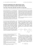
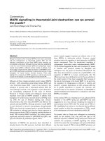
Figure 1
Trend for higher IC levels and IC-induced TNF-α levels in RA compared to control seraTrend for higher IC levels and IC-induced TNF-α levels in RA compared
to control sera. Sera from 15 RA patients and 15 healthy control indi-
viduals were PEG precipitated and added to PBMC cultures and incu-
bated for 20 hours at 37°C with 5% carbon dioxide, after which
supernatants were harvested and TNF-α measured using ELISA. Non-
significant trend toward (a) higher IgG levels and (b) greater TNF-α
induction from RA precipitates as compared with healthy controls were
apparent. Horizontal bars show the median value for each group.
ELISA, enzyme-linked immunosorbent assay; PBMC, peripheral blood
mononuclear cell; PEG, polyethylene glycol; RA, rheumatoid arthritis;
TNF, tumour necrosis factor.
Arthritis Research & Therapy Vol 8 No 3 Mathsson et al.
Page 4 of 10
(page number not for citation purposes)
F(ab')2 directed against the IgG γ chain (A0407, diluted
1:640; DAKO). The detection antibody was a goat F(ab')2
antibody directed against the human IgG γ chain adsorbed
against bovine immunoglobulins (109-056-098, dilution
1:10,000; Jackson ImmunoResearch Europe Ltd, Cambridge,
UK). A well characterized normal human serum was used to
construct a standard curve.
Serum anti-cyclic citrullinated peptide (CCP) was measured
using the Immunoscan RA Mark II assay (Euro-Diagnostica
AB, Malmö, Sweden) and the cutoff was set to 25 U/ml. In a
control group consisting of 99 healthy individuals, two exhib-
ited borderline positive reactivity (30 and 42 U/ml) and one
exhibited high positive reactivity (1,643 U/ml).
Monocyte depletion/enrichment
To investigate the hypothesis that monocytes were the main
responder cells, monocyte enrichment or depletion antibody
cocktails (RosetteSep™ StemCell Technologies, Vancouver,
Canada) were added to heparinized blood and purification
was performed in accordance with the manufacturer's instruc-
tions. This enrichment protocol yields totally untouched mono-
cytes for subsequent functional studies. Depletion and
enrichments were verified by staining with anti-CD14 FITC-
conjugated antibodies followed by flow cytometric analysis.
Cells depleted and enriched for monocytes were diluted in cell
culture medium to the same total cell concentration and the
same volume as used for untreated PBMCs, whereupon dis-
solved PEG precipitates were added to the cells.
FcγR blocking experiments
Anti-FcγRII monoclonal antibody (IV.3 [Fab fragment];
Medarex, Nutley, NY, USA) or anti-FcγRIII (3G8 [F(ab')2 frag-
ment]; Medarex) were added to the cells and left to stand at
4°C for 30 minutes before addition of dissolved PEG precipi-
tates. The antibody concentration used was 1.5 µg/ml; prelim-
inary experiments had shown equivalent blocking effect using
either 1.5 or 4 µg/ml. Antibody IV.3 was previously shown to
react specifically with FcγRIIa [20,21].
Statistical analysis
To neutralize inappropriate impact of outliers, nonparametric
statistics were used throughout the study. Mann Whitney U
test was used for comparison between groups, Spearman's
rank correlation test was used to evaluate correlations
between quantitative variables, and Kruskal-Wallis test was
used to investigate the association between DMARD thera-
pies on the one hand and serum RF and in vitro TNF-α
responses to PEG precipitates on the other. P <0.05 was
considered statistically significant.
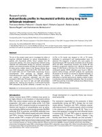
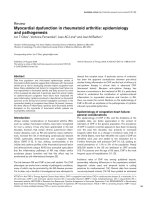
Figure 2
Correlation between SF precipitate induced TNF-α production, IgG lev-els in SF precipitates, and RF (n = 15)Correlation between SF precipitate induced TNF-α production, IgG lev-
els in SF precipitates, and RF (n = 15). Healthy PBMCs were stimu-
lated with PEG precipitates from SF from 15 patients with RA. The
stimulated cells were cultured for 20 hours at 37°C with 5% carbon
dioxide. IgG levels in the SF precipitates correlated with (a) TNF-α pro-
duction after SF PEG stimulation and (b) RF measured in serum. (c)
There was also a nonsignificant positive correlation between RF and
TNF-α induced by SF PEG precipitates. Statistical analyses were per-
formed with nonparametric tests to diminish the effect of outliers.
PBMC, peripheral blood mononuclear cell; PEG, polyethylene glycol;
RA, rheumatoid arthritis; RF, rheumatoid factor; SF, synovial fluid; TNF,
tumour necrosis factor.
Available online />Page 5 of 10
(page number not for citation purposes)
Results
Comparison of sera and synovial fluids from rheumatoid
arthritis patients with healthy control sera
In the first study we compared paired sera and SF from 15 RA
patients with sera from 15 healthy control individuals. We
observed a nonsignificant trend toward higher IgG levels (P =
0.0712) and greater induction of TNF-α (P = 0.0649) by
serum PEG precipitates from RA patients compared with
healthy control individuals (Figure 1a, b). Although the levels of
TNF-α induced by PEG precipitates from serum and parallel
SF samples differed considerably in both directions in individ-
ual pairs, there was no statistically significant difference
between TNF-α induction from RA serum or SF precipitates.
We also found that IgG levels in the SF precipitates were sig-
nificantly higher for RF-positive than for RF-negative RA
patients (P = 0.0033; data not shown). A positive correlation
was established between IgG levels in SF precipitates and
TNF-α production from PBMCs stimulated with SF precipi-
tates (r = 0.604, P = 0.0239; Figure 2a). There was also a
strong positive correlation between IgG levels in the RA SF
precipitates and RF measured in serum (r = 0.729, P =
0.0064; Figure 2b). We also found a link between TNF-α pro-
duced from PBMCs after stimulation with RA SF precipitates
and RF measured in serum (Figure 2c). None of these correla-
tions were evident for PEG precipitates obtained from parallel
RA serum samples (Table 1).
Correlation between tumour necrosis factor-α induction
by synovial fluid precipitates, IgG content in the
precipitates, and rheumatoid factor
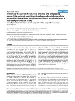
In the second study with paired sera and SF from 32 RA
patients we found the same association as in the first study
between RF, TNF-α production following SF precipitate stim-
ulation and IgG levels in SF precipitates (Figure 3, Table 1).
On splitting the RA patients into RF-positive and RF-negative
subgroups, the former exhibited significantly greater TNF-α
production induced by SF precipitates (P = 0.0004; data not
shown). We did not find any parallel correlations for the serum
precipitates except for a weak correlation between RF and IgG
content in the precipitates (r = 0.388, P = 0.0308; Table 1).
We also tried to measure RF in SF but because of technical
limitations we only got measurable RF values for 59% (19/32)
of the samples. However, in these 19 samples there was a
closer correlation between SF precipitate induced TNF-α and
RF measured in SF (r = 0.667, P = 0.0047; data not shown)
as compared with RF measured in serum (r = 0.284, P =
0.2279 [not significant]). Also, in this second study there was
no significant difference in TNF-α levels induced by PEG pre-
cipitates from RA sera and SF.
Anti-CCP levels correlate with rheumatoid factor but not
with IgG content in PEG precipitates or with PEG
precipitate-induced tumour necrosis factor-α levels
In the second study we measured levels of anti-CCP antibod-
ies in serum samples, of which 26 out of 32 (81%) were anti-
CCP positive. There was a positive correlation between anti-
CCP and RF levels (r = 0.516, P = 0.0041; data not shown)
in the serum samples, but we did not find any associations
between anti-CCP and PEG precipitate-induced TNF-α pro-
duction or IgG content in the precipitates (Table 1).
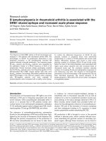
Correlation between tumour necrosis factor-α
production and number of swollen and tender joints
The amount of TNF-α produced after stimulation with SF pre-
cipitates correlated with the number of swollen and tender
joints at the time of sampling (Figure 4a, b). No such correla-
tion was found for stimulations with serum precipitates. We
could not see any correlation between TNF-α production and
age, sex, C-reactive protein, erythrocyte sedimentation rate,
time since last intra-articular steroid injection, disease duration
or medication, including peroral corticosteroids. There was no
statistically significant difference in RF levels between patients
treated with different DMARDs. However, all three patients
treated with Reumacon
®
and four out of seven of the patients
not receiving any DMARD were RF negative.
PEG precipitates from rheumatoid arthritis serum and
synovial fluid induces tumour necrosis factor-α from
monocytes
Paired sera and SF from two RA patients and serum from one
healthy control individual were PEG precipitated and used to
stimulate monocyte depleted, enriched, or unaffected PBMCs.
TNF-α production was totally abolished when 99.9% of the
monocytes were depleted. Conversely, when monocytes in
the PBMC cultures were enriched from 7.5% to 54.7% mono-
cytes, the TNF-α levels induced by serum precipitates were
increased by between 15.5% and 27.4%. For the SF precipi-
tates TNF-α production was increased to a greater extent (by
between 45.4% and 63.1%; data not shown).
Immune complex induced tumour necrosis factor-α
production is partly mediated via FcγRIIa
FcγRIIa and FcγRIII were blocked to investigate receptor
dependency of cytokine production induced by PEG precipi-
tates. TNF-α production induced by the precipitates was
reduced by 55% with blocking of FcγRIIa, but no effect of
blocking FcγRIII was apparent (Figure 5). PEG precipitates
from serum of healthy control individuals did not induce any or
very low amounts of TNF-α and consequently did not exhibit
any effect of FcγR blockade.
Discussion
This, to our knowledge, is the first study to show an associa-
tion between RF, IgG levels in SF ICs, and SF IC induced lev-
els of TNF-α in RA. We also report that IC-induced TNF-α
production is partly mediated via FcγRIIa with monocytes/mac-
rophages as the main or perhaps only responder cells. These
findings support the hypothesis that ICs in joints might provide
a direct link to cytokine-dependent inflammation in RA, at least
in RF-positive patients.
Arthritis Research & Therapy Vol 8 No 3 Mathsson et al.
Page 6 of 10
(page number not for citation purposes)
A stronger association between cytokine induction, IgG levels
and RF was apparent for the SF precipitates than for serum
precipitates, which is in agreement with the general belief that
RF levels in serum reflect inflammation in the joints. RF has
been associated with ICs in several diseases other than RA
[1,5]. RF can also be produced after vaccination in healthy
individuals during the time interval when antibodies and anti-
gen form circulating ICs [4]. RF-producing B cells are present
in the inflamed joints of RA patients [22] and RF measured in
serum might therefore mirror the production of RF in RA joints.
We also attempted to measure RF in SF, but for technical rea-
sons we only achieved measurable values in 59% of the
cases. Nonetheless, in the measurable subgroup of patients
there was a considerably stronger association between SF
PEG precipitate induced TNF-α production and RF in SF as
compared with conventional RF measured in serum. This find-
ing strengthens our hypothesis that serum RF is merely a
reflection of RF produced in the inflamed joints in response to
IgG-containing ICs with TNF-α-inducing properties. Moreover,
our findings of stronger cytokine-inducing properties of ICs
obtained from joints of RF-positive RA patients is consistent
with the fact that seropositive RA is associated with a more
severe disease outcome [2,3].
Anti-CCP antibodies have been shown to be highly specific for
RA [23] and more strongly associated with joint destruction
than RF [24]. As noted in several earlier studies, we saw a pos-
itive correlation between RF and anti-CCP in serum. However,
we did not find any associations between anti-CCP and IC-
induced TNF-α production or IgG levels in the PEG precipi-
tates. Therefore RF per se and not the RF-correlated anti-CCP
levels appear to be associated with IC-induced TNF-α and
consequent joint inflammation.
PEG precipitation is a well recognized technique for the isola-
tion of high-molecular-weight ICs. However, earlier investiga-
tions showed PEG-precipitated sera to contain uncomplexed
immunoglobulins, C3 [25] and a number of serum proteins
including fibronectin and albumin [26], besides IC containing
IgG plus IgA and IgG plus C3. The view that PEG precipitates
are composed only of ICs is therefore too simplistic. Because
our cross-sectional approach employed a large number of ICs
freshly prepared with an aseptic technique, we avoided the
use of alternative, time-consuming techniques such as gel fil-
tration and sucrose gradient centrifugation. To further deter-
mine IC content in our precipitates we measured IgG content
in the precipitates and showed that IC-induced cytokine
Table 1
Correlations between rheumatoid factor or anti-CCP and IgG content in immune complexes and TNF-α inducing properties of
immune complexes
Correlation Study 1 (n = 15) Study 2 (n = 32)
rP rP
RF/SF immune complexes
IgG in SF precipitates/TNF-α induced by SF precipitates 0.604 0.0239 0.503 0.0051
RF/TNF-α induced by SF precipitates 0.404 0.1310 (NS) 0.594 0.001
RF/IgG in SF precipitates 0.729 0.0064 0.360 0.0449
RF/serum immune complexes
IgG in serum precipitates/TNF-α induced by serum precipitates 0.171 NS -0.265 NS
RF/TNF-α induced by serum precipitates 0.332 NS 0.329 NS
RF/IgG in serum precipitates 0.346 NS 0.388 0.0308
Anti-CCP/SF immune complexes
Anti-CCP/TNF-α induced by SF precipitates ND ND 0.043 NS
Anti-CCP/IgG in SF precipitates ND ND 0.083 NS
Anti-CCP/serum immune complexes
Anti-CCP/TNF-α induced by serum precipitates ND ND 0.191 NS
Anti-CCP/IgG in serum precipitates ND ND 0.246 NS
CCP, cyclic citrullinated peptide; ND, not done; NS, not significant; RA, rheumatoid arthritis; RF, rheumatoid factor; SF, synovial fluid; TNF, tumour
necrosis factor.
Available online />Page 7 of 10
(page number not for citation purposes)
induction was dependent on binding to FcγRIIa that, because
of its low affinity, preferably binds ICs over monomeric IgG
[27].
Control experiments have shown that PEG precipitation of
ultracentrifuged NHS or RF positive sera devoid of preformed
ICs do not enhance TNF-α-inducing effects compared with
serum added directly to the cell cultures without prior PEG
precipitation. PEG precipitates from ultracentrifuged RF-neg-
ative NHS or from ultracentrifuged RF-positive sera induce
comparable levels of TNF-α when they are added to responder
PBMC cultures. These findings imply that neither PEG precip-
itation nor RF per se induce IC formation when no ICs are
present initially. PEG precipitation on the other hand enhances
TNF-α production when preformed ICs had been added to
ultracentrifued NHS or RF-positive sera before PEG precipita-
tion. An enhancing effect was also seen when nonaggregated
IgG (Endobulin
®
; Baxter, Vienna, Austria) was added to ultra-
centrifuged RF-positive sera before PEG precipitation, proba-
bly because of minute amounts of dimer IgG in the preparation
acting as small ICs.
In this cross-sectional study SF and serum samples were col-
lected in association with therapeutic arthrocenthesis. Our
finding that IC-induced TNF-α induction in vitro correlates with
the number of swollen and tender joints at the time of sampling
suggests that IC-induced cytokine levels might reflect a gen-
eral quality of joint inflammation in individual patients. We are
currently studying the cytokine inducing properties of paired
SF samples from different joints obtained at the same time
point, as well as paired SF samples from the same joint at dif-
ferent time points; in this way we aim to test the hypothesis
that RF-associated induction of proinflammatory cytokines by
joint ICs is a stable quality over space and time in individual
patients with RA.
In the present study we examined the cytokine inducing effects
of soluble ICs from RA SF. Collagen type II antibodies occur
in a subpopulation of RA patients and these antibodies may
form solid phase ICs at the cartilage surface in RA joints. We
are currently investigating such ICs to obtain information
regarding the similarities and dissimilarities between cytokine
responses to soluble ICs (with hitherto unknown antibody spe-
cificities) obtained in vivo and artificial ICs created using well
known autoantibodies directed against collagen type II [28].
Monocytes/macrophages were shown to be the main or per-
haps only responder cells in the induction of TNF-α in our sys-
tems. The importance of monocytes in IC-driven joint
inflammation is supported by earlier rodent experiments in
which synovial macrophages were shown to play a central role
in IC-induced arthritis models [6-8,29]. In addition, most dis-
ease-modifying drugs in RA are directed at suppressing mono-
cytes and monocyte-derived cytokines [30]. Recent findings
have also highlighted the importance of monocytes/macro-
Figure 3
Correlation between SF precipitate induced TNF-α production, IgG lev-els in SF precipitates, and RF (n = 32)Correlation between SF precipitate induced TNF-α production, IgG lev-
els in SF precipitates, and RF (n = 32). Healthy PBMCs were stimu-
lated with PEG precipitates from SF from 32 patients with RA. The
stimulated cells were cultured for 20 hours at 37°C with 5% carbon
dioxide. IgG levels in the SF precipitates correlated with (a) TNF-α pro-
duction after SF PEG stimulation and (b) with RF measured in serum.
(c) There was also a correlation between RF and TNF-α induced by SF
PEG precipitates. Statistical analyses were performed with nonpara-
metric tests to diminish the effect of outliers. PBMC, peripheral blood
mononuclear cell; PEG, polyethylene glycol; RA, rheumatoid arthritis;
RF, rheumatoid factor; SF, synovial fluid; TNF, tumour necrosis factor.
Arthritis Research & Therapy Vol 8 No 3 Mathsson et al.
Page 8 of 10
(page number not for citation purposes)
phages [9] and monocyte-derived dendritic cells [10] in IC-
induced cytokine production in RA joints.
Earlier studies conducted by Jarvis and coworkers [31,32]
demonstrated cytokine-inducing properties of gel filtrated ICs
from SF of patients with juvenile RA. Pretreatment of these ICs
with native serum decreased subsequent cytokine production
as compared with either pretreatment with heat-inactivated
serum or no pretreatment [32]. These findings and our data on
FcγRIIa-dependent cytokine production together argue that
when ICs become heavily coated with complement, Fc frag-
ments are covered by complement and prevented from inter-
action with Fc receptors, as was proposed by Nilsson [33].
Although complement activation by SF ICs is substantial, for
two reasons we chose to study the effect of our ICs in a
serum-free cell culture system. PEG precipitated ICs are
known to carry covalently bound complement proteins after
complement activation in the joint [25]. The amount of comple-
ment proteins on ICs from different joints therefore differs and
is dependent on access to the classical complement cascade
in the joints of individual patients. By exposing these ICs to a
standardized native serum in vitro, all ICs will induce comple-
ment activation and differences between individual patients
might diminish or disappear. When screening various cell cul-
ture systems we also found that a serum-free medium supple-
mented with Ultroser
®
was superior to serum-containing cell
culture media in sustaining IC-induced cytokine production. It
was thereby also possible to investigate weak IC-induced
responses that were not detected using other cell culture
media formulations.
Although according to the literature RA SF may contain higher
concentrations of ICs than RA serum [34], there was no sig-
nificant difference between TNF-α levels induced by serum or
SF precipitates. To be able to precipitate SF samples, hyaluro-
nidase treatment was needed. Also, a number of joint-specific
proteins such as partly degraded hyaluronic acid might be co-
precipitated with SF ICs in parallel with what has been
described for serum proteins [25,26]. Because of the experi-
mental setup, we chose not to draw any conclusions from
these findings of no difference, but instead we opted to con-
centrate on differences in cytokine responses between PEG
precipitates from body fluids treated equally during the precip-
itation procedure.
Many studies have reported the importance of Fcγ receptors
in the development of experimental arthritis. Thus, knockout
mice lacking the activating FcγRIII have been shown to be pro-
tected from arthritis [13] and knockout mice lacking the inhib-
itory FcγRIIb develop arthritis on a nonarthritis susceptible
background [13,35]. However, the effect of deleting FcγRIIb
has not been consistent [36]. Rodents lack the primate-spe-
cific activating FcγRIIa, which has been shown to be elevated
on RA monocytes compared with healthy control individuals
[15]. Arguments are now accumulating that FcγRIIa might be
a key activating mediator of IC-induced effects in humans and
to act as the functional counterpart of FcγRIII in rodents [14].
In the present study, blocking of FcγRIIa resulted in markedly
reduced IC-induced TNF-α production, indicating that the IC-
induced cytokine production is at least partly mediated via
FcγRIIa. We earlier reported that ICs from, for example,
Figure 4
TNF-α production induced by SF precipitates correlate with the number of swollen and tender jointsTNF-α production induced by SF precipitates correlate with the
number of swollen and tender joints. PBMCs were stimulated with SF
precipitates from 32 RA patients for 20 hours. TNF-α levels in the
supernatants were measured using ELISA and data regarding the
number of swollen and tender joints were collected from the patients'
charts. The numbers of (a) swollen and (b) tender joints correlated with
TNF-α production. Statistical analyses were performed with nonpara-
metric tests to diminish the effect of outliers. ELISA, enzyme-linked
immunosorbent assay; PBMC, peripheral blood mononuclear cell; RA,
rheumatoid arthritis; SF, synovial fluid; TNF, tumour necrosis factor.
Available online />Page 9 of 10
(page number not for citation purposes)
patients with systemic lupus erythematosus and artificial ICs
can induce cytokine production via FcγRIIa, together with a
correlation between IC-induced cytokine production and
monocyte density of FcγRII, but not FcγRI or FcγRIII [17]. We
also observed that ICs from patients with cryoglobulinaemia
induces cytokine production via FcγRIIa [37]. Blom and cow-
orkers [9] recently reported that the expression levels of FcγRII
and Fcγ III are elevated on mature RA macrophages and that
FcγR expression is correlated with IC induced levels of TNF-α
[9]. Collectively, these data indicate an important role for FcγR
expression on monocytes/macrophages in IC-induced inflam-
mation in RA joints, and argue that FcγRIIa blockade is a pos-
sible means to suppress IC-driven inflammation in RA.
However, a role for FcγRIII can not be excluded for two rea-
sons. The anti-FcγRIII antibody 3G8 used in our studies has
been shown occasionally to exert a nonspecific stimulatory
effect on cytokine production [17,28]. Second, levels of
monocyte expression of FcγRIII is low on unstimulated PBMC
monocytes. In our earlier report [17] we investigated FcγR
monocyte surface expression in 10 different PBMC popula-
tions. Whereas FcγRIII/CD16 exhibited low expression
(median fluorescence intensity 39, as comparable with the
nonspecific control antibody), levels of FcγRII/CD32 and
FcγRI/CD64 were substantially higher (median fluorescence
intensities 538 and 1133, respectively; data not shown). The
selective importance of FcγRIII on inflammatory macrophages
with increased FcγRIII surface expression [9] must therefore
be investigated separately.
Conclusion
We demonstrated a clear correlation between RF, IgG levels
in PEG precipitated high-molecular-weight ICs from RA SF,
and TNF-α production induced in vitro by these ICs. This sup-
ports the hypothesis that ICs are formed in the inflamed RA
joint in parallel with RF production. Such ICs may then stimu-
late monocytes/macrophages in the joint to produce TNF-α via
FcγRIIa stimulation. Suppression of monocytes/macrophages
in the joints or blockade of the specific activating FcγRIIa
receptor might therefore be a means to reduce IC-induced
TNF-α production in the joints of seropositive RA patients.
Authors' contributions
LM planned the work, carried out the laboratory work and per-
formed statistical analysis, as well as drafting the manuscript.
JL collected the patient data and samples, and helped to draft
the manuscript. MM helped with the laboratory work, and read
and approved the final manuscript. JR participated in the
design of the study, helped with the statistical analysis, and
helped to draft the manuscript. All authors read and approved
of the final manuscript.
Acknowledgements
This investigation was supported by grants from the Swedish Research
Council, the Swedish Society of Medicine, King Gustav V's 80-years
Fund, the Swedish League Against Rheumatism, the Ugglas Founda-
tion, the Hierta Foundation, the Crafoord Foundation, the Groschinsky
Foundation, the Grönberg Foundation, the Bergvall Foundation, the
Dahlin Foundation, the Carlsson Foundation, the Viberg Foundation, the
Nanna Svartz Foundation and the Swedish Fund for Research without
Animal Experiments.
We thank Assoc Prof R.A Harris for linguistic advice.
References
1. Newkirk MM: Rheumatoid factors: host resistance or autoim-
munity? Clin Immunol 2002, 104:1-13.
2. van Zeben D, Hazes JM, Zwinderman AH, Cats A, van der Voort
EA, Breedveld FC: Clinical significance of rheumatoid factors in
early rheumatoid arthritis: results of a follow up study. Ann
Rheum Dis 1992, 51:1029-1035.
3. Scott DL: Prognostic factors in early rheumatoid arthritis.
Rheumatology (Oxford) 2000:24-29.
4. Tarkowski A, Czerkinsky C, Nilsson LA: Simultaneous induction
of rheumatoid factor- and antigen-specific antibody-secreting
cells during the secondary immune response in man. Clin Exp
Immunol 1985, 61:379-387.
5. Dorner T, Egerer K, Feist E, Burmester GR: Rheumatoid factor
revisited. Curr Opin Rheumatol 2004, 16:246-253.
6. van Lent PL, van den Hoek A, van den Bersselaar L, Dijkstra CD,
van Rooijen N, van den Berg WB: Role of synovial macrophages
in experimental arthritis. Res Immunol 1992, 143:229-234.
7. van Lent PL, van den Hoek AE, van den Bersselaar LA, Spanjaards
MF, van Rooijen N, Dijkstra CD, van de Putte LB, van den Berg
WB: In vivo role of phagocytic synovial lining cells in onset of
experimental arthritis. Am J Pathol 1993, 143:1226-1237.
8. van Lent PL, Holthuysen AE, van den Bersselaar L, van Rooijen N,
van de Putte LB, van den Berg WB: Role of macrophage-like
synovial lining cells in localization and expression of experi-
mental arthritis. Scand J Rheumatol Suppl 1995, 101:83-89.
Figure 5
PEG precipitates from RA sera and SF induces TNF-α production via FcγRIIaPEG precipitates from RA sera and SF induces TNF-α production via
FcγRIIa.Anti-FcγRIIa and anti-FcγRIII antibodies were added to separate
PBMC cultures before addition of PEG precipitates; culture was then
continued for 20 hours. Anti-FcγRIIa antibodies blocked TNF-α produc-
tion in cultures stimulated with PEG precipitates from RA sera and SF.
One out of two experiments is shown. PBMC, peripheral blood mono-
nuclear cell; PEG, polyethylene glycol; RA, rheumatoid arthritis; SF,
synovial fluid; TNF, tumour necrosis factor.
Arthritis Research & Therapy Vol 8 No 3 Mathsson et al.
Page 10 of 10
(page number not for citation purposes)
9. Blom AB, Radstake TR, Holthuysen AE, Sloetjes AW, Pesman GJ,
Sweep FG, van de Loo FA, Joosten LA, Barrera P, van Lent PL, Van
den Berg WB: Increased expression of Fcgamma receptors II
and III on macrophages of rheumatoid arthritis patients
results in higher production of tumor necrosis factor alpha and
matrix metalloproteinase. Arthritis Rheum 2003,
48:1002-1014.
10. Radstake TR, Blom AB, Sloetjes AW, van Gorselen EO, Pesman
GJ, Engelen L, Torensma R, van den Berg WB, Figdor CG, van
Lent PL, et al.: Increased FcgammaRII expression and aberrant
tumour necrosis factor alpha production by mature dendritic
cells from patients with active rheumatoid arthritis. Ann
Rheum Dis 2004, 63:1556-1563.
11. Blom AB, van Lent PL, van Vuuren H, Holthuysen AE, Jacobs C,
van de Putte LB, van de Winkel JG, van den Berg WB: Fc gamma
R expression on macrophages is related to severity and chro-
nicity of synovial inflammation and cartilage destruction dur-
ing experimental immune-complex-mediated arthritis (ICA).
Arthritis Res 2000, 2:489-503.
12. Diaz de Stahl T, Andren M, Martinsson P, Verbeek JS, Kleinau S:
Expression of FcgammaRIII is required for development of
collagen-induced arthritis. Eur J Immunol 2002, 32:2915-2922.
13. Yuasa T, Kubo S, Yoshino T, Ujike A, Matsumura K, Ono M,
Ravetch JV, Takai T: Deletion of fcgamma receptor IIB renders
H-2(b) mice susceptible to collagen-induced arthritis. J Exp
Med 1999, 189:187-194.
14. Tan Sardjono C, Mottram PL, Hogarth PM: The role of Fcgamma-
RIIa as an inflammatory mediator in rheumatoid arthritis and
systemic lupus erythematosus. Immunol Cell Biol 2003,
81:374-381.
15. Wijngaarden S, van de Winkel JG, Jacobs KM, Bijlsma JW, Lafeber
FP, van Roon JA: A shift in the balance of inhibitory and activat-
ing Fcgamma receptors on monocytes toward the inhibitory
Fcgamma receptor IIb is associated with prevention of mono-
cyte activation in rheumatoid arthritis. Arthritis Rheum 2004,
50:3878-3887.
16. Feldmann M, Maini RN: Anti-TNF alpha therapy of rheumatoid
arthritis: what have we learned? Annu Rev Immunol 2001,
19:163-196.
17. Rönnelid J, Tejde A, Mathsson L, Nilsson-Ekdahl K, Nilsson B:
Immune complexes from SLE sera induce IL10 production
from normal peripheral blood mononuclear cells by an Fcgam-
maRII dependent mechanism: implications for a possible
vicious cycle maintaining B cell hyperactivity in SLE. Ann
Rheum Dis 2003, 62:37-42.
18. Pontes-de-Carvalho LC, Lannes-Vieira J, Giovanni-de-Simone S,
Galvao-Castro B: A protein A-binding, polyethylene glycol pre-
cipitation-based immunoradiometric assay. Application to the
detection of immune complexes and C3 in human sera and of
private antigens in cross-reacting parasite extracts. J Immunol
Methods 1986, 89:27-35.
19. Tejde A, Mathsson L, Ekdahl KN, Nilsson B, Ronnelid J: Immune
complex-stimulated production of interleukin-12 in peripheral
blood mononuclear cells is regulated by the complement sys-
tem. Clin Exp Immunol 2004, 137:521-528.
20. Vely F, Gruel N, Moncuit J, Cochet O, Rouard H, Dare S, Galon J,
Sautes C, Fridman WH, Teillaud JL: A new set of monoclonal
antibodies against human Fc gamma RII (CD32) and Fc
gamma RIII (CD16): characterization and use in various
assays. Hybridoma 1997, 16:519-528.
21. Van Den Herik-Oudijk IE, Westerdaal NA, Henriquez NV, Capel PJ,
Van De Winkel JG: Functional analysis of human Fc gamma RII
(CD32) isoforms expressed in B lymphocytes. J Immunol
1994, 152:574-585.
22. Reparon-Schuijt CC, van Esch WJ, van Kooten C, Levarht EW,
Breedveld FC, Verweij CL: Functional analysis of rheumatoid
factor-producing B cells from the synovial fluid of rheumatoid
arthritis patients. Arthritis Rheum 1998, 41:2211-2220.
23. Schellekens GA, Visser H, de Jong BA, van den Hoogen FH,
Hazes JM, Breedveld FC, van Venrooij WJ: The diagnostic prop-
erties of rheumatoid arthritis antibodies recognizing a cyclic
citrullinated peptide. Arthritis Rheum 2000, 43:155-163.
24. Forslind K, Ahlmen M, Eberhardt K, Hafstrom I, Svensson B: Pre-
diction of radiological outcome in early rheumatoid arthritis in
clinical practice: role of antibodies to citrullinated peptides
(anti-CCP). Ann Rheum Dis 2004, 63:1090-1095.
25. Crowley-Nowick PA, Campbell E, Schrohenloher RE, Mestecky J,
Mestecky J, Jackson S: Polyethylene glycol precipitates of
serum contain a large proportion of uncomplexed immu-
noglobulins and C3. Immunol Invest 1996, 25:91-101.
26. Robinson MW, Scott DG, Bacon PA, Walton KW, Coppock JS,
Scott DL: What proteins are present in polyethylene glycol pre-
cipitates from rheumatic sera? Ann Rheum Dis 1989,
48:496-501.
27. Dijstelbloem HM, van de Winkel JG, Kallenberg CG: Inflamma-
tion in autoimmunity: receptors for IgG revisited. Trends Immu-
nol 2001, 22:510-516.
28. Mullazehi M, Mathsson L, Lampa J, Rönnelid J: Surface-bound
anti-type II collagen containing immune complexes induce
production of TNF-alpha, IL-1beta and IL-8 from peripheral
blood mononuclear cells via Fc(gamma)RIIa. A potential
patho-physiological mechanism för humoral anti-collagen
type II immunity in arthritis. Arthritis Rheum 2006 in press.
29. van den Berg WB, van Lent PL: The role of macrophages in
chronic arthritis. Immunobiology 1996, 195:614-623.
30. Barrera P, Boerbooms AM, van de Putte LB, van der Meer JW:
Effects of antirheumatic agents on cytokines. Semin Arthritis
Rheum 1996, 25:234-253.
31. Jarvis JN, Wang W, Moore HT, Zhao L, Xu C: In vitro induction of
proinflammatory cytokine secretion by juvenile rheumatoid
arthritis synovial fluid immune complexes. Arthritis Rheum
1997, 40:2039-2046.
32. Jarvis JN, Xu C, Wang W, Petty HR, Gonzalez M, Morssy N, Wax-
man F, Quintero del Rio A: Immune complex size and comple-
ment regulate cytokine production by peripheral blood
mononuclear cells. Clin Immunol 1999, 93:274-282.
33. Nilsson UR: Deposition of C3b/iC3b leads to the concealment
of antigens, immunoglobulins and bound C1q in complement-
activating immune complexes. Mol Immunol 2001,
38:151-160.
34. Antes U, Heinz HP, Schultz D, Brackertz D, Loos M: C1q-bearing
immune complexes detected by a monoclonal antibody to
human C1q in rheumatoid arthritis sera and synovial fluids.
Rheumatol Int 1991, 10:245-250.
35. Kleinau S, Martinsson P, Heyman B: Induction and suppression
of collagen-induced arthritis is dependent on distinct
fcgamma receptors. J Exp Med 2000, 191:1611-1616.
36. Kagari T, Tanaka D, Doi H, Shimozato T: Essential role of Fc
gamma receptors in anti-type II collagen antibody-induced
arthritis. J Immunol 2003, 170:4318-4324.
37. Mathsson L, Tejde A, Carlson K, Hoglund M, Nilsson B, Nilsson-
Ekdahl K, Ronnelid J: Cryoglobulin-induced cytokine production
via FcgammaRIIa: inverse effects of complement blockade on
the production of TNF-alpha and IL-10. Implications for the
growth of malignant B-cell clones. Br J Haematol 2005,
129:830-838.