Báo cáo khoa học: "A multidisciplinary approach for the treatment of GIST liver metastasis" docx
Bạn đang xem bản rút gọn của tài liệu. Xem và tải ngay bản đầy đủ của tài liệu tại đây (1.1 MB, 4 trang )
BioMed Central
Page 1 of 4
(page number not for citation purposes)
World Journal of Surgical Oncology
Open Access
Case report
A multidisciplinary approach for the treatment of GIST liver
metastasis
Pejman Radkani*, Marcelo M Ghersi, Juan C Paramo and Thomas W Mesko
Address: Department of Surgery, Section of Surgical Oncology, Mount Sinai Medical Center, Miami Beach, Florida, USA
Email: Pejman Radkani* - ; Marcelo M Ghersi - ; Juan C Paramo - ;
Thomas W Mesko -
* Corresponding author
Abstract
Background: Advanced gastrointestinal stromal tumors (GISTs) can metastasize and recur after
a long remission period, resulting in serious morbidity, mortality, and complex management issues.
Case presentation: A 67-year-old woman presented with epigastric fullness, mild jaundice and
weight loss with a history of a bowel resection 7 years prior for a primary GIST of the small bowel.
The finding of a heterogeneous mass 15.5 cm in diameter replacing most of the left lobe of the liver
by ultrasonography and CT, followed by positive cytological studies revealed a metastatic GIST.
Perioperative optimization of the patient's nutritional status along with biliary drainage, and portal
vein embolization were performed. Imatinib was successful in reducing the tumor size and
facilitating surgical resection.
Conclusion: A well-planned multidisciplinary approach should be part of the standard
management of advanced or metastatic GIST.
Background
Gastrointestinal stromal tumors (GISTs) are neoplasms of
the gastrointestinal tract. Despite their less aggressive
pathologic nature, GISTs can metastasize and recur after a
long remission period. Such cases may produce serious
morbidity, mortality, and complex management issues
for the treating physician. We hereby report the case of a
patient who presented with an isolated metastatic GIST to
the liver that was successfully treated with a multidiscipli-
nary approach including imatinib therapy, portal vein
embolization, and hepatic lobectomy.
Case presentation
A 67-year-old woman presented with epigastric fullness,
mild jaundice and a 12-pound weight loss over a period
of 3 months. The patient had a history of a bowel resec-
tion 7 years prior to presentation for an unknown malig-
nancy. On physical examination, her abdomen was soft
with a palpable and non-tender mass in the mid-epigas-
trium. Initial work-up including ultrasonography revealed
a large liver lesion, follow-up CT confirmed the presence
of a heterogeneous mass 17.5 cm in diameter replacing
most of the left lobe of the liver (Figure 1a, 1b) with
marked compression of the right biliary tree. Initial Liver
function testes showed:
total billirubin: 4, direct billirubin: 3.93, alkaline phos-
phatase: 942, AST: 124, ALT: 156. The addition laboratory
values were within normal limit.
The patient was admitted to the hospital for additional
work-up. A percutaneous transhepatic cholangiogram
Published: 9 May 2008
World Journal of Surgical Oncology 2008, 6:46 doi:10.1186/1477-7819-6-46
Received: 23 October 2007
Accepted: 9 May 2008
This article is available from: />© 2008 Radkani et al; licensee BioMed Central Ltd.
This is an Open Access article distributed under the terms of the Creative Commons Attribution License ( />),
which permits unrestricted use, distribution, and reproduction in any medium, provided the original work is properly cited.
World Journal of Surgical Oncology 2008, 6:46 />Page 2 of 4
(page number not for citation purposes)
was performed, with placement of a right biliary
drainage catheter for decompression. The bilirubin
and liver function tests at the day before drainage
placement were as follow: total billirubin: 4 direct bil-
lirubin: 3.93 ALK: 942 AST: 124 ALT: 156. Two days
later the labs were as follow: total billirubin: 3.45
direct billirubin: 3.28, alkaline phosphatase: 788 AST:
117 ALT: 123, and 30 days later was: total billirubin:
0.7 direct billirubin: 0.30 alkaline phosphatase: 130
AST: 28 ALT: 25. A core liver biopsy was also done at
the time, which demonstrated atypical spindle cells.
Immuno-histochemical studies yielded positive
CD117, vimentin and actin stains, all consistent with
GIST. It was later established that the patient had pre-
viously undergone a small bowel resection for a pri-
mary GIST. Upper and lower endoscopy as well as
small bowel series were subsequently performed.
These revealed no tumors of the GI tract, suggesting
the liver mass was a late and isolated metastatic man-
ifestation of the prior GIST tumor.
A multidisciplinary and staged treatment course was rec-
ommended. Side effects and benefits of using Imatinib
drug were considered by our tumor board, and the patient
was started at a dose of 600 mg per day to reduce tumor
size. The patient was followed regularly for the next few
months as an outpatient. Her jaundice resolved and the
biliary catheter was successfully removed four months
after placement. A significant clinical improvement was
noted, with resolution of the patient's initial symptoms
and a 7-pound increase in body weight. Frequent abdom-
inal CT scans showed a hepatic mass that diminished in
size, but stabilized after 6 months of imatinib therapy at a
diameter of 11 cm (Figure 2a, 2b).
The patient then underwent portal vein embolization
(PVE) in hopes of promoting hypertrophy of the right
lobe and further atrophy of the tumor-laden left hepatic
lobe, in preparation for surgical resection.
Two months following PVE, while still on imatinib, the
patient underwent an uncomplicated left hepatic lobec-
tomy with cholecystectomy (Figure 3). Intraoperative
ultrasonography showed a hypertrophied right liver lobe,
and a 11 cm tumor involving liver segments 2, 3, 4A an
4B. Pathologic examination corroborated the diagnosis of
metastatic GIST with margins of resection free of tumor.
The patient tolerated the procedure well and was sent
home after a 14-day hospitalization. The postoperative
course was complicated by the formation of a subhepatic
abscess that was successfully treated with drainage cathe-
ters and systemic antibiotics. Imatinib was discontinued
approximately one month after surgery for a total of one
year of therapy. Follow-up CT 6 months after surgery
demonstrated no residual neoplastic disease (Figure 4a,
4b). At fourteen-months follow up, the patient was found
to be doing very well with no evidence of recurrent dis-
ease.
Discussion
Gastrointestinal stromal tumors are the most common
mesenchymal neoplasms of the GI tract. They have an
overall incidence of 3000–5000 cases per year in the
United States [1-3]. It is thought that these tumors differ-
entiate from intestinal pacemaker cells, also known as
interstitial cells of Cajal [1]. They affect mostly males
between the ages of 50 and 70, and are usually found inci-
dentally at early stages [1-4]. Large or advanced lesions
may present with a variety of clinical findings, including
bleeding, abdominal pain, early satiety, bowel obstruc-
tion, or perforation.
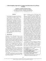
computerized tomography A) and B); mass in the left lobe of the liver has decreased in size with respect to the prior studyFigure 2
computerized tomography A) and B); mass in the left lobe of
the liver has decreased in size with respect to the prior
study.
Computerized tomography A) and B); evaluation of the liver demonstrated a large inhomogeneous mass with multiple areas of cystic component within the left lobe of the liverFigure 1
Computerized tomography A) and B); evaluation of the liver
demonstrated a large inhomogeneous mass with multiple
areas of cystic component within the left lobe of the liver.
The mass measured 17.6 × 14 cm. Mild dilatation of the int-
rahepatic biliary radicals in the right lobe liver.
World Journal of Surgical Oncology 2008, 6:46 />Page 3 of 4
(page number not for citation purposes)
GISTs are usually detected by endoscopy, CT or MRI per-
formed for abdominal symptoms. The gold standard for
diagnosing GISTs is pathological tissue examination,
which normally demonstrates atypical splindle cells. A
positive stain for CD117 carries a specificity of 95% for
these tumors, and will unequivocally establish a diagnosis
[1-7].
When GISTs originate in the small bowel, they behave in
a more aggressive manner [8]. The most common site for
metastases is the liver and the peritoneal cavity, but can
also occur in bone, skin, soft tissues, and lymph nodes [5].
Negative prognostic factors for aggressiveness and recur-
rence include tumor size, a high mitotic index, or an
unknown site of origin [9,10]. Recurrence usually occurs
19–26 months after surgery [3,11,12]. For this reason, the
National Comprehensive Cancer Network suggests rou-
tine follow-up CT scans of the abdomen and pelvis every
3–6 month for the first 3–5 years after resection [7].
The only definitive treatment for GISTs is surgical resec-
tion. This can be done laparoscopically in some cases, or
with the traditional open approach. The mainstay of sur-
gical therapy in primary or metastatic disease is to achieve
a complete resection with negative margins [7]. Conven-
tional chemotherapy and radiation therapy may have
minor adjunctive benefits in unresectable or metastatic
GISTs [2]. Imatinib mesylate (Gleevec
®
), a selective inhib-
itor of tyrosine kinase, has revolutionized the manage-
ment of this disease in recent years. Imatinib has a
significant shrinking effect on GISTs, and can be used
when primary GISTs have attained a very large size or are
in unfavorable locations, increasing the risk of positive
resection margins [1]. Imatinib has also become the first
line of treatment for recurrent and/or metastatic GISTs, as
described for the patient in this case report [13]. Imatinib
is generally very well tolerated; and most patients can tol-
erate treatment without interruption. The more common
side effects of Imatinib mesylate include [14,15]: nausea,
vomiting, diarrhea, and muscle cramps. It is common to
see a decrease in the neutrophil and platelet counts espe-
cially during the first month of therapy [16]. The drug
should be stopped to allow recovery if the absolute neu-
trophil count (ANC) falls to <1,000/microL and/or the
platelet count to <50,000/microL during the first months
of therapy [17]. Our patient had no evidence of these side
effects during therapy. More recently, sunitinib malate
(Sutent
®
), a multikinase inhibitor has been approved by
the Food and Drug Administration for treatment of GISTs
that are refractory to imatinib [18].
Resection of GIST liver metastases may be curative when
the primary disease has been eradicated and negative sur-
gical resection margins are attained. However, a large
tumor burden in the hepatic parenchyma may prohibit
resection given the risk of insufficient remaining liver tis-
sue and subsequent postoperative liver failure [19]. An
option to counteract this phenomenon is the use of portal
vein embolization (PVE) in cases of unilobular involve-
ment of the liver. First used in the 1980s, selective PVE
induces atrophy of a selected liver region as well as a com-
pensatory hypertrophy of the remaining liver parenchyma
[20-23]. Preoperative PVE is recommended if less than
30% to 40% of normal the liver is expected to remain and
be functional after resection [24,25].
Conclusion
This case, to our best knowledge, represents the first in the
literature describing a multidisciplinary approach for the
successful management of a large metastasic GIST to the
liver. We attribute the success of this case to a well thought
out management plan set forth by a dedicated tumor
Computerized tomography A) and B); no evidence of meta-static diseaseFigure 4
Computerized tomography A) and B); no evidence of meta-
static disease.
left lobe of the liver, with falciform ligament gallbladder and xiphoid processsFigure 3
left lobe of the liver, with falciform ligament gallbladder and
xiphoid process.
Publish with BioMed Central and every
scientist can read your work free of charge
"BioMed Central will be the most significant development for
disseminating the results of biomedical research in our lifetime."
Sir Paul Nurse, Cancer Research UK
Your research papers will be:
available free of charge to the entire biomedical community
peer reviewed and published immediately upon acceptance
cited in PubMed and archived on PubMed Central
yours — you keep the copyright
Submit your manuscript here:
/>BioMedcentral
World Journal of Surgical Oncology 2008, 6:46 />Page 4 of 4
(page number not for citation purposes)
board utilizing advanced and evidence-based therapeutic
modalities. Timing and resource utilization were key fac-
tors in the management of this patient. Perioperative opti-
mization of the patient's nutritional status and state of
health with biliary drainage was helpful. The pharmaco-
logic effect of imatinib reduced the tumor size and
improved the surgical resectability. Additionally, PVE
facilitated the operation and promoted healthy liver tissue
hypertrophy. Lastly, careful operative technique and ded-
icated follow up allowed for a good surgical outcome in
this patient. A well-planned multidisciplinary approach
should be part of the standard management of advanced
or metastatic GISTs.
Competing interests
The authors declare that they have no competing interests.
Authors' contributions
TM, JP and MG designed the study. PR carried out the data
and bibliographic research and drafted the manuscript.
MG carried out the picture acquisition, manuscript revi-
sion and editing process. TM and JP did the last manu-
script revision and the editing process.
Acknowledgements
Written consent of the patient was obtained for publication of this case
report.
The authors would like to thank Antonio Martinez, MD, from the depart-
ment of pathology at Mount Sinai Medical Center for his help and contribu-
tion in the pathology aspects of this manuscript.
References
1. Gold JS, DeMatteo RP: Combined surgical and medical therapy,
the gastrointestinal stromal tumor model. Ann Surg 2006,
244:176-184.
2. DeMatteo RP, Lewis JJ, Leung D, Mudan SS, Woodruff JM, Brennan
MF: Two hundred gastrointestinal stromal tumors: recur-
rence patterns and prognostic factors for survival. Ann Surg
2000, 231:51-58.
3. Langer C, Gunawan B, Schüler P, Huber W, Füzesi L, Becker H: Prog-
nostic factors influencing surgical management and out-
come of gastrointestinal stromal tumours. Br J Surg 2003,
90:332-339.
4. Miettinen M, El-Rifai W, HL Sobin L, Lasota J: Evaluation of malig-
nancy and prognosis of gastrointestinal stromal tumors: a
review. Hum Pathol 2002, 33:478-483.
5. Miettinen M, Lasota J: Gastrointestinal stromal tumors; review
on morphology, molecular pathology, prognosis, and differ-
ential diagnosis. Arch Pathol Lab Med 2006, 130:1466-1478.
6. Dematteo RP, Maki RG, Antonescu C, Brennan MF: Targeted
molecular therapy for cancer: the application of STI571 to
gastrointestinal stromal tumor. Curr Probl Surg 2003,
40:144-193.
7. Zwan SM Van Der, Dematteo RP: Gastrointestinal stromal
tumors: 5 years later. Cancer 2005, 104:1781-1788.
8. DeMatteo RP: The GIST of targeted cancer therapy: a tumor,
a mutated gene (c-kit), and a molecular inhibitor (STI571).
Ann Surg Oncol 2002, 9:831-839.
9. Emory TS, Sobin LH, Lukes L, Lee DH, O'Leary TJ: Prognosis of gas-
trointestinal smooth-muscle (stromal) tumors: dependence
on anatomic site. Am J Surg Pathol 1999, 23:82-87.
10. Berman J, O'Leary TJ: Gastrointestinal stromal tumor work-
shop. Hum Pathol 2001, 32:578-582.
11. Crosby JA, Catton CN, Davis A, Couture J, O'Sullivan B, Kandel R,
Swallow CJ: Malignant gastrointestinal stromal tumors of the
small intestine: a review of 50 cases from a prospective data-
base.
Ann Surg Oncol 2001, 8:50-59.
12. Pierie JP, Choudry U, Muzikansky A, Yeap BY, Souba WW, Ott MJ:
The effect of surgery and grade on outcome of gastrointesti-
nal stromal tumors. Arch Surg 2001, 136:383-389.
13. Blackstein ME, Rankin C, Fletcher C, Heinrich M, Benjamin R, von
Mehren M, Blanke C, Fletcher JA, Borden E, Demetri G: Clinical
benefit of imatinib in patients with metastatic gastrointesti-
nal stromal tumors (GIST) negative for the expression of
CD117 in the S0033 trial. J Clin Oncol 2005, 23(16 suppl9010
[ />item.34d60f5624ba07fd506fe310ee37a01d/
?vgnid=76f8201eb61a7010VgnVCM100000ed730ad1RCRD&vmview
tail_view&confID=34&abstractID=34173].
14. Guilhot F: Indications for imatinib mesylate therapy and clin-
ical management. Oncologist 2004, 9:271-281.
15. Schiffer CA: BCR-ABL tyrosine kinase inhibitors for chronic
myelogenous leukemia. N Engl J Med 2007, 357:258-265.
16. Deininger MW, O'Brien SG, Ford JM, Druker BJ: Practical manage-
ment of patients with chronic myeloid leukemia receiving
imatinib. J Clin Oncol 2003, 21:4255-4256.
17. Quintas-Cardama A, Kantarjian H, O'Brien S, Garcia-Manero G, Rios
MB, Talpaz M, Cortes J: Granulocyte-colony-stimulating factor
(filgrastim) may overcome imatinib-induced neutropenia in
patients with chronic-phase chronic myelogenous leukemia.
Cancer 2004, 100:2592-2597.
18. Maki RG: Recent advances in therapy for gastrointestinal
stromal tumors. Curr Oncol Rep 2007, 9:165-169.
19. Hao CY, Ji JF: Surgical treatment of colorectal cancer: strate-
gies and controversies. Eur J Surg Oncol 2006, 32:473-483.
20. Kokudo N, Makuuchi M: Current role of portal vein emboliza-
tion and hepatic artery chemoembolization. Surg Clin North Am
2004, 84:643-657.
21. Jaeck D, Bachellier P, Nakano H, Oussoultzoglou E, Weber JC, Wolf
P, Greget M: One or two-stage hepatectomy combined with
portal vein embolization for initially nonresectable colorec-
tal liver metastases. Am J Surg 2003, 185:221-229.
22. Kawasaki S, Makuuchi M, Kakazu T, Miyagawa S, Takayama T, Kosuge
T, Sugihara K, Moriya Y: Resection for multiple metastatic liver
tumors after portal embolization. Surgery 1994, 115:674-677.
23. de Baere T, Roche A, Elias D, Lasser P, Lagrange C, Bousson V: Pre-
operative portal vein embolization for extension of hepatec-
tomy. Hepatology 1996, 24:1386-1391.
24. Kubota K, Makuuchi M, Kusaka K, Kobayashi T, Miki K, Hasegawa K,
Harihara Y, Takayama T:
Measurement of liver volume and
hepatic functional reserve as a guide to decision-making in
resectional surgery for hepatic tumors. Hepatology 1997,
26:1176-1181.
25. Madoff DC, Abdalla EK, Vauthey JN: Portal vein embolization in
preparation for major hepatic resection: evolution of a new
standard of care. J Vasc Intrv Radiol 2005, 16:770-790.