Báo cáo khoa học: "Conservatively treated glassy cell carcinoma of the cervix" pot
Bạn đang xem bản rút gọn của tài liệu. Xem và tải ngay bản đầy đủ của tài liệu tại đây (500.28 KB, 3 trang )
BioMed Central
Page 1 of 3
(page number not for citation purposes)
World Journal of Surgical Oncology
Open Access
Case report
Conservatively treated glassy cell carcinoma of the cervix
Gabriella Ferrandina*
1,2
, Vanda Salutari
1
, Marco Petrillo
2
, Arnaldo Carbone
3
and Giovanni Scambia
2
Address:
1
Department of Oncology, Catholic University, Campobasso, Rome, Italy,
2
Gynecologic Oncology Unit, Catholic University, Rome, Italy
and
3
Institute of Human Pathology, Catholic University, Campobasso, Rome, Italy
Email: Gabriella Ferrandina* - ; Vanda Salutari - ; Marco Petrillo - ;
Arnaldo Carbone - ; Giovanni Scambia -
* Corresponding author
Abstract
Background: Very little data about the conservative treatment of early stage glassy cell cervical
cancer have been reported.
Case presentation: A 30-year old patient, nulligravida was admitted to the Gynecologic
Oncology Unit of the Catholic University of Campobasso for irregular post-coital vaginal bleeding.
The patients was staged as having FIGO stage IB1 (tumor diameter = 2 cm) squamous cervical
cancer. After extensive counseling of the patient and her family, laparoscopic pelvic
lymphadenectomy and cold knife conization were performed. The final diagnosis was FIGO Stage
IB1 glassy cell carcinoma. Currently, after a follow-up of 38 months, she has no evidence of disease.
Conclusion: We reported a case of early stage glassy cell cancer patient, who was conservatively
treated by conization and laparoscopic pelvic lymphadenectomy.
Background
Over the past decade, the treatment of cervical cancer has
evolved registering a gradual abandonment of radical sur-
gery in favor of more conservative approaches: this
becomes even more relevant considering that approxi-
mately 15% of all cervical cancers, and 45% of surgically
treated stage IB cervical cancers occur in women < 40 years
of age [1]. These figures are expected to increase due to the
widespread use of cervical cancer screening which results
in overall younger age and an earlier stage of disease at
diagnosis. In addition, more and more frequently women
defer childbearing, so that an increasing number of
women would be diagnosed cervical cancer before having
started or completed their reproductive program. Among
the uterus preserving techniques, radical vaginal trachelec-
tomy (RVT) with laparoscopic pelvic lymphadenectomy
[2] has gained acceptance over the years by the gyneco-
logic oncology community due to the favorable results in
terms of oncological and obstetrical outcome [3].
Among the strict criteria employed in the selection of
cases who can potentially be offered uterus preserving
approaches, tumor histology per se seems not to be a rele-
vant factor [4], with the exception of rare histological
types such as adenosquamous, neuroendocrine tumors or
glassy cell carcinomas which have been generally associ-
ated with a higher risk of recurrence [5,6], and considered
a contraindication to conservative treatment [7,8]. In par-
ticular, glassy cell carcinomas first described by Glücks-
mann and Cherry [9] in the uterine cervix, are typically
composed of malignant cells showing a moderate amount
of cytoplasm with "ground glass" appearance, distinct cell
Published: 28 August 2008
World Journal of Surgical Oncology 2008, 6:92 doi:10.1186/1477-7819-6-92
Received: 5 June 2008
Accepted: 28 August 2008
This article is available from: />© 2008 Ferrandina et al; licensee BioMed Central Ltd.
This is an Open Access article distributed under the terms of the Creative Commons Attribution License ( />),
which permits unrestricted use, distribution, and reproduction in any medium, provided the original work is properly cited.
World Journal of Surgical Oncology 2008, 6:92 />Page 2 of 3
(page number not for citation purposes)
membranes stained with eosin or periodic acid-Schiff, and
large nuclei with prominent nucleoli. These tumors have
been considered since the beginning as an uncommon
variant of poorly differentiated adenosquamous carci-
noma [9], endowed with resistance to radiation therapy
and unfavorable prognosis [10].
To our knowledge, only three cases of glassy cell carcino-
mas undergoing conservative treatment by laparoscopic
pelvic lymphadenectomy and radical vaginal trachelec-
tomy have been reported [11].
Here, we report the case of a stage IB1 cervical glassy cell
carcinoma patient, who was safely treated with cold knife
conization plus laparoscopic pelvic lymphadenectomy.
Case presentation
A 30-year old patient, nulligravida was admitted in March
2005, to the Gynecologic Oncology Unit of the Catholic
University of Campobasso, for irregular post-coital vagi-
nal bleeding. Her medical history was unremarkable. Her
gynecological history was negative with menarche at the
age of 12 years, and regular menses until 6 months before
the occurrence of the symptoms.
Gynaecological examination revealed a normal size
uterus, and no adnexal masses. A circumscribed, ulcerated
lesion (maximum diameter = 2 cm) was documented in
the posterior esocervix. Parametria and vagina appeared
uninvolved. Colposcopy-guided biopsy and curettage of
endocervical canal were performed revealing an invasive
squamous cell cervical carcinoma with areas of poor dif-
ferentiation. Transabdominal and transvaginal ultra-
sound examination documented the presence of a normal
size uterus showing normal echogenicity with the excep-
tion of a vascularized hypoechogenic area (18 × 14 × 11
mm) located in the cervix.
Staging evaluation including chest X-ray, total body CT
scan, and pelvic magnetic resonance imaging (MRI) doc-
umented the presence of a tumor mass (maximum diam-
eter = 2 cm) located in the uterine cervix, and no enlarged
lymph nodes. Examination under anesthesia revealed an
ulcerated lesion of maximum diameter of 2 cm, without
vaginal and parametrial involvement. Squamous cell car-
cinoma antigen levels were negative. The patient was
staged as having FIGO stage IB1 cervical cancer.
After extensive counseling of the patient and her family,
she opted for a conservative approach. Open laparoscopy
was carried out: peritoneal washing and a careful inspec-
tion of the adnexae and intra abdominal organs was per-
formed. Systematic pelvic lymphadenectomy was
performed up to internal iliac lymph nodes, and they
returned as negative at frozen section examination. Sev-
eral biopsies of the vaginal walls were obtained; these
were negative for disease on frozen section. A cold knife
conization was performed, and frozen section analysis
showed that the lateral and deep margins of the tissue
specimen were uninvolved. The biopsy of the endocervi-
cal canal also resulted negative at frozen section.
At definitive pathological examination, a nodular lesion
of maximum diameter of 2.0 cm (width extension)
located in the cone (height = 2 cm, width = 3 cm), was
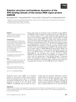
detected. Microscopic examination revealed a tumor com-
posed of nests of large cells with large eosinophilic cyto-
plasm presenting a ground-glass appearance (Figure 1).
Cell membranes were easily recognizable, and tumor
nuclei appeared large, presenting prominent nucleoli, and
also areas of abundant eosinophil infiltration were
present. The tumor showed a stromal invasion of 8 mm
out of 1.7 stromal thickness. The lateral and deep margins
of the cone were uninvolved for at least 9 mm. All perito-
neal biopsies, as well as pelvic lymph nodes (n = 18) were
negative. No lymphovascular space involvement was
observed. The final diagnosis was FIGO Stage IB1 poorly
differentiated carcinoma with > 90% of the tumor repre-
sented by neoplastic cells with glassy cell features. A sec-
ond pathologist, blinded to the first's impression
confirmed the diagnosis. Given the rarity of this histolog-
ical type and its prognostic features, therapeutic options
including radical trachelectomy, hysterectomy, or adju-
vant treatment were carefully discussed with the patient,
who nevertheless decided to undergo only strict follow-up
procedures. The patient was then followed with gyneco-
Glassy cell carcinoma of the cervix: the undifferentiated, glassy cells display large nuclei with prominent nucleoli and granular cytoplasmFigure 1
Glassy cell carcinoma of the cervix: the undifferenti-
ated, glassy cells display large nuclei with prominent
nucleoli and granular cytoplasm. Areas of abundant
eosinophils infiltration are present. (Hematoxylin & Eosin,
magnification: 200×).
World Journal of Surgical Oncology 2008, 6:92 />Page 3 of 3
(page number not for citation purposes)
logical examination, pap smear, and colposcopy every 3
months for the first 2 years, and every 6 months thereafter,
and was also requested to perform chest x-ray and pelvic
MRI every year. Currently, after a follow up of 38 months,
she has no evidence of disease.
Cervical stenosis was documented after 21 months since
surgery, and was easily managed by cannulation of the
cervical canal under anesthesia.
Discussion
We report a case of early stage glassy cell cancer in a
patient, who was conservatively treated by conization and
laparoscopic pelvic lymphadenectomy. Indeed, among
the fertility preservation approaches to early stage cervical
carcinoma, RVT has gained much attention because of the
recognized oncologic efficacy and safety. Intra- and post-
operative complications have been reported to be approx-
imately 4% and 12% of cases, respectively [8], and even
less radical procedures such as conization plus laparo-
scopic pelvic lymphadenectomy have been investigated in
selected cases of stage IB1 squamous cell carcinoma < 2
cm diameter [7]. While the fertility preserving procedures
are widely accepted for tumors with squamous histologi-
cal type, and also adenocarcinomas, which per se should
not be considered a contraindication to conservative treat-
ment, some concerns have been raised for rare histologi-
cal types such as adenosquamous, neuroendocrine or
glassy cell carcinomas. In particular, conservatively treated
neuroendocrine and adenosquamous tumors have been
reported to carry out a very unfavorable prognosis [5,6].
On the other hand, very few data about early stage glassy
cell cervical cancer have been reported: of 3 cases treated
with laparoscopic pelvic lymphadenectomy and RVT, all
were reported as having no evidence of disease at time of
publication [11]. No case of early stage glassy cell carci-
noma treated with conization plus laparoscopic pelvic
lymphadenectomy has been reported until now.
Despite the extensive counseling about the possibility to
perform trachelectomy or adjuvant treatment after final
diagnosis, our patient decided only to undergo strict fol-
low-up procedures, and is currently without evidence of
disease after 38 months since initial diagnosis.
Conclusion
We report a case of an early stage glassy cell cervical carci-
noma patient, who was successfully treated with coniza-
tion and laparoscopic pelvic lymphadenectomy. Given
the rarity of this tumor histological type, and the paucity
of data about its natural history, which has been reported
to be similar to other histological types only with the
employment of multimodal treatment strategies [12],
caution should be taken to i) carefully evaluate the
patients' fertility potential; ii) extensively counsel the
patients about the risk/benefit of a conservative treatment;
iii) investigate the patients' compliance to undergo strict
follow-up procedures.
Competing interests
The authors declare that they have no competing interests.
Authors' contributions
GF conceived of the study, participated in its design and
drafting. VS participated in the design of the study and
collected the clinical data. MP participated in the design of
the study and collected the clinical data. AC carried out
the histopathological evaluation. GS conceived of the
study, and participated in its design and coordination and
helped to draft the manuscript. All authors read and
approved the final manuscript.
Consent
Written informed consent was obtained from the patient
for publication of this case report and any accompanying
images. A copy of the written consent is available for
review by the Editor-in-Chief of this journal.
References
1. Chan PG, Sung HY, Sawaya GF: Changes in cervical cancer inci-
dence after three decades of screening US women less than
30 years old. Obstet Gynecol 2003, 102(4):765-73.
2. Dargent D, Brun JL, Roy M, Mathevet P, Remy I: La trachelectomie
elargie (T.E.) une alternative a' l'hysterèctomie radicale
dans le traitement des cancers infiltrants dèveloppeès sur la
face externe du col utèrin. J Obstet Gynecol 1994, 2:285-292.
3. Plante M, Roy M: Fertility preserving options for cervical can-
cer. Oncology 2006, 20:479-88.
4. Schlaerth JB, Spirtos NM, Schlaerth AC: Radical trachelectomy
and pelvic lymphadenectomy with uterine preservation in
the treatment of cervical cancer. Am J Obstet Gynecol 2003,
188:29-34.
5. Lea JS, Coleman RL, Garner EO, Duska LR, Miller DS, Schorge JO:
Adenosquamous histology predicts poor outcome in low-
risk stage IB1 cervical adenocarcinoma. Gynecol Oncol 2003,
91:558-562.
6. Weed JC, Graff AT, Shoup B, Tawfik O: Small cell undifferenti-
ated (neuroendocrine) carcinoma of the uterine cervix. J Am
Coll Surg 2003, 197:44-51.
7. Rob L, Charvat M, Robova H, Pluta M, Strnad P, Hrehorcak M, Skapa
P: Less radical fertility-sparing surgery than radical trache-
lectomy in early cervical cancer. Int J Gynecol Cancer 2007,
17:304-310.
8. Beiner ME, Covens A: Surgery insight: radical vaginal trachelec-
tomy as a method of fertility preservation for cervical can-
cer. Nature Practice Clin Oncol 2007, 4:353-361.
9. Glücksmann A, Cherry C: Incidence, histology, and response to
radiation of mixed carcinomas (adenoacanthomas) of the
uterine cervix. Cancer 1956, 9:971-979.
10. Lotocki RJ, Krepart GV, Paraskevas M, Vadas G, Heywood M, Fung
FK: Glassy cell carcinoma of the cervix. Gynecol Oncol 1992,
44:254-259.
11. Plante M, Renaud MC, Francois H, Roy M: Vaginal radical trache-
lectomy: an oncologically safe fertility preserving surgery.
An updated series of 72 cases and review of the literature.
Gynecol Oncol 2004,
94:614-623.
12. Gray HJ, Garcia R, Tamimi HK, Koh WJ, Goff BA, Greer BE, Paley PJ:
Glassy cell carcinoma of the cervix revisited. Gynecol Oncol
2002, 85:274-277.