Báo cáo khoa học: "Extrapulmonary small cell sarcinoma: involvement of the brain without evidence of extracranial malignancy by serial PET/CT scans" ppsx
Bạn đang xem bản rút gọn của tài liệu. Xem và tải ngay bản đầy đủ của tài liệu tại đây (466.27 KB, 5 trang )
BioMed Central
Page 1 of 5
(page number not for citation purposes)
World Journal of Surgical Oncology
Open Access
Case report
Extrapulmonary small cell sarcinoma: involvement of the brain
without evidence of extracranial malignancy by serial PET/CT scans
Christopher N Hueser*
1
, Nghi C Nguyen
2
, Medhat Osman
2
,
Necat Havlioglu
3
and Anjali J Patel
4
Address:
1
Department of Internal Medicine, Division of Hematology and Oncology, St. Louis University Hospital, St. Louis, MO 63110, USA,
2
Department of Nuclear Medicine, St. Louis University Hospital, St. Louis, MO 63110, USA,
3
Department of Pathology, St. Louis University
Hospital, St. Louis, MO 63110, USA and
4
Department of Anesthesia and Critical Care, St. Louis University Hospital, St. Louis, MO 63110, USA
Email: Christopher N Hueser* - ; Nghi C Nguyen - ; Medhat Osman - ;
Necat Havlioglu - ; Anjali J Patel -
* Corresponding author
Abstract
Background: Extrapulmonary small cell carcinoma (EPSCC) involving the brain is a rare
manifestation of an uncommon tumor type.
Case presentation: We report a 59 year-old Caucasian female diagnosed with an EPSCC
involving the left parietal lobe without detectable extracranial primary tumor followed by serial
positron emission tomography/computed tomography (PET/CT) imaging. Histopathological
examination at both initial presentation and recurrence revealed small cell carcinoma. Serial PET/
CT scans of the entire body failed to reveal any extracranial [
18
F]2-fluoro-2-deoxy-D-glucose
(FDG) avid lesions at either diagnosis or follow-up.
Conclusion: Chemotherapy may show a transient response in the treatment of EPSCC. Further
studies are needed to help identify optimal treatment strategies. Combination PET/CT technology
may be a useful tool to monitor EPSCC and assess for an occult primary malignancy.
Background
First described by Duguid and Kennedy in 1930 [1,2]
EPSCC is recognized as a clinicopathologic entity distinct
from small cell lung carcinoma [3-5]. Small cell carcino-
mas arising outside the lung have been reported in almost
every organ of the body [5-7]. Primary locations include
the head and neck, salivary glands, thyroid, larynx, tra-
chea, thymus, pleura, esophagus, stomach, intestines, rec-
tum, pancreas, gallbladder, cervix, uterus, breast, prostate,
urinary bladder, and skin [8]. The most common site of
presentation differs according to case series [1,6,7]. Only
one case of an EPSCC involving the brain is documented
in the literature [1].
An estimated one thousand new cases of EPSCC occur
yearly, with an overall incidence between 0.1% and 0.4%
of all cancers [4,9]. Approximately 2.5% of small cell car-
cinomas present at extraplumonary sites [3,4,8]. Since
there is no national or international tumor registry, many
cases are not reported, and the true incidence may be
underecognized [9,10]. It is postulated that EPSCC origi-
nate from totipotent stem cells that can differentiate into
various cell types [9].
The histologic criteria for EPSCC and small cell lung can-
cer (SCLC) are the same, namely uniform small cells with
dense nuclei, inconspicuous nucleoli and sparse cyto-
Published: 25 September 2008
World Journal of Surgical Oncology 2008, 6:102 doi:10.1186/1477-7819-6-102
Received: 15 November 2007
Accepted: 25 September 2008
This article is available from: />© 2008 Hueser et al; licensee BioMed Central Ltd.
This is an Open Access article distributed under the terms of the Creative Commons Attribution License ( />),
which permits unrestricted use, distribution, and reproduction in any medium, provided the original work is properly cited.
World Journal of Surgical Oncology 2008, 6:102 />Page 2 of 5
(page number not for citation purposes)
plasm [10]. The presence of cytoplasmic argyrophilia or
neurosecretory granules further substantiates the diagno-
sis [10,11].
Staging criteria for EPSCC is the same as that for SCLC.
Limited disease (LD) is defined as a localized tumor with
or without regional lymph node involvement; any exten-
sion beyond the loco-regional boundaries is defined as
extensive disease (ED) [2,10].
Clinically these tumors represent a rare, heterogeneous
group of neoplasms [12] characterized by their aggressive
nature, early dissemination and propensity to recur
[3,6,9].
Recent studies have demonstrated that extensive disease,
poor performance status and an increased white blood
cell count are the major prognostic factors that correlate
with mortality [4,10].
Optimal management is not well characterized because of
the rarity of these tumors, and the lack of randomized
clinical trials to guide treatment; hence, there are no
standard treatment regimens. Most of the data are extrap-
olated from treatment of small cell carcinoma of the lung.
In this report, we present the clinicopathologic features
and serial PET/CT evaluation of an EPSCC of the brain.
Case presentation
We report a 59 year-old Caucasian female diagnosed with
EPSCC of the left parietal lobe without evidence of an
extracranial primary tumor. The patient presented with a
three-week history of progressive deterioration of right
upper extremity coordination and motor strength. A stag-
ing PET/CT scan and CT scans of the chest, abdomen, and
pelvis, prior to surgery, were performed that revealed a left
parietal lobe mass with an intense, FDG-avid rim anteri-
orly. Neither study showed evidence of metastasis in the
lungs or elsewhere in the body. A magnetic resonance
image (MRI) of the brain revealed a 4.4 × 4.2 × 4.5 cm left
parietal mass.
The patient was treated with intravenous steroids and sub-
sequently underwent an MRI-guided sterotatic left parietal
craniotomy with complete resection of the tumor one day
after admission. Samples of the resection were sent for
pathological review. At diagnosis the complete blood
count and complete metabolic profile were within normal
limits. The patient did not experience either immediate or
late post-surgical complications and was discharged to a
rehabilitation facility for post-operative recovery and
improvement of her performance status for future chem-
otherapy.
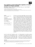
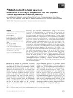
Pathologic examination of the parietal lobe resection was
consistent with small cell carcinoma (figure 1). The tumor
was reported as high-grade with nuclear pleomorphism,
sparse cytoplasm and large areas of necrosis. The cells
showed strong reactivity for synaptophysin and focally for
thyroid transcription factor-1 (TTF-1). The tumor cells
were negative for S-100, glial fibrillary acidic protein
(GFAP), cytokeratin AE1/AE3 keratin, anti-cytokeratin
(CAM 5.2) and chromogranin (figure 1).
Two months after initial resection surveillance MRI of the
brain revealed recurrence of a left parietal tumor. The
patient developed profound, progressive neurological
deterioration consisting of hemiparesis and expressive
aphasia. At this point she underwent a second complete
resection of the tumor. Pathology again revealed a small
cell carcinoma with an immunoprofile identical to that of
the original specimen.
The patient experienced profound improvement in her
neurological status after the second resection although her
ECOG status was 2. She was discharged to a neuro-reha-
bilitation facility for recovery and optimization of per-
formance status.
Light microscopy and immuostaining of patient's tumorFigure 1
Light microscopy and immuostaining of patient's tumor. A) Syaptophysin immunostaining, 150×. B) Hematoxylin and
eosin staining, 150×. C) Thyroid Transcription Factor-1 (TTF-1) immunostaining, 300×.
World Journal of Surgical Oncology 2008, 6:102 />Page 3 of 5
(page number not for citation purposes)
Four months following the second resection a 1.7 cm left
parietal lesion was identified on MRI and chemotherapy
with topotecan was initiated. This patient received six
cycles of topotecan at the standard dose of 1.5 mg/m
2
on
days 1 through 5 of a 21-day cycle. Overall, the patient tol-
erated therapy well with only grade 2 nausea and grade 2
anemia. Response to therapy was assessed by monthly
MRI scans of the brain. After two months of therapy a
reduction in tumor size greater than 50% was observed.
The disease eventually progressed and was referred to hos-
pice care 2 months following progression.
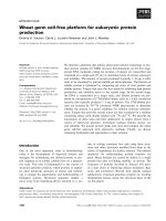
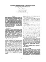
On repeat PET/CT scans, following the second resection
and 9 months after diagnosis (figure 2), there was absence
of FDG-avidity in the left parietal lobe. Approximately
one year after diagnosis of EPSCC, her disease progressed
and the patient chose to enter hospice care.
Discussion
The prognosis of EPSCC is similar to that of SCLC with
fewer than 13% of patients surviving 5 years [10]
although some patients have enjoyed prolonged survival
and even cure [9]. Median survival for all patients has
been reported to be 9.2 to 14 months [3,6]. The median
survival for patients with LD is 19.8 months, while for
patients with ED the median survival is 7 months [3]. The
clinical course of this tumor is very aggressive, with a ten-
dency for early systemic spread and recurrence after treat-
ment.
Extrapulmonary small cell carcinoma is a rare, aggressive
tumor for which there is no standard treatment guidelines
[13]. Some authors suggest that optimal management of
patients with EPSCC-limited disease consists of both local
modalities (surgery or radiotherapy) and systemic therapy
[10,14]. The chemotherapeutic regimens used for EPSCC
are similar to those utilized for SCLC. Combination cispl-
atin and etoposide (EP) is a commonly used regimen for
EPSCC with a response rates reported, in extensive dis-
ease, as 50% to 70%. It remains the cornerstone of therapy
in SCLC [4,5,15]. In the first line setting single agent topo-
tecan and paclitaxel have shown to be possible therapeu-
tic options. Neither of these agents has been compared in
a randomized phase III trial to EP [15]. Both surgery and
radiotherapy have been employed for local control with
varying degrees of success [3,10].
Since EPSCCs have responded well to agents active against
SCLC, it was decided to initiate therapy in this patient.
Topotecan was chosen because it was felt that the patient
would not tolerate a platinum based regimen due to her
poor performance status. Topotecan is a semi-synthetic
derivative of camptothecin that specifically targets topoi-
somerase-I. It has shown clinical activity against SCLC
[16]. The use of topotecan may be particularly appropriate
for patients in which palliation of symptoms is the pri-
mary goal of therapy.
Immunohistochemistry can help diagnose EPSCC as
these tumors stain positive for chromogranin A and TTF-
1. Ordonez reported that TTF-1 lacks specificity to distin-
guish primary versus metstatic lesions [17]. Other studies
have reported that TTF-1 may be useful in differentiating
small cell from other extrapulmonary neuorendocrine
tumors [18,19]. Extrapulmonary small cell carcinoma
stains positive for TTF-1 while other extrapulmonary neu-
orendocrine tumors do not [18]. In contrast, a study by
Prok demonstrated that in 16 of 43 patients with meta-
static carcinoma of unknown primary to the brain, TTF-1
stained positive [20]. Positive staining for TTF-1 should be
factored into each clinical setting when determining
whether the tumor is an EPSCC or a metastatic lesion.
Whole-body PET imaging with FDG is used in the diagno-
sis, staging, and follow-up of many cancers with accura-
cies ranging from 80% to 90% [21]. PET/CT is still in its
No extracranial FDG-avid lesions to suggest malignancy are seen by PET/CTFigure 2
No extracranial FDG-avid lesions to suggest malignancy are seen by PET/CT. A) PET/CT, prior to craniotomy,
showing anterior rim enhancement of the left parietal tumor. B) PET/CT revealing a photopenic area following second resec-
tion of the left parietal tumor. C) Follow-up PET/CT at 9 months.
World Journal of Surgical Oncology 2008, 6:102 />Page 4 of 5
(page number not for citation purposes)
infancy; however, several studies published over the last
few years demonstrate that PET/CT transforms image
fusion from primarily a research tool to everyday clinical
practice. In addition, these studies prove that PET/CT has
a higher diagnostic accuracy than PET alone, or CT alone,
or visually correlated PET and CT. Furthermore, PET/CT
frequently provides statistically significant improvements
over PET or CT alone in staging and restaging of different
cancers [22]. Clearly, there are more data on the use of
PET/CT in lung cancer than any other type of malignancy.
As the staging procedures for SCLC do not differ from
those for non-small cell lung cancer (NSCLC) the primary
role of PET/CT imaging is to delineate limited disease
from extensive disease. There are relatively few indications
for PET/CT scanning in SCLC as there is usually extensive
disease at presentation. Nonetheless, it has been shown
that whole-body PET is superior to conventional staging
in the detection of all involved sites, thus it is a highly val-
uable tool for staging SCLC [23]. Further, dual-modality
PET/CT is able to detect more primary tumors than PET,
CT and PET and CT side-by-side in the diagnosis of carci-
noma of unknown primary with less patient radiation
exposure [24]. PET/CT can help localize the primary in
CUP in approximately 40% of all cases, even after a thor-
ough work-up with a variety of other investigations [25].
To the best of our knowledge, FDG PET/CT imaging of
EPSCC involving the brain has never been reported
before.
It is possible that the patient had an occult bronchial pri-
mary tumor that was beyond the limits of detection by
PET/CT. There was an interval of nine months between
the first and final PET/CT scans; this is a considerable
amount of time for a bronchial primary to manifest. Dur-
ing this period the tumor recurred twice in the brain and
serial PET/CT scans did not reveal extracranial malignancy
at diagnosis or at later dates. As such we are of the strong
opinion that the tumor did arise outside the lung.
Conclusion
Our patient's response to therapy yielded a transient clin-
ical response without profound toxicity. Further studies
are needed to help identify optimal treatment strategies in
this rare tumor type. Dual-modality PET/CT technology
may be a useful tool to monitor EPSCC and may help in
our understanding of this rare entity.
Abbreviations
CAM 5.2: anti-cytokeratin; ED: extensive disease; EPSCC:
extrapulmonary small cell carcinoma; FDG: [
18
F]2-fluoro-
2-deoxy-D-glucose; GFAP: glial fibrillary acidic protein;
LD: limited disease; MRI: magnetic resonance imaging;
NSCLC: non-small cell lung cancer; PET/CT: positron
emission tomography/computed tomography; SCLC:
small cell lung carcinoma; TTF-1: thyroid transcription
factor-1
Competing interests
The authors declare that they have no competing interests.
Authors' contributions
CH, NN, MO, NH, AP conception and design, acquisition
of data, analysis and interpretation of data, have been
involved in drafting the manuscript, revising it critically
for important intellectual content and have given final
approval of the version to be published.
Acknowledgements
The reporting of this case was approved by the Ethics committee of St.
Louis University as the consent of the patient or the next of kin could not
be obtained.
References
1. Galanis E, Frytak S, Lloyd R: Extrapulmonary Small Carcinoma.
Cancer 1997, 79:1729-1736.
2. Remick SC, Hafez GR, Carbone PP: Extrapulmonary small-cell
carcinoma: A review of the literature with emphasis on ther-
apy and outcome. Medicine 1987, 66:457-471.
3. Kim KO, Lee HY, Chun SH, Shin SJ, Kim MK, Lee KH, Hyun MS, Bae
SH, Ryoo HM: Clinical overview of extrapulmonary small cell
carcinoma. J Korean Med Sci 2006, 21:833-7.
4. Haider K, Shahid RK, Finch D, Sami A, Ahmad I, Yadav S, Alvi R, Pop-
kin D, Ahmed S: Extrapulmonary small cell cancer: A Canadian
province's experience. Cancer 2006, 107:2262-2268.
5. Re GL, Canzonieri V, Bo DV, Barzan L, Zancanaro C, Trovo M:
Extrapulmonary small cell carcinoma: A single-institution
experience and review of the literature. Ann Oncol 1994,
5(10):909-913.
6. Kim JH, Lee S, Park J, Kim HY, Lee SI, Nam EM, Park JO, Kim K, Jung
CW, Im YH, Kang WK, Lee MH, Park K: Extrapulmonary small-
cell carcinoma: A single-institution experience. Jpn J Clin Oncol
2004, 34:250-254.
7. Henricus FM, Heijden van der, Heijdra Y: Extrapulmonary Small
Carcinoma. Southern Medical Journal 2005, 98:345-349.
8. Lobins R, Floyd J: Semin Oncol: Small cell carcinoma of
unknown primary. Seminars in Oncology 2007, 34:39-42.
9. Shahab N, Mirza IA, Doll D: Extrapulmonary Small Cell Carci-
noma. Seminars in Oncology 2007, 34:1-2.
10. Shamelian SOA, Nortier JWR: Extrapulmonary small-cell carci-
noma: report of three cases and update on therapy and prog-
nosis. Neth J Med 2000, 56:51-5.
11. Frazier SR, Kaplan P, Loy TS: The pathology of extrapulmonary
small cell carcinoma. Semin Oncol 2007, 34:30-38.
12. Orhan B, Yalcin S, Evrensel T, Yerci O, Manavoglu S: Successful
treatment of cranial metastases of extrapulmonary small
cell carcinoma with chemotherapy alone.
Medical Oncology
1998, 15:66-69.
13. Gaast A van der, Verwey J, Prins E, Splinter TAW: Chemotherapy
as treatment of choice in extrapulmonary undifferentiated
small cell carcinomas. Cancer 1990, 65:422-424.
14. Casas F, Ferrer R, Ferrus B, Casals J, Bieta A: Primary small cell
carcinoa of the esophagus: a review of the literature with
emphasis on therapy and prognosis. Cancer 1997,
80:1366-1372.
15. Rosti G, Carminati O, Monti M, Tamberi S, Marangolo M: Chemo-
therapy advances in small cell lung cancer. Annals of Oncology
2006, 17(Supplement 5):v99-v102.
16. O'Brien M, Eckardt J, Ramlau R: Recent Advances with Topote-
can in the Treatment of Lung Cancer. Oncologist 2007,
12:1194-1204.
17. Ordonez NG: Value of thyroid transcription factor-1 immu-
nostaining in distinguishing small cell carcinomas from other
small cell carcinomas. Am J Surg Pathol 2000, 24:1217-1223.
Publish with BioMed Central and every
scientist can read your work free of charge
"BioMed Central will be the most significant development for
disseminating the results of biomedical research in our lifetime."
Sir Paul Nurse, Cancer Research UK
Your research papers will be:
available free of charge to the entire biomedical community
peer reviewed and published immediately upon acceptance
cited in PubMed and archived on PubMed Central
yours — you keep the copyright
Submit your manuscript here:
/>BioMedcentral
World Journal of Surgical Oncology 2008, 6:102 />Page 5 of 5
(page number not for citation purposes)
18. Agoff SN, Lamps LW, Phillip AT, Amin MB, Schmidt RA, True LD,
Folpe AL: Thyroid transcription factor-1 is expressed in
Extrapulmonary small cell carcinomas but not in other
Extrapulmonary neuroendocrine tumors. Mod Pathol 2000,
13:238-242.
19. Kaufmann O, Deitel M: Expression of thyroid transcription fac-
tor-1 in pulmonary and Extrapulmonary small cell carcino-
mas and other neuroendocrine carcinomas of various
primary sites. Histopathology 2000, 36:415-420.
20. Prok AL, Prayson RA: Thyroid transcription factor-1 is useful in
identifying brain metastases of pulmonary origin. Annals of
Diagnostic Pathology 2006, 10:67-71.
21. Czernin J, Phelps ME: Positron emission tomography scanning:
current and future applications. Annu Rev Med 2002, 53:89-112.
22. Czernin J, Allen-Auerback M, Schelbert HR: Improvements in
Cancer Staging with PET/CT: Literature-Based Evidence as
of September 2006. J Nucl Med 2007, 48(Suppl 1):78S-88S.
23. Kamel EM, Zwahlen D, Wyss MT, Stumpe KDM, von Schulthess K,
Steinert HC: Whole-body
18
F-FDG PET improves the man-
agement of patients with small-cell lung cancer. J Nucl Med
2003, 44:1911-1917.
24. Gutzeit A, Antoch G, Kuhl H, Egelhof T, Fischer M, Hauth E, Goedhe
S, Backisch A, Debatin J, Freundenberg L: Unknown Primary
Tumors: Detection with Dual-Modality PET/CT-An Initial
Experience. Radiology 2005, 234:227-234.
25. Freudenberg LS, Rosenbaum-Krumme SJ, Bockisch A, Eberhardt W,
Frilling A: Cancer of unknown primary. Recent Results Cancer Res
2008, 170:193-202.