Báo cáo khoa học: "Inflammatory myofibroblastic tumor of epididymis: a case report and review of literature" potx
Bạn đang xem bản rút gọn của tài liệu. Xem và tải ngay bản đầy đủ của tài liệu tại đây (593.25 KB, 6 trang )
BioMed Central
Page 1 of 6
(page number not for citation purposes)
World Journal of Surgical Oncology
Open Access
Case report
Inflammatory myofibroblastic tumor of epididymis: a case report
and review of literature
Pankaj P Dangle*
1
, Wenle Paul Wang
2
and Kamal S Pohar
3
Address:
1
The James Cancer Hospital and Solove Research Institute, Ohio State University and Comprehensive Cancer Center, Columbus Ohio,
43210, USA,
2
Department of Pathology, The Ohio State University, Columbus Ohio, 43210, USA and
3
Department of Urology, The James Cancer
Hospital and Solove Research Institute, Ohio State University and Comprehensive Cancer Center, Columbus Ohio, 43210, USA
Email: Pankaj P Dangle* - ; Wenle Paul Wang - ;
Kamal S Pohar -
* Corresponding author
Abstract
Background: Epididymal inflammatory myofibroblastic tumor, also known by various other
synonyms is a rare benign disease. Only eight cases have been reported to date. The most common
presentation is a scrotal mass of variable duration. For a scrotal mass it is difficult to distinguish a
benign or malignant etiology, in addition to the origin whether from testis or epididymis. As a result
the definitive diagnosis can only be established by surgical exploration.
Case presentation: We report the ninth case of epididymal IMT who based on clinical and
radiological findings underwent radical orchidectomy, with the histology suggestive of inflammatory
myofibroblastic tumor. At 4 years follow up the patient is free of disease recurrence.
Conclusion: IMT though rare should be considered in the differential diagnosis of epididymal
mass. Clinically it is often difficult to distinguish the origin of mass and even though the disease has
benign nature and course it is crucial to counsel patients for orchidectomy as definitive diagnosis
is established on surgical exploration.
Background
Inflammatory Myofibroblastic tumor (IMT) of epidi-
dymis is a distinct but rare entity. IMT is also described by
other synonyms, more commonly as inflammatory pseu-
dotumor. Extra genitourinary and genitourinary sites are
well documented with various proposed etiological theo-
ries [1]. Epididymal IMT is rare and only eight cases are
reported in the literature [2-8]. The most common
reported presentation of epididymal IMT is lump in the
scrotum. Due to its uncertain etiology many of these
patients have been offered antibiotics with no clinical
response. We describe a case of a young healthy male with
a painless indurated scrotal mass with possible involve-
ment of the testicle. Based on patient's age and clinical
findings the lump was suspected to be a testicular tumor
and therefore was subjected to radical orchidectomy. We
present our case and review of literature for epididymal
IMT.
Case presentation
A 22 year old healthy Caucasian male noticed a swelling
and a palpable mass in the right scrotum for a period of
one week. Patient denied any history of fever, trauma, ure-
thral discharge and any previous history of recurrent uri-
nary tract or sexually transmitted infections. There was no
past history of exposure to tuberculosis. Physical examina-
Published: 11 November 2008
World Journal of Surgical Oncology 2008, 6:119 doi:10.1186/1477-7819-6-119
Received: 11 July 2008
Accepted: 11 November 2008
This article is available from: />© 2008 Dangle et al; licensee BioMed Central Ltd.
This is an Open Access article distributed under the terms of the Creative Commons Attribution License ( />),
which permits unrestricted use, distribution, and reproduction in any medium, provided the original work is properly cited.
World Journal of Surgical Oncology 2008, 6:119 />Page 2 of 6
(page number not for citation purposes)
tion revealed a nontender indurated solid mass in the
lower pole of right testicle possibly also involving the
epididymis.
Scrotal ultrasound demonstrated a solid heterogeneous
mass involving right testicle with possible extratesticular
extension into the epididymis. Quantitative serum Beta -
human chorionic gonadotropin, alpha-fetoprotein and
LDH (lactate dehydrogenase) were within normal limits.
With the presumed diagnosis of testicular tumor a right
radical orchidectomy was performed. On gross pathologic
examination the mass was abutting the tunica albugenia
but further examination revealed being confined to epidi-
dymis with normal testicular parenchyma. Histology of
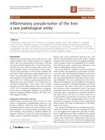
the mass (4 × 2.2 × 1.8 cm) demonstrated a spindle
myoepithelial and polygonal cell proliferation with
intense lymphoplasmacytic infiltrate. (Fig. 1) The mass
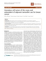
also revealed scattered neutrophils with positive immu-
nostaining for smooth muscle actin, vimentin (Fig. 2),
CD3, CD20, CD68 and AE1/AE3 but was negative for
ALK-1 (Fig. 3) and CD 138. There was presence of numer-
ous T cells, B cells and macrophages but absence of atypi-
cal epithelial cells. The lesion also lacked presence of
sperm, Michaelis Gutman bodies, GMS (Grocott's silver)
and AFB (acid fast bacilli) stain for any fungal or acid fast
organism respectively. The histological and staining pat-
tern was consistent with inflammatory myofibroblastic
tumor of the epididymis. The reagents, their source, pre-
treatment, dilution and incubation times are listed in
table 1.
The patient recovered well with no evidence of any recur-
rence at the site of resection or other sites after 4 years of
follow-up.
Based on such an unusual and rare finding a thorough
Medline search revealed eight additional patients with
similar presentation of scrotal lump. All patients had
exploration of the scrotal mass due to its solid heteroge-
nous features on ultrasound and clinical examination. All
patients underwent either excision of mass or radical
orchidectomy.
Discussion
Inflammatory Myofibroblastic tumor (IMT) is a well
described disease and can occur in many organs such as
lung, skin, soft tissues, breast, gastrointestinal tract, pan-
creas, oral cavity, bone and central nervous system. How-
ever various sites in genitourinary tract have also been
reported but less commonly [1], epididymis is least com-
mon with only 8 cases (20 to 73 years) being reported to
date [2-8].
Only those tumors with spindle myoepithelial cell prolif-
eration and lymphocytic infiltrate qualify for IMT. Various
synonyms like inflammatory pseudo tumor, plasma cell
granuloma, plasma cell pseudo tumor, atypical Myofi-
broblastic tumor and post operative spindle nodule are
used interchangeably [1]. In spite of this the term inflam-
matory myofibroblastic tumor is preferred as inflamma-
tory pseudo tumor has been applied to diverse entities like
reparative pseudosarcomatous lesion of lower genitouri-
nary tract [9], infectious etiology like mycobacterium
avium intracellulare and Epstein Barr virus (EBV) [10,11].
The post operative spindle cell nodule [1] denotes to spin-
Table 1: Immunohistochemical reagents used in our case.
Antibody specificity Vendor/Source Cat. Number/Clone Pretreatment Dilution Incubation time(minutes)
CD3 Dako A0452/Rabbit TRS pH6/30 sec in Pressure
cooker
1 in 400 30
CD20 Dako M0755/L26 TRS pH6/30 sec in Pressure
cooker
1 in 200 30
CD68 Dako M0814/KP1 TRS pH6/30 sec in Pressure
cooker
1 in 3000 30
SMA Dako M0851/1A4 No 1 in 400 30
ALK-1 Dako M7195/ALK-1 TRS pH6/30 sec in Pressure
cooker
1 in 50 30
CD138 Dako M7228/MI15 TRS pH6/25 min in steamer 1 in 90 30
Vimentin Dako M0725/V9 TRS pH6/25 min in steamer 1 in 200 30
World Journal of Surgical Oncology 2008, 6:119 />Page 3 of 6
(page number not for citation purposes)
dle cell proliferation with easily identifiable mitotic fig-
ures deposited in a less conspicuous myxoid background
where as a classic IMT describes a lesion characterized by
spindle cell proliferation in a loose, edematous myxoid
stroma associated with granulation tissue type and a
mixed acute and chronic inflammatory cells composed of
lymphocytes, plasma cells, eosinophils with occasional
neutrophils and mast cells.
The pathophysiology of IMT is not well understood, vari-
ous etiologies have been proposed including a reparative
process related to delayed chronic response to remote or
undetected trauma [1]. Infectious etiologies such as
Epstein Barr virus, mycobacterium avium intracellulare
and herpes virus 8 have also been suggested to be associ-
ated as an etiological agent with IMT [10-12]. However no
such similar association of EBV as an etiological agent has
been demonstrated with epididymal IMT [4]. Cytogenetic
studies show that some IMT (mediastinal and abdominal)
lesions have genetic clonal abnormality at chromosome
region 2p22–24 with breakage in band p22–24, with spe-
cific involvement of 2p23, suggesting a neoplastic change
[13]. In some of the IMTs an anaplastic lymphoma kinase
(ALK) gene on 2p23 has been implicated in pathogenesis
of this lesion. A fluorescence in situ hybridization with a
probe flanking the ALK gene at 2p23 demonstrated trans-
location of ALK gene. An immunohistochemical staining
for ALK showed positive cytoplasmic staining in the
myofibroblastic cells [13,14]. Two case reports [7,8]
including ours have studied ALK immunostaining on
epididymal tissue with none staining positive for ALK.
Patients with IMT can present with fever, night sweats,
weight loss, malaise or abnormal laboratory parameters
such as elevated ESR (erythrocyte sedimentation rate),
anemia, leukocytosis and site specific symptoms [15].
However patients with IMT of epididymis rarely present
with above symptoms but most commonly with a palpa-
ble mass of variable duration ranging from 3 weeks to 5
years [2,4]. The mass is clinically often indistinguishable
from the testis. One patient described in the literature,
clinically had multiple [5] extra testicular masses, with 3
Low magnification of IMT (100×)Figure 1
Low magnification of IMT (100×). Spindle cells mixed
with inflammatory cells. The spindle cells are epithelioid,
mixed with chronic inflammatory cells. The myoepithelial
cells are loosely arranged. There is increased vascularity in
the IMT.
(Immunostains) – Immunostaining showing spindle myoepithelial cells positive for smooth muscle actin and vimentinFigure 2
(Immunostains) – Immunostaining showing spindle myoepithelial cells positive for smooth muscle actin and
vimentin.
World Journal of Surgical Oncology 2008, 6:119 />Page 4 of 6
(page number not for citation purposes)
in the body of the epididymis, 1 at head of the epididymis
and 1 in tunica vaginalis on subsequent exploration [2].
Our patient presented with 1 week history of a palpable
mass with no precedent history of trauma and recurrent
urinary or sexually transmitted infections. A summary of
all reported eight cases and our case has been presented in
table 2. Based on the clinical examination the differential
diagnosis of such a mass is testicular tumor, adenomatoid
carcinoma, paratesticular sarcoma, epididymal adenocar-
cinoma.
The diagnosis of IMT is based on the histological features
of spindle myoepithelial cell proliferation, lymphocytic
and inflammatory infiltration. Other immunomarkers
could substantiate the diagnosis of IMT. Immunomarkers
such as vimentin, actin and CD 68 are positive in 25%
cases [1]. A similar finding was noted by Brauers and Lam
et al [3,4] in epididymal IMT, with immunostaining being
positive for vimentin, actin, CD 68 and α1-anti chymot-
rypsin. In our patient histology stained positive for vimen-
tin, smooth muscle actin and CD 68 but negative for ALK-
1 and CD138.
Various non surgical treatment options have been pro-
posed at sites other than genitourinary tract including
cyclosporine, corticosteroid, methotrexate, antibiotics
[16-18] and radiation [19] with variable success. Sponta-
neous regression has also been reported [15]. Surgical
excision is definitive to exclude malignant etiology for
scrotal masses. Our patient and most patients [4-8]
described in the literature had orchidectomy as a final
treatment. Though Cooperman et al [2] described local
excision of clinically evident extra testicular masses with a
normal testicle confirmed on ultrasound, the frozen sec-
tion of these masses excluded presence of malignancy.
Similarly Brauers et al [3] report epididymectomy for a
clinically palpable 1 cm mass with normal testis on exam-
ination. Lam et al [4] however performed orchidectomy
for a firm scrotal mass clinically indistinguishable from
testis. In our patient based on clinical examination and
ultrasound, it was difficult to justify local excision due to
difficulty in differentiating whether the mass was separate
from testis.
The abdominal and retroperitoneal variant presents with
more aggressive pattern compared to their extra abdomi-
nal counterparts, with recurrence rate of 23–37% [15,20].
The true potential for metastasis as reported by Coffin et
al [15] in their series of 84 patients is unclear whereas
Meis and Enzinger [20] reported cases with metastasis.
The reason for such inconsistent finding is uncertain,
whether it represents multifocal disease is unclear at
present [15,20]. However recurrence of epididymal IMT
has not been reported to date. Our patient is free of any
recurrent disease at previous site of excision or other dis-
tant sites at end of 4 years of follow-up.
Conclusion
IMT though rare should be considered in the differential
diagnosis of epididymal mass. Clinically it is often diffi-
cult to distinguish the origin of mass either from testis or
epididymis. Radiological studies are unable to differenti-
ate benign or malignant nature and as a result definitive
diagnosis is established on surgical exploration. Depend-
ing on the gross characteristics and frozen section of clin-
ically distinct masses, either a local excision or radical
orchidectomy is offered. Thus even though the disease has
benign nature and course it is crucial to counsel patients
for orchidectomy as definitive diagnosis is established on
surgical exploration.
Consent
Written informed consent was obtained from the patient
for publication of this case report and any accompanying
images. A copy of the written consent is available for
review by the Editor-in-Chief of this journal.
Competing interests
The authors declare that they have no competing interests.
Authors' contributions
PPD was involved in conception and design, acquisition
of data, data analysis, and interpretation, manuscript
drafting and final approval. WPW was involved in acqui-
sition of data, data analysis, provided pathologic imaging,
(Immunostain) – Immunostaining negative for ALK-1Figure 3
(Immunostain) – Immunostaining negative for ALK-
1.
World Journal of Surgical Oncology 2008, 6:119 />Page 5 of 6
(page number not for citation purposes)
interpretation of data and final approval. KSP was
involved in conception and design, acquisition of data,
data analysis, and interpretation, manuscript drafting and
final approval.
References
1. Weiss SW, Goldblum JR: Enzinger and Weiss's soft tissue tumors 4th
edition. St Louis: C.V Mosby; 2001:274-384.
2. Cooperman R, White B, Zincke JP, Kardon D, Andrawis R: Extrat-
esticular inflammatory myofibroblastic tumor. J Urol 2003,
169:1473.
3. Brauers A, Striepecke E, Mersdof A, Sohn M, Fiuzesi L: Inflamma-
tory pseudotumor of the epididymis. Eur Urol 1997, 32:253.
4. Lam KY, Chan KW, Ho MHM: Inflammatory pseudotumor of
epididymis. Br J urol 1995, 75:255.
5. Orosz Z, Besznyak I: Diffuse inflammatory pseudotumor of tes-
tis, the epididymis and the spermatic cord. Pathol Oncol Res
1995, 1(1):75-79.
6. Chan KW, Chan KL, Lam KY: Inflammatory pseudotumor of the
epididymis and Epstein-Barr virus; a study of two cases.
Pathology 1997, 29:100-101.
7. Kapur P, Treat K, Chuang AT, Hoang M: Pathologic quiz case:
paratesticular mass in a young man. Inflammatory myofi-
broblastic tumor of the paratestis. Arch Pathol Lab Med 2004,
128(4):589-590.
8. Megremis S, Papamitsaki E, Ieromonachou P, Zois E: Inflammatory
myofibroblastic tumor of the paratestis: Sonographic
appearance with pathologic correlation. J Ultrasound Med 2007,
26(9):1227-30.
9. Albores-Saavedra J, Manivel JC, Essenfeld H, Dehner LP, Drut R,
Gould E, Rosai J: Pseudosarcomatous myofibroblastic prolifer-
ation in the urinary bladder of children. Cancer 1990, 66:1234.
10. Umlas J, Federman M, Crawford C, et al.: A spindle cell pseudotu-
mor resulting from atypical Mycobacterium Avium -Intrac-
ellulare in patients with acquired immunodeficiency
syndrome (AIDS). Am J Surg Pathol 1991, 12:1181.
11. Arber DA, Weiss LM, Chang KL: Detection of Epstein Barr Virus
in inflammatory pseudotumor. Semin Diagn Pathol 1998, 15:155.
12. Gomez-Roman JJ, Ocejo-Vinyal JF, Sanchez-Valesco P, Leyva-Cobian
F, Val-Bernal FJ: Presence of Human Herpes virus 8 DNA
Table 2: Brief summary of cases reported in the literature.
Reference Age Presentation Immunomarkers Treatment Follow-up
Positive Negative
Orosz et al [5] 63 Left Scrotal mass α-smooth muscle
actin, muscle specific
actin, vimentin, kappa
and lambda chain
Desmin, S-100,
Factor VIII-related
antigen,
Radical
Orchidectomy
_
Lam et al [4] 43 Rt Scrotal mass Vimentin, smooth
muscle actin
Desmin, cytokeratin Initial antibiotic,
Surgical excision as
definitive treatment
At 6 months follow-
up no recurrence
Chan et al [6] 43 Rt. Scrotal mass Polyclonality of
plasma cells for Light
chains
Radical
Orchidectomy
_
20 Left Scrotal mass Polyclonality of
plasma cells for Light
chains
Excision of lump
from tail of
epididymis
_
Brauers
et al [3]
73 Left Scrotal mass Vimentin, α1anti-
chymotrypsin, CD
68, α-smooth muscle
actin
Desmin, myoglobin,
myosin
Epididymectomy _
Cooperman et al [2] 30 Rt. Scrotal mass _ _ Excision of masses _
Kapur et al [7] 36 Rt. Scrotal mass and
rt. Inguinal
lymphadenopathy
Vimentin, smooth
muscle actin,
Cytokeratin (AE1/
AE3), muscle specific
actin, desmin, CD34
ALK, inhibin
Radical
Orchidectomy
_
Stylianos
et al [8]
45 Left Scrotal mass Smooth muscle cell
specific actin, Desmin
CD34, S-100,
cytokeratin, AE1/
AE3, ALK
Radical
Orchidectomy
No recurrence at 3
year follow-up
Our case 22 Rt. Scrotal mass Vimentin, smooth
muscle actin, CD3,
CD20, CD 68, AE1/
AE3
ALK-1, CD 138 Radical
Orchidectomy
No recurrence at 4
year follow-up
Publish with BioMed Central and every
scientist can read your work free of charge
"BioMed Central will be the most significant development for
disseminating the results of biomedical research in our lifetime."
Sir Paul Nurse, Cancer Research UK
Your research papers will be:
available free of charge to the entire biomedical community
peer reviewed and published immediately upon acceptance
cited in PubMed and archived on PubMed Central
yours — you keep the copyright
Submit your manuscript here:
/>BioMedcentral
World Journal of Surgical Oncology 2008, 6:119 />Page 6 of 6
(page number not for citation purposes)
sequence in renal transplantation associated pleural Kaposi
Sarcoma. Arch Pathol La Med 1999, 123:1269.
13. Griffin CA, Hawkins AL, Dvorak C, Henkle C, Ellingham T, Perlman
EJ: Recurrent involvement of 2p23 in inflammatory myofi-
broblastic tumors. Cancer Res 1999, 59:2776.
14. Coffin CM, Hussong J, Perkins S, et al.: ALK and p80 expression in
inflammatory myofibroblastic tumor. Mod Pathol 1999, 13:8A.
15. Coffin CM, Patel A, Perkins S, Elenitoba-Johnson KS, Perlman E, Grif-
fin CA: Extra pulmonary inflammatory myofibroblastic
tumor (inflammatory pseudotumor): a clinicopathologic and
immunohistochemical study of 84 cases. Am J Surg Pathol 1995,
19(8):859-72.
16. Nishimaki T, Matsuzaki H, Sato Y, Kondo Y, Kasukawa R:
Cyclosporine for inflammatory pseudotumor. Int Med 1992,
31:404.
17. Shah SS, Lowder CY, Schmitt MA, Wilke WS, Kosmorsky GS, Meisler
DM: Low dose methotrexate therapy for occular inflamma-
tory disease. Ophthalmology 1992, 99(9):1419-1423.
18. Weinberg PB, Bromberg PA, Askin FB: Recurrence of plasma cell
granuloma 11 years after initial resection. South Med J 1987,
80:519-21.
19. Imperato JP, Folkman J, Sagerman RH, Cassady J: Treatment of
plasma cell granuloma of the lung with radiation therapy: a
report of two cases and review of literature. Cancer 1986,
57:2127.
20. Meis JM, Enzinger FM: Inflammatory fibrosarcoma of the
mesentery and retroperitoneum. A tumor closely simulat-
ing inflammatory pseudotumor. Am J Surg Pathol 1991,
15(12):1146-56.