Báo cáo khoa học: "Lung adenocarcinoma presenting as obstructive jaundice: a case report and review of literature" ppsx
Bạn đang xem bản rút gọn của tài liệu. Xem và tải ngay bản đầy đủ của tài liệu tại đây (1.31 MB, 6 trang )
BioMed Central
Page 1 of 6
(page number not for citation purposes)
World Journal of Surgical Oncology
Open Access
Review
Lung adenocarcinoma presenting as obstructive jaundice: a case
report and review of literature
Stephanos Pericleous
1
, Samrat Mukherjee
2
and Robert R Hutchins*
2
Address:
1
Department of HPB Surgery, Imperial College, Hammersmith Hospital campus, Du Cane Road, London, UK and
2
Department of HPB
Surgery, Royal London Hospital, Whitechapel, London, UK
Email: Stephanos Pericleous - ; Samrat Mukherjee - ;
Robert R Hutchins* -
* Corresponding author
Abstract
Background: Lung cancer is known to metastasize to the pancreas with several case reports
found in the literature, however, most patients are at an advanced stage and receive palliative
treatment.
Case presentation: We describe the case of a 56 year old male patient who presented with a
picture of obstructive jaundice. Investigations revealed an obstructing lesion in the pancreas and a
further lesion in the lung with benign appearances. The patient underwent a pancreatectomy and,
unexpectedly, the histology of the resected specimen demonstrated metastatic adenocarcinoma of
bronchogenic origin. He was referred to a cardiothoracic team who proceeded to resect the
patient's thoracic lesion before administration of adjuvant chemotherapy. The patient was reviewed
18 months post operatively and remains symptom free with no clinical or radiological evidence of
recurrence. We were unable to identify any previous case reports (of lung adenocarcinoma) with
such a presentation which were ultimately treated with resection of both lesions.
Conclusion: Similar situations are bound to arise again in the future and we believe that this
report could demonstrate that there is a case for aggressive surgical management in a highly
selected group of patients: those with NSCLC and a synchronous solitary pancreatic deposit.
Background
That a variety of malignant tumours can metastasise to the
pancreas is well documented. Several case reports have
reported patients with lung cancer whose clinical presen-
tation was that of obstructive jaundice [1].
Most patients presenting in this manner are at an
advanced stage with widespread disease, and are usually
managed symptomatically. This generally involves pallia-
tive chemotherapy and/or radiotherapy coupled with
other measures to relieve the biliary obstruction such as
biliary stent insertion. In the few cases where operative
intervention is considered, it is usually limited to a biliary
bypass to relieve the jaundice.
We describe an unusual presentation where an adenocar-
cinoma of the lung with a synchronous solitary metastatic
deposit in the pancreas (not visible on CT) was treated
with operative resection of both lesions. The uniqueness
of this case is enhanced by the fact that both lesions were
identified preoperatively although their nature was not.
Published: 11 November 2008
World Journal of Surgical Oncology 2008, 6:120 doi:10.1186/1477-7819-6-120
Received: 19 April 2008
Accepted: 11 November 2008
This article is available from: />© 2008 Pericleous et al; licensee BioMed Central Ltd.
This is an Open Access article distributed under the terms of the Creative Commons Attribution License ( />),
which permits unrestricted use, distribution, and reproduction in any medium, provided the original work is properly cited.
World Journal of Surgical Oncology 2008, 6:120 />Page 2 of 6
(page number not for citation purposes)
Case presentation
A 56 year old male lawyer presented to his local hospital
complaining of a recent change in his urine colour (to
bright orange) and general malaise. The patient suffered
from moderate bronchiectasis and asthma for which he
took inhalers (fluticasone propionate, salmeterol and
ipratropium bromide). He was also known to be hyper-
tensive (controlled on diltiazem) and suffered from severe
eczema. He had never been a smoker but his daily con-
sumption of alcohol amounted to 1.5 bottles of wine.
Initial workup revealed deranged liver function tests and
relevant tumour markers were raised (Ca 19-9 181 kU/l,
CEA 25.8 μg/l). A subsequent abdominal ultrasound
showed biliary dilatation to the level of the pancreas. This
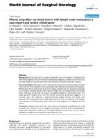
was confirmed on an MRCP. However CT (64 slice fine
cut spiral pancreas protocol CT) and MRI examinations
failed to reveal any pancreatic mass (figure 1). An ERCP
which followed confirmed the lower CBD stricture with
features of external compression and a plastic biliary stent
was inserted.
The patient was then referred to our unit for further treat-
ment. The working diagnosis at this stage was a pancreatic
tumour and the patient underwent staging with a view to
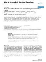
a pancreatic resection. Unusually, as part of the initial
workup, the patient had had a CT of his thorax, showing
a right lung lesion, thought to be benign, on a background
of known chronic respiratory disease (figure 2). A FDG-
CT scan abdomenFigure 1
CT scan abdomen. Stent visible in bile duct.
CT scan chestFigure 2
CT scan chest. Lesion in the right lung.
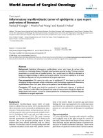
FDG PET scanFigure 3
FDG PET scan. Lesion in the right lung.
World Journal of Surgical Oncology 2008, 6:120 />Page 3 of 6
(page number not for citation purposes)
PET scan was performed to delineate the lung lesion fur-
ther (figure 3). This scan was reported as positive, thus
raising the possibility of:
• A lung primary with pancreatic metastasis
• Synchronous pancreatic and lung primaries
• A pancreatic primary with lung metastasis
CT guided biopsy of the lung lesion was performed, the
histology of which showed reactive changes but no evi-
dence of malignancy. As such and in view of the patient's
background of respiratory disease the PET scan was inter-
preted as demonstrating reactive changes. Given the pres-
entation, tumour markers, imaging appearances and
biopsy results the working diagnosis remained that of a
pancreatic cancer with no evidence of metastatic disease.
The patient proceeded to a pylorus preserving pancreati-
coduodenectomy (PPPD). There was no evidence of intra-
abdominal spread at laparotomy. The head of the pan-
creas contained a palpable mass. This was resected in rou-
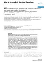
tine fashion. The histology of the resected specimen was a
single poorly differentiated adenocarcinoma (figure 4)
(11 mm in maximum dimension) staining strongly posi-
tive to TTF-1 and CK7 (figure 5), and negative staining for
CK20 and PSA. The tumour did not approach any of the
resection margins or surfaces. Also, none of the surround-
ing 16 lymph nodes had any evidence of disease.
In view of the reported immunohistochemical profile,
coupled with the identification of a lung lesion, the
tumour was interpreted as metastatic adenocarcinoma of
bronchial origin rather than as a primary pancreatic
lesion. As a result the patient was referred to a thoracic sur-
geon for consideration of removal of the lung lesion. Six
weeks later the patient underwent a mini thoracotomy
where a 2 × 3 cm lesion was identified in the medial seg-
ment of the upper lobe of the right lung. The segment was
removed along with hilar and mediastinal lymph nodes
for staging. Histology of this specimen reported a lung
adenocarcinoma with complete excision and no lymph
node involvement.
Three weeks after his lung resection the patient was started
on adjuvant chemotherapy with gemcitabine and carbo-
platin. This regime was continued for 6 months. The
patient was seen eighteen months from presentation.
Clinically he remained symptom free and a follow-up CT
of his chest and abdomen revealed no evidence of recur-
rence.
Discussion
Pancreatic cancer is one of the leading causes of cancer
deaths ranking 4th in the US and 6th in Europe [2]. How-
ever, little attention is devoted to secondary deposits of
other tumours to the pancreas. Retrospective studies on
pancreatectomy procedures have reported that metastatic
disease represents merely 3% or so of resected malignant
pancreatic masses [3,4]. As such they are often mistaken as
pancreatic primaries and only recognised for what they
truly are in retrospect on histological examination [5].
Some 98% of patients with a malignant process who
present with obstructive jaundice will do so as a result of
a primary pancreatic cancer [6]. On the other hand,
autopsy statistics suggest that the pancreas is a more fre-
High magnification view of lesion resected from the pancreas (haematoxylin and eosin)Figure 4
High magnification view of lesion resected from the
pancreas (haematoxylin and eosin).
High magnification view of lesion resected from the pancreas (immunohistochemical staining with TTF-1)Figure 5
High magnification view of lesion resected from the
pancreas (immunohistochemical staining with TTF-
1).
World Journal of Surgical Oncology 2008, 6:120 />Page 4 of 6
(page number not for citation purposes)
quent site for metastatic disease, albeit on a subclinical
scale. The incidence of secondary pancreatic tumours is up
to 16% of autopsy studies [7], with a wide variation of pri-
mary cancers responsible. Patients who present with a
clinical picture which relates directly to disease in the pan-
creas at presentation will tend to do so with the symptoms
of obstructive jaundice or pancreatitis [8]. More often
than not these patients prove to have advanced disease
which is only amenable to palliative treatment.
Lung cancer metastasizes to many sites, but most fre-
quently to bone, the liver and the adrenal glands [9,10].
Approximately one third of patients will present with
symptoms relating to extra thoracic spread [10]. The pan-
creas is considered to be an infrequent target to which
lung cancer will metastasize to. Figures are reported in the
range of 0–12% [11-13]. The majority of those which do
are of SCLC histological subtype [14]. Rarer still, at pres-
entation, is for lung cancer to present with a clinical pic-
ture of jaundice due to synchronous metastatic
adenocarcinoma [1]. In those cases where it does, this is
more likely to be due to widespread hepatic disease than
to extrahepatic biliary obstruction [15]. A larger subgroup
of patients with lung cancer will develop a metachronous
pancreatic metastasis, which will usually be identified on
follow-up investigations. One recent case report pub-
lished in March 2008 reports the first case of lung adeno-
carcinoma with a metachronous isolated deposit in the
pancreas and no evidence of other disease. This case was
treated with biliary stenting and palliative chemotherapy
[16].
Of secondary deposits discovered in the pancreas, lung
cancer makes up (along with renal cell carcinoma, breast
and gastric cancer) a high percentage (table 1) [7,17-36].
Indicative published figures are 14.2% (49 of 311 second-
ary tumours) [7], 17.0% (18 of 108)[18] and 18.2% (4 of
22) [17]. The large majority of cancer patients with meta-
static disease to the pancreas are treated with palliative
intent as patients usually present with widespread disease.
Where surgery is contemplated, it is usually limited to
bypass procedures in patients with obstructive jaundice.
There have been reports where patients with this presenta-
tion have undergone more major procedures such as pan-
creatic resection[37], but this has tended to be in
ignorance of the fact that the aetiology of the obstruction
was of metastatic origin, as was in our case. There are sev-
eral publications advocating the consideration of a pan-
creatic resection in selected cases. One of these is a
literature review by Minni et al, where 333 cases with sec-
ondary deposits in the pancreas were reviewed. Of these,
234 had treatment information of which 150 (64.1%)
underwent pancreatic resections [3]. More than 25 differ-
ent histologic types are reported 45.0% of which were
renal cell, 14.7% lung, 7.5% breast and 6.6% colonic car-
cinomas. In a series of twelve patients with a variety of dif-
ferent metastatic tumours to the pancreas, Le Borgne et al
[38], suggest that a more aggressive surgical approach
should be considered, especially in patients with meta-
chronous ampullary and pancreatic deposits from renal
cell carcinomas, sarcomas and carcinoid tumours. They
reported 35% survival rate at 2 years and 17% at 4 years.
Stage IV NSCLC has a poor prognosis. Median survival
with best supportive care is reported as 3.6 months (range,
2.4 to 4.9 months) whilst platinum based chemotherapy
regimes increase this statistic to 6.5 months (range, 4.7 to
8.5 months). This patient is alive and disease free 18
months following presentation. It is accepted practice
today to consider selected patients with solitary intracra-
nial deposits for resection [39-41]. Also it has been sug-
gested repeatedly that a survival benefit may be achieved
by surgical treatment of solitary extracranial spread of
NSCLC [42-46]. The experience and information availa-
ble for the surgical treatment of metastatic disease from
the lung exclusively to the pancreas is very limited and few
guidelines are available on the appropriate management
of such cases. Most series describe treatment which, from
the outset had a palliative intent. Hiotis et al [47], how-
ever, report three cases of patients with metachronous
(information from personal correspondence with author)
NSCLC metastatic disease to the pancreas who underwent
Table 1: Summary of world literature on pancreatic metastases
from lung cancer
Lung cancer histology subtype
Small Cell Lung Cancer (22)
Adenocarcinoma
1
(4)
Large Cell (2)
Squamous Cell (2)
Anaplastic bronchial (1)
'Lung Cancer'
2
(4)
Presenting symptoms
Obstructive Jaundice
1
(15)
Acute Pancreatitis (13)
No Symptoms
3
(5)
Gastrointestinal bleed (1)
Not Available (1)
Treatment Received
4
Palliative Chemotherapy (13)
Biliary stent (8)
Palliative Operation (4)
Best Supportive Care (7)
Pancreatic Resection (6)
Adjuvant Chemotherapy (2)
Exploratory laparotomy (1)
Includes our case.
2
No further information from authors
3
Includes
patients who were identified on surveillance.
4
Some patients received
more than one treatment.
papers reviewed: [6,8,16,17,19-38,47]
World Journal of Surgical Oncology 2008, 6:120 />Page 5 of 6
(page number not for citation purposes)
pancreatectomies with curative intent. All patients devel-
oped recurrence.
Conclusion
In the majority of cancers, synchronous presentation gen-
erally carries a worse prognosis than a metachronous one.
Our case is an example of a synchronous metastatic
deposit resected (albeit) inadvertently. However, resec-
tion of both lesions has led to long-term disease-free sur-
vival. Therefore we believe that this report demonstrates
that in selected cases consideration should be given not
just to palliation but to potentially curative surgery
whether it be synchronous or more likely metachronous
presentation of metastatic lung cancer to the pancreas.
This is very different from what has been described previ-
ously where very few operations with curative intent have
been carried out, in particular on patients with NSCLC.
List of abbreviations
CT: Computed Tomography; MRCP: Magnetic Resonance
Cholangiopancreatography; ERCP: Endoscopic Retro-
grade Cholangiopancreatography; CBD: Common Bile
Duct; FDG-PET: Fluorodeoxyglucose – Positron emission
tomography; NSCLC: Non-small cell lung carcinoma;
TTF-1: Thyroid Transcription Factor-1; PSA: Prostate Spe-
cific Antigen; CK7, CK20: Cytokeratin 7, Cytokeratin 20.
Consent
Written consent was sought and obtained from the
patient prior to publication of this article.
Competing interests
The authors declare that they have no competing interests.
Authors' contributions
SP operated on the patient, conducted the collection of
the data and the literature and conceived the case report.
SM was involved in collection of literature and drafting
the article. RRH was the principal investigator, operated
on the patient collected data and was involved in the
drafting of the article.
All the authors have read and approved the final manu-
script.
References
1. Smith HJ: Extrahepatic bile duct obstruction in primary carci-
noma of the lung: incidence, diagnosis, and non-operative
treatment. J Natl Med Assoc 1980, 72:215-220.
2. Michaud DS: Epidemiology of pancreatic cancer. Minerva Chir
2004, 59:99-111.
3. Minni F, Casadei R, Perenze B, Greco VM, Marrano N, Margiotta A,
Marrano Dl: Pancreatic metastases: observations of three
cases and review of the literature. Pancreatology 2004,
4:509-520.
4. Roland CF, van Heerden JA: Nonpancreatic primary tumors
with metastasis to the pancreas. Surg Gynecol Obstet 1989,
168:345-347.
5. Doring C, Lindlar F: [Clinically a primary lung carcinoma – dur-
ing autopsy metastasis of a pancreatic cancer]. Med Welt
1969, 8:407-411.
6. Z'graggen K, Fernandez-del CC, Rattner DW, Sigala H, Warshaw AL:
Metastases to the pancreas and their surgical extirpation.
Arch Surg 1998, 133:413-417.
7. Cubilla AlFPJ: Tumors of the Exocrine Pancreas 1980, 137:.
8. Kim KH, Kim CD, Lee SJ, Lee G, Jeen YT, Lee HS, Chun HJ, Song CW,
Um SH, Lee SW, Choi JH, Ryu HS, Hyun JH: Metastasis-induced
acute pancreatitis in a patient with small cell carcinoma of
the lung. J Korean Med Sci 1999, 14:107-109.
9. Abrams HL, Spiro R, Goldstein N: Metastases in carcinoma; anal-
ysis of 1000 autopsied cases. Cancer 1950, 3:74-85.
10. Beckles MA, Spiro SG, Colice GL, Rudd RM: Initial evaluation of
the patient with lung cancer: symptoms, signs, laboratory
tests, and paraneoplastic syndromes. Chest 2003,
123:97S-104S.
11. Galluzzi S, Payne PM: Bronchial carcinoma: a statistical study of
741 necropsies with special reference to the distribution of
blood-borne metastases. Br J Cancer 1955, 9:511-527.
12. Jereczek B, Jassem J, Karnicka-Młodkowska H, Badzio A, Mos-
Antkowiak R, Szczepek B, Chojak E, Dziadziuszko R, Lisowska B,
Malak K: Autopsy findings in small cell lung cancer. Neoplasma
1996, 43:133-137.
13. Lankisch PG, Lohr A, Kunze E: [Acute metastasis-induced pan-
creatitis in bronchial carcinoma]. Dtsch Med Wochenschr 1987,
112:1335-1337.
14. Maeno T, Satoh H, Ishikawa H, Yamashita YT, Naito T, Fujiwara M,
Kamma H, Ohtsuka M, Hasegawa S: Patterns of pancreatic
metastasis from lung cancer. Anticancer Res 1998, 18:2881-2884.
15. Johnson DH, Hainsworth JD, Greco FA: Extrahepatic biliary
obstruction caused by small-cell lung cancer. Ann Intern Med
1985, 102:487-490.
16. Perfetti V, Markopoulos K, Maffe GC, Picheo R, Corazza GR: Juxta-
papillary pancreatic metastasis with obstructive jaundice as
isolated recurrence of lung adenocarcinoma. Dig Liver Dis
2008, 40:230-231.
17. Moussa A, Mitry E, Hammel P, Sauvanet A, Nassif T, Palazzo L, Malka
D, Delchier JC, Buffet C, Chaussade S, Aparicio T, Lasser P, Rougier
P, Lesur G: Pancreatic metastases: a multicentric study of 22
patients. Gastroenterol Clin Biol 2004, 28:872-876.
18. Nakamura E, Shimizu M, Itoh T, Manabe T: Secondary tumors of
the pancreas: clinicopathological study of 103 autopsy cases
of Japanese patients. Pathol Int 2001, 51:686-690.
19. Crippa S, Angelini C, Mussi C, Bonardi C, Romano F, Sartori P, Uggeri
F, Bovo G: Surgical treatment of metastatic tumors to the
pancreas: a single center experience and review of the liter-
ature. World J Surg 2006, 30:1536-1542.
20. Jeong IB, Kim SM, Lee TH, Im EH, Huh KC, Kang YW, Choi YW: Pan-
creatic metastasis and obstructive jaundice in small cell lung
carcinoma. Korean J Intern Med 2006, 21:132-135.
21. Liratzopoulos N, Efremidou EI, Papageorgiou MS, Romanidis K,
Minopoulos GJ, Manolas KJ: Extrahepatic biliary obstruction due
to a solitary pancreatic metastasis of squamous cell lung car-
cinoma. Case report. J Gastrointestin Liver Dis 2006,
15:73-75.
22. Chowhan NM, Madajewicz S: Management of metastases-
induced acute pancreatitis in small cell carcinoma of the
lung. Cancer 1990, 65:1445-1448.
23. Evans AT: Necrotising pancreatitis and diabetes associated
with disseminated small cell carcinoma of lung. Scott Med J
1988, 33:377.
24. Hall M, Bundred NJ, Hall AW: Oat cell carcinoma of the bron-
chus and acute pancreatitis. Eur J Surg Oncol 1987, 13:371-372.
25. Kubota T, Ikezoe T, Harada R, Nakata H, Kobayashi M, Taguchi H:
[Pancreatic metastasis from lung cancer: report of an
autopsy case]. Nihon Kokyuki Gakkai Zasshi 2003, 41:917-921.
26. Moazzam N, Mir A, Potti A: Pancreatic metastasis and extrahe-
patic biliary obstruction in squamous cell lung carcinoma.
Med Oncol 2002, 19:273-276.
27. Niccolini DG, Graham JH, Banks PA: Tumor-induced acute pan-
creatitis. Gastroenterology 1976, 71:142-145.
28. Noseda A, Gangji D, Cremer M: Acute pancreatitis as presenting
symptom and sole manifestation of small cell lung carci-
noma. Dig Dis Sci 1987, 32:327-331.
Publish with BioMed Central and every
scientist can read your work free of charge
"BioMed Central will be the most significant development for
disseminating the results of biomedical research in our lifetime."
Sir Paul Nurse, Cancer Research UK
Your research papers will be:
available free of charge to the entire biomedical community
peer reviewed and published immediately upon acceptance
cited in PubMed and archived on PubMed Central
yours — you keep the copyright
Submit your manuscript here:
/>BioMedcentral
World Journal of Surgical Oncology 2008, 6:120 />Page 6 of 6
(page number not for citation purposes)
29. Papagiannis A, Zarogoulidis K, Delis D, Patakas D: A 52-year-old
man with a lung mass and acute abdominal pain. Chest 2000,
117:894-896.
30. Sakar A, Kara E, Aydede H, Ayhan S, Celik P, Yorgancioglu A: A case
of a small cell lung carcinoma presenting with jaundice due
to pancreatic metastasis. Tuberk Toraks 2005, 53:181-184.
31. Schmitt JK: Pancreatitis and diabetes mellitus with metastatic
pulmonary oat-cell carcinoma. Ann Intern Med 1985,
103:638-639.
32. Schwarz RE, Chu PG, Grannis FW Jr: Pancreatic tumors in
patients with lung malignancies: a spectrum of clinicopatho-
logic considerations. South Med J 2004, 97:811-815.
33. Seo PJ, Kim DM, Kang MS, Lee SI, Kim HJ: [A case of metastasis-
induced acute pancreatitis improved by chemotherapy].
Korean J Gastroenterol 2005, 46:409-412.
34. Stewart KC, Dickout WJ, Urschel JD: Metastasis-induced acute
pancreatitis as the initial manifestation of bronchogenic car-
cinoma. Chest 1993, 104:98-100.
35. Wernecke K, Peters PE, Galanski M: Pancreatic metastases: US
evaluation. Radiology 1986, 160:399-402.
36. Woo JS, Joo KR, Woo YS, Jang JY, Chang YW, Lee J 2nd, Chang R:
Pancreatitis from metastatic small cell lung cancer success-
ful treatment with endoscopic intrapancreatic stenting.
Korean J Intern Med 2006, 21:256-261.
37. Kotan C, Er M, Ozbay B, Uzun K, Barut I, Ozgoren E: Extrahepatic
biliary obstruction caused by small-cell lung cancer: a case
report. Acta Chir Belg 2001, 101:190-192.
38. Le BJ, Partensky C, Glemain P, Dupas B, de Kerviller B: Pancreati-
coduodenectomy for metastatic ampullary and pancreatic
tumors. Hepatogastroenterology 2000, 47:540-544.
39. Patchell RA, Tibbs PA, Walsh JW, Dempsey RJ, Maruyama Y, Kryscio
RJ, Markesbery WR, Macdonald JS, Young B: A randomized trial of
surgery in the treatment of single metastases to the brain. N
Engl J Med 1990, 322:494-500.
40. Hu C, Chang EL, Hassenbusch SJ 3rd, Allen PK, Woo SY, Mahajan A,
Komaki R, Liao Z: Nonsmall cell lung cancer presenting with
synchronous solitary brain metastasis. Cancer 2006,
106:1998-2004.
41. Koutras AK, Marangos M, Kourelis T, Partheni M, Dougenis D, Icon-
omou G, Vagenakis AG, Kalofonos HP: Surgical management of
cerebral metastases from non-small cell lung cancer. Tumori
2003, 89:292-297.
42. Luketich JD, Martini N, Ginsberg RJ, Rigberg D, Burt ME: Successful
treatment of solitary extracranial metastases from non-
small cell lung cancer. Ann Thorac Surg 1995, 60:1609-1611.
43. Ambrogi V, Tonini G, Mineo TC: Prolonged survival after extrac-
ranial metastasectomy from synchronous resectable lung
cancer. Ann Surg Oncol 2001, 8:663-666.
44. Hirano Y, Oda M, Tsunezuka Y, Ishikawa N, Watanabe G: Long-
term survival cases of lung cancer presented as solitary bone
metastasis. Ann Thorac Cardiovasc Surg 2005, 11:401-404.
45. Shimizu K, Nagai K, Yoshida J, Nishimura M, Hayashi R, Yokose T:
Successful management of solitary malar metastasis from
lung cancer. Lung Cancer 2002, 36:337-339.
46. Kim KS, Na KJ, Kim YH, Ahn SJ, Bom HS, Cho CK, Kim HJ, Kim YI,
Lim SC, Kim SO, Oh IJ, Song SY, Choi C, Kim YC: Surgically
resected isolated hepatic metastasis from non-small cell
lung cancer: a case report. J Thorac Oncol 2006, 1:494-496.
47. Hiotis SP, Klimstra DS, Conlon KC, Brennan MF: Results after pan-
creatic resection for metastatic lesions. Ann Surg Oncol 2002,
9:675-679.