Báo cáo khoa học: "Bilateral gluteal metastases from a misdiagnosed intrapelvic gastrointestinal stromal tumor" docx
Bạn đang xem bản rút gọn của tài liệu. Xem và tải ngay bản đầy đủ của tài liệu tại đây (1.21 MB, 6 trang )
BioMed Central
Page 1 of 6
(page number not for citation purposes)
World Journal of Surgical Oncology
Open Access
Case report
Bilateral gluteal metastases from a misdiagnosed intrapelvic
gastrointestinal stromal tumor
Dritan Pasku*
1
, Apostolos Karantanas
2
, Elpida Giannikaki
3
, Maria Tzardi
3
,
Emmanouil Velivassakis
1
and Pavlos Katonis
1
Address:
1
Department of Orthopaedic and Traumatology, University Hospital of Heraklion, Crete, Greece,
2
Department of Radiology, University
Hospital of Heraklion, Crete, Greece and
3
Department of Pathology, University Hospital of Heraklion, Crete, Greece
Email: Dritan Pasku* - ; Apostolos Karantanas - ;
Elpida Giannikaki - ; Maria Tzardi - ;
Emmanouil Velivassakis - ; Pavlos Katonis -
* Corresponding author
Abstract
Background: The location of gastrointestinal stromal tumors (GIST) outside of the
gastrointestinal system is a rare event.
Case presentation: A 56-year old woman presented with a GIST of the pelvis was misdiagnosed
and treated as a uterine leiomyosarcoma. The diagnosis was made after the CD117 (KIT) positivity
in the biopsy of the excised bowel mass four years from the first presentation. During this period
she presented a bilateral muscle and subcutaneous metastasis in the gluteal area.
Conclusion: The correct diagnosis of the extra-gastrointestinal stromal tumor is a challenge even
for experienced pathologists. CD117 (KIT) positivity is the most important immunohistochemical
feature in the histological diagnosis. To our knowledge a metastatic EGIST (extra-gastrointestinal
stromal tumor) to the skeletal muscle bilaterally has not been described previously in the English
medical literature.
Background
Gastrointestinal stromal tumors (GISTs) are the most fre-
quent mesenchymal neoplasms that may occur in any seg-
ment of the gastrointenstinal tract. In the earlier medical
literature GISTs were diagnosed as smooth muscle tumors
(leiomyoma, leiomyosarcoma and leiomyoblastoma) or
tumors of the peripheral nerves (neurofibroma and
schwannoma). Nevertheless, the application of immuno-
chemistry has reclassified them. GISTs are mostly found in
the stomach (60–70%) and in the small intestine (20–
30%). The location in the esophagus and in the colon is
about 5% [1,2]. Extra-gastrointestinal stromal tumors
(EGISTs) may occur in the mesentery, omentum and ret-
roperitoneun in about 5% of all cases [3,4].
The GISTs are usually positive for CD117 (KIT), which is
the specific defining immunohistochemical feature for
this group of tumors. The accurate surgical resection and
the postoperative therapy with the single-agent KIT inhib-
itor imatinib mesilate (Gleevec, Glivec) is the gold stand-
ard treatment for GISTs. Post-operative recurrence is
observed in 40–90% of the cases treated with surgery
alone [5].
Published: 30 December 2008
World Journal of Surgical Oncology 2008, 6:139 doi:10.1186/1477-7819-6-139
Received: 17 September 2008
Accepted: 30 December 2008
This article is available from: />© 2008 Pasku et al; licensee BioMed Central Ltd.
This is an Open Access article distributed under the terms of the Creative Commons Attribution License ( />),
which permits unrestricted use, distribution, and reproduction in any medium, provided the original work is properly cited.
World Journal of Surgical Oncology 2008, 6:139 />Page 2 of 6
(page number not for citation purposes)
The differential diagnosis between GISTs and other simi-
lar tumors is a challenge. GISTs usually metastasize to the
liver and lung, but in recent medical literature an intracra-
nial metastasis from a perisacral GIST has been reported
[6]. To the best of our knowledge, metastasis to skeletal
muscles and subcutaneous fat has never been described
for EGISTs. We present herein, the natural history and glu-
teal bilateral soft tissue secondary deposits from a misdi-
agnosed and treated only surgically intrapelvic GIST.
Case presentation
A 56-year old woman was presented to the orthopaedic
department with a 3-month history of a painless mass in
the upper external area of the left gluteus.
Two years, previously she underwent an abdominal hys-
terectomy because of an enlarging mass found in the pel-
vis, located between the uterus and the recto-sigmoid
area. CA 125 was moderately elevated at 61.9 U/ml. The
resected mass was bilobular (9 × 7 × 6, 5 cm and 5, 2 × 5
× 5 cm) with hard and partially soft elements. Multiple
intramural uterine leiomyomas were also identified. The
tumor was diagnosed as "a leiomyosarcoma" composed
of cellular bundles of spindle cells with medium grade
atypia and presence of giant cells. The range of mitosis was
very high, up to 50/10 H.P.F (high power field). Immuno-
histochemical analysis with avidin-biotin-peroxidase
complex showed positive reaction for vimentin, smooth
muscle actin and desmin (Fig 1 and 2). The patient was
treated post-operatively with radiotherapy and chemo-
therapy (Gemcitabine and docetaxel).
One year after the hysterectomy, a single metastasis was
diagnosed during a routine follow up in the left upper
lung lobe treated surgically with lobectomy. The histolog-
ical diagnosis was compatible with the known primary
tumor. One year later, the patient was presented in the
orthopaedic department with a painless mass in the left
gluteus area. The clinical examination revealed a focal
lump in the left gluteal area. The subsequent magnetic res-
onance imaging study, showed a mass with central necro-
sis located in the subcutaneous fatty tissue, in close
proximity to the gluteal muscles. In addition, a second
small lesion with similar findings was detected in the right
gluteal muscle (Fig 3).
A diagnosis of soft tissue metastasic disease was suggested
and a surgical resection of both masses was undertaken.
The post-operative period was uneventful. The histologi-
cal diagnosis confirmed the presence of a spindle cell sar-
coma with central necrosis and morphologic features
similar to the patient's previous sarcoma (Fig 4). A routine
abdominal CT-Scan showed changes of the previous hys-
terectomy and an edematous appearance of the sigmoid
colon wall, as well as presacral fat edema attributed to pre-
vious radiotherapy.
Four years after the hysterectomy the patient was pre-
sented at the emergency department with symptoms of
intestinal obstruction. During laparotomy, a mass of 10.5
× 4.5 × 4 cm was identified infiltrating the bowel wall and
protruding into the sigmoid lumen. A colectomy com-
bined with colostomy was performed. Microscopically,
Microscopic photogragh of the first tumor, showing interlac-ing bundles of spindle cells with medium atypia (×200, H&E)Figure 1
Microscopic photogragh of the first tumor, showing
interlacing bundles of spindle cells with medium aty-
pia (×200, H&E).
Microscopic photograph of the first tumor showing giant cells and abundant mitosis (×200, H&E)Figure 2
Microscopic photograph of the first tumor showing
giant cells and abundant mitosis (×200, H&E).
World Journal of Surgical Oncology 2008, 6:139 />Page 3 of 6
(page number not for citation purposes)
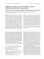
Metastatic disease from extragastrointestinal GISTFigure 3
Metastatic disease from extragastrointestinal GIST. a) The axial T1-w MR image shows an intermediate signal intensity
ovoid-shaped mass, lateral to the gluteus maximus muscle, within the subcutaneous fat (arrow). An intact fat plane separated
the mass from the muscle. b) The fat suppressed T2-w TSE MR image, shows the high signal intensity of the central mass and
the intermediate signal intensity of the anterior wall (open arrow). A second smaller lesion with similar imaging characteristics
is shown in the right gluteus maximus muscle (thin arrow). c) The contrast enhanced fat suppressed T1-w SE MR image shows
the peripheral enhancement of the wall (open arrow). The central non enhancing component presumably corresponds to
necrosis. Ring-like enhancement is also shown in the smaller lesion (thin arrow).
World Journal of Surgical Oncology 2008, 6:139 />Page 4 of 6
(page number not for citation purposes)
the resected mass composed of interlacing bundles of
spindle and epithelioid mesenchymal cells with morpho-
logical features similar to the previously described
tumors. The immunohistochemical analysis of the cells
showed positive for vimentin and a focal positivity for
actin (α-SMA). This time, a CD117(c-KIT) immunohisto-
chemical stain was performed and the neoplastic cells
showed extensive positivity. A diagnosis of a mesenchy-
mal stromal tumor was established (Fig 5 and 6). A retro-
spective analysis of the original tumor performing
immunhistochemistry, showed a focal positivity of the
neoplastic cells for CD117(c-kit) (Fig 5)
Based on these features, the primary pelvic tumor was
most probably an EGIST originally misdiagnosed as leio-
myosarcoma. Post-operatively the patient underwent
therapy with Imatinib Mesylate (Gleevec, Novartis) and
one year after surgery is now free of symptoms.
Discussion
GISTs are mesenchymal tumors of the gastrointestinal
tract, believed to originate from interstitial cells of Cajal or
related stem cells. Cajal cells are intermediates between
the gastrointestinal (GI) autonomic nervous system and
the smooth muscle cells regulating GI motility and auto-
nomic nerve function [7,8]. Gastrointenstinal mesenchy-
mal tumors (GIMTs) are classified below: a) myogenic
tumors that differentiate into smooth muscle cells such as
leiomyoma (LM) and leimyosarcoma (LMS), b) neuro-
genic tumors that differentiate into neurocytes such as
schwannoma and c) GIST that differentiate into other cell
types. In general, about 80% of GIMT are GIST, 15% are
myogenic tumors and 5% are neurogenic tumors [1,6]
The incidence of GISTs is estimated to be 20/1.000.000
persons per year. The median age of the manifestation
ranges between 55 and 65 years of age. These tumors are
very rare in individuals before the age of 40 and some
studies show a small male predominance [1,2,6].
Microscopic photograph, showing a metastatic lesion in the muscle (×200, H&E)Figure 4
Microscopic photograph, showing a metastatic lesion
in the muscle (×200, H&E).
Microscopic photograph, showing the tumor infiltrating the large bowel wall (×100, H&E)Figure 5
Microscopic photograph, showing the tumor infiltrat-
ing the large bowel wall (×100, H&E).
Immunohistochemical stain for CD 117, showing positivity of the tumor cells in the large bowel (×400)Figure 6
Immunohistochemical stain for CD 117, showing pos-
itivity of the tumor cells in the large bowel (×400).
World Journal of Surgical Oncology 2008, 6:139 />Page 5 of 6
(page number not for citation purposes)
In general the GISTs have a wide clinical manifestation
that depends upon the location and the size of the tumor.
In colorectal area the tumor may be manifested with
lower GI bleeding, perforation, obstruction and pain. The
stomach is the most common site of GIST benign tumors,
whereas most esophageal and colonic GISTs are malig-
nant [1]. Approximately 20 to 25% of gastric and 40%–
50% of small intestinal GISTs, are clinically malignant [9].
GISTs usually metastasize to the liver and lung but in
recent publications there have been reports to the skin
and brain as sites of metastases [6]. Other case reports
described involvement of the omentum, the mesentery
and the retroperitoneum, secondary to their GI tract orig-
inal location [3,9]. Metastases to the bone and soft tissue
are very rare. Miettinen et al. reported humeral metastasis
and paraspinal soft tissue involvement in two patients
with colonic GIST [2].
In the case presented herein, the tumor was misdiagnosed
and remained untreated for about 4 years. Our hypothesis
is that the tumor could have had escaped complete resec-
tion from gynecologists either due to small adhesions
within the recto-sigmoid area or due to minimal involve-
ment of the gut wall. Histologically a GIST tumor is very
similar to leiomyosarcoma (LMS) but in our case non c-
KIT positivity was not checked and the tumor was origi-
nally diagnosed as uterine leiomyosarcoma. The patient
was treated with chemotherapy and radiotherapy but the
GISTs are refractory to both [1]. Therefore, we present the
natural history of an EGIST localized in pelvis with pul-
monary and soft tissue metastasis and a late manifestation
from the area of the resected primary.
The differential diagnosis between leiomysarcoma and
GIST remains a challenge. Of a total of 133 rectal and anal
GISTs identified in the Armed Forces Institute of Pathol-
ogy at Washington and in the Haartman Institute of the
university of Helsinki, 80 tumors (60%) had been origi-
nally diagnosed from other centers as leiomyosarcoma
(LMSs), 29 tumors (21.8%) as smooth muscle tumors of
uncertain malignant potential, 21 tumors (15.8%) as lei-
omyoma (LMs) and only 3 tumors (2.25%) as GISTs [8].
Immunohistochemically, the majority of GI mesechymal
tumors are GISTs and are strongly c-KIT positive (96–
100%) and in particular, esophageal and rectal ones are
nearly consistently CD34-positive (95–100%) [1,6].
Our case may be considered as EGIST. Traditionally the
EGIST are mesenchymal tumors with similar clinico-
pathologic and genetic profile localized in the omentum,
mesentery and retroperitoneum comprising less than 5%
of GISTs [1,2]. In the last few years, few cases have been
reported in unusual anatomic location [10,11]. Others
suggest that true EGISTs are extremely rare, less than 1,5%,
and probably are extramural gastric (omental) or small
intestinal (mesenteric) in origin [1]. One of the criteria for
diagnosing EGISTs is the recognition of minor association
or adhesions with a neighboring gut segment [12]. The
prognosis of EGIST, as all GISTs tumors, depends on the
mitotic activity, the tumor size, but also the age and loca-
tion [1,2,5]. Gastric tumors have a less aggressive behavior
than intestinal tumors. Size larger than 5 cm is also more
malignant and a mitotic count over 50/50 HPF demon-
strate a high-grade malignancy. Based on these observa-
tions our patient had a very aggressive EGIST with high
probability for metastasis.
To the best of our knowledge, this is the first case of
EGISTs to metastasize bilaterally in the gluteal region. In
a retrospective study of 118 metastasis to soft tissues over
a period of 30 year, regardless of the primary, 5 cases were
located in the gluteal region, all from carcinomas and
melanoma. In only one case located to the abdominal
wall, the primary tumor was GIST of the small bowel [13].
Skeletal muscle is resistant to both primary and metastatic
cancer. Previous reports have cited various mechanisms as
reasons for muscle resistance to malignancy [14-16].
Weiss found that, cancer cells survive best in denervated
muscle compared with electrically stimulated muscles
[17]. This finding suggests that most cancer cells die soon
after haematogenous spread to muscle because of an
inhospitable mechanical, pH, and a metabolic environ-
ment in normally functioning muscles. Muscles that are
injured may have a different mechanical, pH, or meta-
bolic environment that is more favourable to the survival
of metastatic cancer cells. We propose that, because of the
post-operative radiotherapy, similarly to the muscle
injury, the metabolic changes of the gluteal area included
in the field of the radiotherapy, probably created a favour-
able environment for the development of the soft tissue
metastasis.
In conclusion, EGISTs are rare tumours of abdominal cav-
ity with potentially high malignancy and metastatic
capacity, exhibiting clinical and histological difficulty for
a correct diagnosis. Metastatic disease may occur in the
soft tissues. Early recognition and prompt diagnosis, will
allow the proper treatment to be initiated.
Consent
Written informed consent was obtained from the patient
for publication of this case report and accompanying
images. A copy of the written consent is available for
review by the Editor-in-Chief of this journal.
Competing interests
The authors declare that they have no competing interests.
Publish with BioMed Central and every
scientist can read your work free of charge
"BioMed Central will be the most significant development for
disseminating the results of biomedical research in our lifetime."
Sir Paul Nurse, Cancer Research UK
Your research papers will be:
available free of charge to the entire biomedical community
peer reviewed and published immediately upon acceptance
cited in PubMed and archived on PubMed Central
yours — you keep the copyright
Submit your manuscript here:
/>BioMedcentral
World Journal of Surgical Oncology 2008, 6:139 />Page 6 of 6
(page number not for citation purposes)
Authors' contributions
DP, EV and PV initiated and co-wrote the paper and per-
formed the surgical excision of the gluteal metastases. AK
analysed the MR images, prepared the illustrations and
legend, and performed the proof editing. EG and MT
examined the specimen and prepared the histological
illustrations.
References
1. Miettinen M, Lasota J: Gastrointestinal stromal tumors-defini-
tion, clinical, histological, immunohistochemical and molec-
ular genetic features and differential diagnosis. New findings
on their biology. Virchows Arch 2001, 438:1-12.
2. Miettinen M, Sarlomo-Rikala M, Sobin LH, Lasota J: Gastrointestinal
stromal tumors and leiomyosarcomas in the colon. A clin-
icopathologic, immunohistochemical and molecular genetic
study of 44 cases. Am J Surg Pathol 2000, 24:1339-1352.
3. Miettinen M, Momhan JM, Sarlomo-Rikala M, Kovatich A, Carr NJ,
Emory TS, Sobin LH: Gastrointestinal stromal tumors/smooth
muscle tumors/GISTs in the omentum and mesentery-Clin-
icopathologic and immunohistochemical study of 26 cases.
Am J Surg Pathol 1999, 22:1109-1118.
4. Ferchichi L, Kourda N, Zermani R, Aouem J, Zaouche A, Abdjellil Z,
Najah N, Baltagi Ben Jilani S: Extragastrointestinal stromal
tumors: a report of 4 cases. Ann Chir 2006, 131:271-275.
5. DeMatteo RP, Lewis JJ, Leung D, Mudan SS, Woodruff JM, Brennan
MF: Two hundred gastrointestinal stromal tumors: recur-
rence paterns and prognostic factors for survival. Ann Surg
2000, 231:51-58.
6. Kaku S, Tanaka T, Ohtuka T, Seki K, Sawauchi S, Numoto RT,
Murakami S, Komine K, Abe T: Perisacral gastrointestinal stro-
mal tumor with intracranial metastasis. Case report. Neurol
Med Chir (Tokyo) 2006, 46:254-257.
7. Huizinga JD, Thuneberg L, Kluppel M, Malysz J, Mikkelsen HB, Bern-
stein A: W/kit gene required for interstitial cells of cajal and
for intestinal pacemaker activity. Nature 1993, 373:347-349.
8. Miettinen M, Furlong M, Sarlomo-Rikala M, Burke A, Sobin LH, Lasota
J: Gastrointestinal stromal tumors, Intramural leiomyomas
and leiomyosarcomas in the rectum and anus. A clinico-
pathologic, immunohistochemical and molecular genetic
study of 144 cases. Am J Surg Pathol 2001, 25:1121-1133.
9. Miettinen M, Lasota J: Gastrointestinal stromal tumors: Review
on morphology, molecular pathology, prognosis and differ-
ential diagnosis. Arch Pathol Lab Med 2006, 130:1466-1478.
10. Lam MM, Corless CL, Goldblum JR, Heinrich MC, Downs-Kelly E,
Rubin BP: Extragastrointestinal stromal tumors presenting as
vulvovaginal/rectovaginal septal masses: a diagnostic pitfall.
Int J Gynecol Pathol 2006, 25:288-292.
11. Peitsidis P, Zarganis P, Trichia H, Vorgias G, Smith JR, Akrivos T:
Extragastrointestinal stromal tumor mimicking a uterine
tumor. A rare clinical entity. Int J Gynecol Cancer 2008,
18:1115-8.
12. Agaimy A, Wunsch PH: Gastrointestinal stromal tumours: a
regular origin in the muscularis propria, but an extremely
diverse gross presentation. A review of 200 cases to critically
re-evaluate the concept of so-called extra-gastrointestinal
stromal tumours. Langenbecks Arch Surg 2006, 391:322-9.
13. Plaza JA, Perez-Montiel D, Mayerson J, Morrison C, Suster S: Metas-
tases to soft tissue: a review of 118 cases over a 30-year
period. Cancer 2008, 112:193-203.
14. Acinas Garcia O, Fernandez FA, Satue EG, Buelta L, Val-Bernal JF:
Metastasis of malignant neoplasms to skeletal muscle. Rev
Esp Oncol 1984, 31:57-67.
15. Williams JB, Youngberg RA, Bui-Mansfield LT, Pitcher JD: MR imag-
ing of skeletal muscle metastases. AJR Am J Roentgenol 1997,
168:555-557.
16. Pretorius ES, Fishman EK: Helical CT of skeletal muscle metas-
tases from primary carcinomas. AJR Am J Roentgenol 2000,
174:401-404.
17. Weiss L: Biomechanical destruction of cancer cells in skeletal
muscle: a rate-regulator for hematogenous metastasis. Clin
Exp Metastasis 1989, 7:483-491.