Báo cáo y học: "Clinical and immunological effects of Rituximab in patients with lupus nephritis refractory to conventional therapy: a pilot study" doc
Bạn đang xem bản rút gọn của tài liệu. Xem và tải ngay bản đầy đủ của tài liệu tại đây (588.75 KB, 9 trang )
Open Access
Available online />Page 1 of 9
(page number not for citation purposes)
Vol 8 No 3
Research article
Clinical and immunological effects of Rituximab in patients with
lupus nephritis refractory to conventional therapy: a pilot study
Mónica Vigna-Perez
1
, Berenice Hernández-Castro
1
, Octavio Paredes-Saharopulos
1
,
Diana Portales-Pérez
2
, Lourdes Baranda
1,3
, Carlos Abud-Mendoza
3
and Roberto González-Amaro
1
1
Departamento de Inmunología, Facultad de Medicina, UASLP, San Luis Potosí, SLP, México
2
Laboratorio de Inmunología Celular y Molecular, Facultad de Ciencias Químicas, UASLP, San Luis Potosí, SLP, México
3
Unidad Regional de Reumatología y Osteoporosis, Hospital Central Dr Ignacio Morones Prieto, San Luis Potosí, SLP, México
Corresponding author: Roberto González-Amaro,
Received: 11 Dec 2005 Revisions requested: 16 Jan 2006 Revisions received: 9 Feb 2006 Accepted: 10 Apr 2006 Published: 5 May 2006
Arthritis Research & Therapy 2006, 8:R83 (doi:10.1186/ar1954)
This article is online at: />© 2006 Vigna-Perez et al.; licensee BioMed Central Ltd.
This is an open access article distributed under the terms of the Creative Commons Attribution License ( />),
which permits unrestricted use, distribution, and reproduction in any medium, provided the original work is properly cited.
Abstract
We studied the clinical and immunological effects of Rituximab
(anti-CD20) therapy in patients with lupus nephritis. In an open
clinical trial, 22 patients with active systemic lupus
erythematosis and renal involvement (mainly class III and IV
according to the WHO classification) that was refractory to
conventional therapy were studied. In all these patients,
Rituximab (0.5 to 1.0 g at days 1 and 15) was added to the
immunosuppressive therapy and its therapeutic effect was
evaluated. In addition, the levels and function of regulatory T
lymphocytes and the apoptosis of immune cells were assessed.
We found a significant reduction in disease activity (p < 0.05,
MEX-SLEDAI index), and proteinuria (p < 0.05) at days 60 and
90 of Rituximab therapy. Although most patients showed
improvement in creatinine clearance and erythrocyturia, no
significant changes in these parameters were detected. In most
patients (20/22), B cell depletion was observed, but no clear-
cut effect of Rituximab on complement levels or auto-antibody
titers was detected (p > 0.05 in all cases). One patient died at
day 70 with invasive histoplasmosis. No important adverse
effects of Rituximab therapy were registered in other patients. A
significant enhancement in the levels of different CD4+
regulatory cells (T
REG
, Th3, Tr1), but not CD8+ Ts lymphocytes,
was observed at day 30. This increase was sustained for T
REG
cells at day 90, and accompanied by an improvement in their
regulatory function. In addition, we observed an unexpected
increase in the apoptosis of T cells at day 30. Interestingly, the
enhancement in the suppressive function of T
REG
cells was not
observed in the two patients that showed the poorest clinical
response to Rituximab. We conclude that the data obtained in
this open clinical trial suggest that Rituximab is a promising
candidate for randomized controlled trials in patients with lupus
nephritis refractory to the conventional immunosuppressive
therapy. The effects of Rituximab on regulatory cells and
apoptosis of T lymphocytes are interesting and its possible role
in the putative effect of this biological agent in systemic lupus
erythematosis deserves additional studies.
Introduction
Systemic lupus erythematosus (SLE) is an autoimmune dis-
ease of unknown etiology that affects multiple tissues and
organs [1]. The main immunological alterations observed in
these patients are the loss of tolerance to self-antigens with
polyclonal activation of B lymphocytes, production of different
auto-antibodies, and an altered function of T cells [2-4]. In
addition, several abnormalities of B lymphocytes, including an
increased Ca
2+
influx, and a decreased fraction of naïve B cells
with enhanced levels of circulating plasmablasts have been
described [5,6]. Furthermore, B lymphocytes from SLE
patients show an increased synthesis of certain cytokines (for
example, IL-10), and have an important role as antigen pre-
senting cells, inducing the activation of CD4+ auto-reactive T
cells, which in turn allow the activation and differentiation of B
cells, and the production of high affinity auto-antibodies [5,7].
Thus, it is very likely that the hyperactivity of B cells and auto-
antibody production in SLE depend on the interactions of self
antigens with B cell surface immunoglobulin receptors, and
the cognate interaction of these cells with helper T
GITRL = glucocorticoid-induced TNF receptor family-related ligand; FITC = fluorescein isothiocyanate; IL = interleukin; mAb = monoclonal antibody;
PBMNC = peripheral blood mononuclear cells; SLE = systemic lupus erythematosus; TGF = transforming growth factor; TNF = tumor necrosis factor;
Tr1 = type-1 T regulatory; T
REG
= T regulatory.
Arthritis Research & Therapy Vol 8 No 3 Vigna-Perez et al.
Page 2 of 9
(page number not for citation purposes)
lymphocytes. Several subsets of CD4+ cells with immuno-reg-
ulatory activity have been described, including T regulatory
(T
REG
) lymphocytes, and type-1 T regulatory (Tr1) cells [8,9].
T
REG
lymphocytes are characterized by the expression of CD4
and by high levels of the α chain of IL-2 receptor (CD25) as
well as by their lack of responsiveness to antigenic stimulation
[8,10]. These CD4+CD25
bright
CTLA-4+ cells arise from thy-
mus as natural regulatory cells and exert their activity by cell-
to-cell contact as well as by inducing the differentiation of
CD4+CD25- lymphocytes into regulatory cells [6,10,11]. On
the other hand, Tr1 lymphocytes express CD4 and synthesize
IL-10 [9]. These regulatory cells are antigen specific lym-
phocytes derived from conventional CD4+CD25- naive pre-
cursors upon exposure to antigen and IL-10 [9,12]. Additional
regulatory cells have been described, including the
CD8+CD28- suppressor (Ts) lymphocytes, and the CD4+
Th3 cells, which are characterized by the synthesis of trans-
forming growth factor (TGF)-β [13,14].
In recent years, different groups have explored the role of reg-
ulatory T cells in the pathogenesis of human autoimmune dis-
eases, including multiple sclerosis, and rheumatoid arthritis
[15,16]. In addition, the possible effect of biological therapy
with anti-tumor necrosis factor (TNF)-α agents in rheumatoid
arthritis patients on the levels and function of T
REG
cells has
been recently explored by our and another group [17,18]. In
the case of SLE, it has been reported that CD8+ suppressor
cells have a defective function [19]. However, in the case of
CD4+CD25+ Treg cells, there are only quantitative studies in
patients with SLE, and no functional studies have been per-
formed [20,21].
Table 1
Main clinical data of SLE patients studied
Patient Age (years)/gender Disease duration
(years)
Main clinical
manifestation
Nephritis stage/
duration
a
Disease activity
b
Previous therapy Current therapy
c
1 41/F 15 Ou, Ar, Le, Ly IV/8 12 GC, Cyc, Aza GC, Aza
2 24/M 4 Er, Ph, Le IV/4 10 GC, Aza, Mmf, CsA GC, Mmf
3 36/F 6 Er, Ph, Ou, Ar, Le, Ly III/6 14 GC, Aza, CsA GC, Aza, Mtx
4 19/M 5 Le IV/4 8 GC, Cyc, Mmf GC
5 56/F 12 Er, Ph, Ou, Se, Ar IV/3 15 GC, Cyc, Mmf, Aza GC, Mmf
6 9/M 1 Er, Ph, Se, Ar IV/1 12 GC, Mmf, Mtx, Aza, Cy GC, Aza, Mtx, Mmf
7 26/F 11 Er, Ph, Ou, Se, Ar, Le IV/4 10 GC, Mtx, Aza GC, Mtx, Aza
8 31/F 16 Ph, Ou, Se, Ar, CNS IV/8 10 GC, Cyc, Lfm, Aza, Mtx, Mmf GC, Aza, Mtx, Mmf, Cyc
9 23/F 3 Ph, Ou, Se, Ar, CNS IV/3 12 GC, Mmf, Aza, Cyc, Mtx GC, Mtx, Mmf
10 32/F 6 Er, Ph, Ou, Ar, Ly IV/6 8 GC, Mtx, Aza, Cyc Aza
11 24/F 2 Er, Ph, Ou, Se III/2 9 GC, Mtx, Aza Aza, Mtx, Mmf
12 27/F 13 Er, Ph, Ou, Se, Ly IV/13 19 GC, Mmf, Cyc, Aza GC, Aza, Mtx, Mmf
13 43/F 6 Er, Ph, Le IV/6 9 GC, Mtx, Aza, Pdn Mtx
14 21/F 8 Er, Ph, Ou, Se, Ly V/8 9 GC, Mtx, Aza, Cyc, Mmf None
15 36/F 1 Er, Ph, Ly V/1 10 GC, Aza Aza
16 25/F 1 Er, Ou, Se, Ly, CNS IV/1 11 GC, Aza, Mtx GC
17 33/F 8 Er, Ph, Ou, Se, Ar, Le IV/8 10 GC, Aza, Mtx, Mmf, Cq GC, Aza, Mtx, Mmf
18
d
28/F 3 Er, Ph, Ou, Se, Le, Ly IV/3 9 GC, Cyc, Aza, Mmf, Mtx, Cq GC, Aza, Mtx, Mmf
19 40/F 4 Er, Ph, Ou, Se, Ar IV/4 10 GC, Aza, Mtx Aza
20 19/F 1 A, Le, Ly IV/1 10 GC, Aza, Mtx, Mmf GC, Aza, Mmf
21 24/F 4 Er, Ph, Le IV/3 10 GC, Aza, Mtx, Mmf, Cyc GC, Aza, Mtx
22 22/F 1 Ou, Ar, Le, Se IV/1 10 GC, Aza, Mtx, Mmf, Cq GC, Aza, Mtx, Mmf
a
According to the WHO classification [33]; duration is in years.
b
According to the MEX-SLEDAI index.
c
Immunosuppressive therapy to which
Rituximab was added.
d
This patient had invasive histoplasmosis of the lungs with massive hemorrhage and mucormycosis of a coronary artery; she
died at day 70 of Rituximab therapy. Ar, arthritis; Aza, azathioprine; Cfm, cyclophosphamide; Cq, chloroquine; CNS, central nervous system disease;
Er, erythema; GC, glucocorticoids; Le, leukopenia; Lfm, leflunomide; Ly, severe lymphopenia (lymphocyte count <1,000 cells/mm
3
); Mmf,
mycophenolate mofetil; Mtx, methotrexate; N, nephritis; Ou, oral ulcers; Ph, photosensitivity; Se, serositis.
Available online />Page 3 of 9
(page number not for citation purposes)
B cell depletion therapy has been recently assayed in patients
with different autoimmune diseases, including SLE [22-24]. In
this regard, it has been found that patients with SLE that
receive the mouse/human chimeric anti-CD20 monoclonal
antibody (mAb) Rituximab show clinical improvement and dim-
inution of the abnormalities found in B lymphocytes [25-27].
Rituximab specifically binds to the B cell-specific antigen
CD20, a cell surface protein believed to function in B cell cycle
initiation and differentiation [28]. CD20 protein is expressed
on immature and mature B lymphocytes, but not in early B cells
precursors or plasma cells [29]. Rituximab is a very effective B
cell depleting agent in vivo, inducing the lysis of these cells
mediated by complement and by Fc receptor-bearing cyto-
toxic cells as well as by inducing their programmed cell death
[30,31]. This B cell depleting agent does not have an impor-
tant effect on serum immunoglobulin levels, and SLE patients
that receive it show a variable diminution in auto-antibody titers
[25-27]. It is feasible, therefore, that Rituximab exerts addi-
tional effects on the immune system that account for its effect
on disease activity in SLE. In this regard, it has been described
that Rituximab therapy is associated with a diminution of
expression of CD40L (CD154) and other activation markers of
T lymphocytes [32].
In this work, we have performed an open clinical trial with
Rituximab in SLE patients, and evaluated their clinical
response as well as its effect on the levels and function of reg-
ulatory T lymphocytes and the apoptosis of immune cells.
Materials and methods
Patients
Twenty-two patients (19 females and 3 males) with a diagno-
sis of SLE according to the criteria of the American College of
Rheumatology were studied. The main clinical data of these
patients are given in Table 1. All patients had active disease
with renal involvement, which was classified according to the
Figure 1
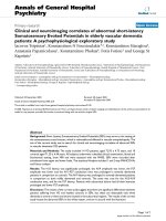
Effect of Rituximab therapy on different laboratory and clinical parame-ters in patients with systemic lupus erythematosusEffect of Rituximab therapy on different laboratory and clinical parame-
ters in patients with systemic lupus erythematosus. Twenty-two sys-
temic lupus erythematosus patients with active disease and renal
involvement received Rituximab (0.5 to 1.0 g at days 0 and 15), and the
(a) disease activity (MEX-SLEDAI index), (b) 24 hour proteinuria, (c)
erythrocyturia, and (d) creatinine clearance were measured at days 0,
60, and 90.
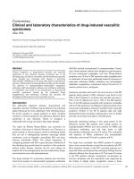
Figure 2
Effect of Rituximab therapy on the levels of peripheral blood regulatory T cellsEffect of Rituximab therapy on the levels of peripheral blood regulatory
T cells. In 17 systemic lupus erythematosus patients that received
Rituximab (0.5 to 1.0 g at days 0 and 15), the levels of (a)
CD4+CD25
bright
and (c) CD4+CTLA-4+ cells, and CD4+ lymphocytes
synthesizing (b) TGF-β+ or (d) IL-10 was assessed in peripheral blood
by flow cytometry before (day 0), and at days 30 and 90 of Rituximab
therapy, as described in Materials and methods. The inset in (a) shows
how the expression of CD25 by activated CD8+ cells (left histogram)
was employed to define CD4+CD25
bright
cells (right histogram). The
arithmetic mean is indicated with a horizontal line, and the standard
deviation with a vertical line. In all cases, the percentage of positive
cells is referred to the gate of lymphocytes, defined by side and forward
scatter characteristics.
Arthritis Research & Therapy Vol 8 No 3 Vigna-Perez et al.
Page 4 of 9
(page number not for citation purposes)
World Health Organization [33]. In most of them, nephropathy
was refractory to the administration of conventional immuno-
suppressive drugs, and two patients had a disease relapse
with massive proteinuria despite this type of therapy.
Rituximab was added to the immunosuppressive therapy at a
dose of 0.5 to 1.0 g on days 1 and 15. No changes in the ther-
apy with immunosuppressive drugs, including doses of gluco-
corticoids, were made during the study. Six patients were not
receiving glucocorticoids when Rituximab administration was
started.
The clinical response to Rituximab was evaluated by routine
laboratory tests, and clinical examination at days 30, 60 and
90. Complete renal response was defined as normal serum
creatinine, inactive urine sediment, and urinary proteinuria
<500 mg/24 h. Partial remission was defined as >40%
improvement in the renal parameters that were abnormal at the
onset of the study. Immune effects of Rituximab therapy were
evaluated at day 30, and 90. The appearance of important
adverse effects, defined as a severe infectious process or sig-
nificant clinical manifestations associated with the administra-
tion of intravenous biological agents was registered. In all
cases, informed consent was obtained, and the local Univer-
sity ethics committee approved this study.
Blood samples and cell isolation
Peripheral blood samples were obtained from all patients
before (day 0) and at 30 and 90 days after Rituximab therapy.
Peripheral blood mononuclear cells (PBMNCs) were isolated
by Ficoll-Hypaque density gradient centrifugation and cell via-
bility (trypan blue dye exclusion) was always greater than 95%.
CD4+ lymphocytes were purified using a MACS LS separa-
tion column (Miltenyi Biotec, Bergisch Gladbach, Germany). In
brief, PBMNCs were incubated with a biotinylated antibody
cocktail for 15 minutes at 4°C, washed, incubated with anti-
Biotin Micro Beads (Miltenyi) for 20 minutes at 4°C and
washed. Then, CD4+ lymphocytes were negatively selected
using a MACS LS separation column (Miltenyi). For isolation
of CD4+CD25+ cells, CD4+ lymphocytes were incubated
with anti-CD25 microbeads for 15 minutes at 4°C, followed by
positive selection using an additional separation column.
CD4+CD25- lymphocytes were also recovered. Cell purity
was always greater than 90%, as assessed by flow cytometry
analysis.
Quantification of regulatory T cells
PBMNCs were double immunostained for CD4 and CD25 or
CD4 and CTLA-4 using specific fluorescein isothiocyanate
(FITC)- or phycoerythrin (PE)-labeled mAb (BD Pharmingen,
San Diego, CA, USA), and then analyzed with a FACSCalibur
flow cytometer using the Cell Quest software (Becton Dickin-
son, San Jose, CA, USA). For the detection of CTLA-4, cells
were previously fixed with p-formaldehyde, and permeabilized
with saponin. Results were expressed as the percent of
CD4+CD25
bright
and CD4+CTLA-4+ lymphocytes. As sug-
gested by Cao et al. [34], CD4+CD25
bright
cells were defined
as those CD4+ lymphocytes showing higher expression of
CD25 than autologous CD8+ activated in vitro with phytohe-
magglutinin (PHA).
The percent of CD4+ lymphocytes synthesizing IL-10 was
determined using the appropriate commercial kit (Miltenyi), fol-
lowing the instructions of the manufacturer. In this assay, cells
were first incubated with a catch reagent (a bi-specific dimeric
antibody) that binds to a cell surface antigen of leukocytes and
is also able to react with the IL-10 secreted by the cell. Then,
cells were incubated for 60 minutes at 37°C, washed and
labeled with FITC-, and PE-tagged anti-CD4 and anti-IL-10
mAbs. Finally, double positive cells were detected with a FAC-
SCalibur flow cytometer, and results were expressed as the
percent of Tr1 lymphocytes. In additional experiments, CD4+
cells with membrane-bound TGF-β were detected by staining
PBMNCs with anti-CD4, and anti-TGF-β labeled mAbs and
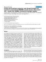
Figure 3
Rituximab effect on the suppressive function of T regulatory (T
REG
) cells in patients with systemic lupus erythematosusRituximab effect on the suppressive function of T regulatory (T
REG
) cells in patients with systemic lupus erythematosus. CD4+CD25+ T cells were
isolated and then mixed or not with autologous CD4+CD25- lymphocytes in the presence of phytohemagglutinin, and cultured for 72 hours. Then,
cell proliferation was determined by
3
H-methyl-thymidine incorporation. These assays were performed before (day 0), and at days 30 and 90 of
Rituximab therapy, as described in Materials and methods. All experiments were run in triplicate, and the results expressed as the percent of cell pro-
liferation (CD4+CD25- lymphocytes cultured without CD4+CD25+ cells = 100%). * p < 0.05
Available online />Page 5 of 9
(page number not for citation purposes)
flow cytometry analysis. Finally, Ts lymphocytes (CD8+CD28)
were also detected by two-color flow cytometry analysis using
specific mAbs.
Detection of apoptosis
Fresh isolated PBMNCs were fixed and stained by the TUNEL
technique using the Apo-Direct kit (BD Pharmingen). After the
fluorescent dUTP nick end labeling, cells were analyzed by
flow cytometry. In additional experiments, PBMNCs from the
same sample were stained with annexin V labeled with FITC
and propidium iodide and analyzed in a FACSCalibur flow
cytometer. In addition, annexin V staining was combined with
mAbs specific for CD3 or CD19. Results were expressed as
the percent of positive apoptotic cells.
Cell proliferation assays
Non-regulatory CD4+CD25- T cells (1 × 10
5
) were mixed or
not with CD4+CD25+ regulatory T cells (1 × 10
4
) in the pres-
ence of PHA (5.0 µg/ml) and cultured for 72 hours in complete
culture medium in 96 well plates.
3
H-methyl-thymidine (1.0
µCi/well, specific activity 5.0 Ci/mM; New England Nuclear,
Boston, MA, USA) was added for the last 12 hours of culture,
and at the end of incubation cells were harvested and prolifer-
ation was determined using a liquid scintillation counter. All
these experiments were run in triplicate and the results were
expressed as the percent of cell proliferation, according to the
following formula: % cell proliferation = (cpm of CD4+CD25-
plus CD4+CD25+ cells/cpm of CD4+CD25- cells alone) ×
100.
Statistical analysis
Data were compared with the Sigma STAT software (SPSS
Inc., Chicago, IL, USA) using T paired and correlation (rho
Spearman's coefficient) tests, with a level of significance of p
< 0.05.
Results
As previously reported [24-26], we found that Rituximab ther-
apy induced improvements in different clinical parameters of
most SLE patients included in this study. Disease activity
(MEX-SLEDAI index) significantly diminished in 90% of
patients at days 60 and 90 of Rituximab therapy (Figure 1a). In
all patients, the dose of glucocorticoids remained unchanged
during the study, and in six cases these drugs were not admin-
istered (Table 1). Proteinuria levels also significantly dimin-
ished, showing an important reduction in most patients (Figure
1b), in some cases as early as day 15, but in other patients at
days 60 or 90 of Rituximab therapy (data not shown). How-
ever, a substantial enhancement of proteinuria was observed
in two patients during Rituximab therapy. Although an improve-
ment in creatinine clearance was observed upon Rituximab
administration in 72% of patients studied (Figure 1c), the
Figure 4
Effect of Rituximab therapy on the apoptosis of peripheral blood mono-nuclear cells (PBMNCs) in patients with systemic lupus erythematosusEffect of Rituximab therapy on the apoptosis of peripheral blood mono-
nuclear cells (PBMNCs) in patients with systemic lupus erythematosus.
Freshly isolated PBMNCs from patients with systemic lupus erythema-
tosus were fixed, stained using the TUNEL technique, and analyzed by
flow cytometry before (day 0), and at days 30 and 90 of Rituximab ther-
apy. Data from all samples studied are shown in (a), and representative
histograms of flow cytometry analysis are shown in (b). The arithmetic
mean is indicated with a horizontal line and the standard deviation with
a vertical line. Numbers in (b) correspond to the percent of positive
cells.
Figure 5
Analysis of apoptosis of subsets of peripheral blood mononuclear cells in patients with systemic lupus erythematosus under Rituximab therapyAnalysis of apoptosis of subsets of peripheral blood mononuclear cells
in patients with systemic lupus erythematosus under Rituximab therapy.
Freshly isolated mononuclear cells from systemic lupus erythematosus
patients were double labeled with annexin V-FITC plus antibodies spe-
cific for (a) T cells or (b) B cells, and analyzed by flow cytometry, as
described in Materials and methods. As indicated, detection of apop-
totic cells was performed before (day 0) and after (days 30 and 90)
administration of Rituximab. The arithmetic mean is indicated with a hor-
izontal line, and the standard deviation with a vertical line. (c) Repre-
sentative histograms of flow cytometry analysis of CD3+ and CD19+
cells at day 30.
Arthritis Research & Therapy Vol 8 No 3 Vigna-Perez et al.
Page 6 of 9
(page number not for citation purposes)
change in this parameter of renal function was not significant
(p > 0.05, Wilcoxon sum rank test). Likewise, even though
most patients showed reduction in erythrocyturia, this change
was not statistically significant (Figure 1d). Based on these
results, five patients showed a complete remission of renal dis-
ease, whereas seven patients showed a partial renal response.
Six additional patients exhibited improvement in one or several
renal parameters, but they could not be classified as having a
partial or complete remission. Finally, two patients that did not
show improvement of any renal function parameter had renal
failure at days 60 and 90.
On the other hand, there was a great variability in autoantibody
titers (anti-nuclear, anti-dsDNA, anti-phospholipid antibodies),
and serum complement levels (CH
50
, C3, C4) upon Rituximab
therapy either at days 30, 60, or 90 with no significant
changes (p > 0.05 in all cases, data not shown). In most
patients (20/22), B cell depletion (<5.0 B cells/mm
3
) was
induced by Rituximab. Finally, neither serious infectious proc-
esses nor important clinical manifestations associated with the
administration of the biological agent were observed in 21 out
of 22 patients included in the study. However, patient number
18 (Table 1) was admitted to our hospital at day 70 of Rituxi-
mab therapy with severe metabolic acidosis associated to dia-
betes mellitus and pneumonia; 48 hours later, this patient had
severe respiratory failure with diffuse pulmonary infiltrates and
died. At necropsy, invasive histoplasmosis in the lungs with
massive hemorrhage was observed as well as mucormycosis
in a coronary artery.
Different immunological effects of the Rituximab therapy were
assessed in most of the patients (17 out of 22) included in this
study. We first determined the levels of T
REG
cells in the
peripheral blood of these patients after and before Rituximab
therapy. Before starting anti-CD20 administration, we found a
variable number of CD4+CD25
bright
cells in the 17 patients
studied, which was significantly increased at days 30 and 90
of Rituximab therapy (p < 0.05 compared to day 0, t paired
test; Figure 2a). Likewise, the levels of other T cells with regu-
latory phenotype were enhanced upon Rituximab therapy.
CD4+TGF-β+ lymphocytes, which very likely correspond to
Th3 cells, significantly increased after administration of Rituxi-
mab (Figure 2b). A similar phenomenon was observed, at day
30 but not day 90, for CD4+CTLA-4+ and CD4+IL-10+ lym-
phocytes (Figure 2c,d), a phenotype that corresponds to Tr1
cells. In contrast, no significant changes were observed in the
levels of Ts (CD8+CD28-) cells upon Rituximab therapy (p >
0.05, data not shown). No significant correlation between the
enhancement in the levels of regulatory T cells and the
improvement of different clinical parameters was detected
(rho Spearman's coefficient; data not shown).
We then studied the effect of Rituximab administration on the
suppressor activity of T
REG
cells. We found that before starting
Rituximab therapy, CD4+CD25+ lymphocytes from patients
with SLE showed a variable capability to inhibit the prolifera-
tion of autologous CD4+CD25- cells
(CD4+CD25+:CD4+CD25- ratio 1:10) stimulated with PHA
(Figure 3). At days 30 and 90 of Rituximab therapy, a modest
but significant increase in the regulatory function (with a
decreased cell proliferation of CD4+CD25- cells) of T
REG
cells
was observed (p < 0.05 compared to day 0, t paired test; Fig-
ure 3). Similar results were obtained, but with higher levels of
inhibition, when the ratio of regulatory:responder cells was 1:1
(data not shown). As in the case of the levels of regulatory T
cells, we did not detect any significant correlation between the
enhancement in the suppressive function and the improve-
ment of different clinical parameters (data not shown). How-
ever, the two patients (9 and 11) that had the poorest clinical
response to Rituximab administration did not show any
improvement in the suppressive function of their T
REG
cells
(Figure 3). Nevertheless, a modest increase in the levels of
CD4+ regulatory T cells was observed in these patients.
We also analyzed the possible in vivo effect of Rituximab on
the apoptosis of PBMNCs in the patients included in this
study. Interestingly, we found an important and significant
increase in the percent of TUNEL+ cells at day 30 of Rituxi-
mab therapy (p < 0.05, t paired test; Figure 4). The presence
of apoptotic cells in these samples was confirmed by annexin
V labeling and propidium iodide staining (p < 0.05, data not
shown). Two-color flow cytometry analysis using anti-CD3 and
anti-CD19 mAbs plus annexin V-FITC staining showed a sig-
nificant enhancement in the programmed cell death of T lym-
phocytes (Figure 5a,c). As expected, very low levels of B
lymphocytes, and consequently of apoptotic B cells, were
detected in most cases at day 30 of Rituximab administration
(Figure 5b,c). When these assays were performed at day 90,
we did not further detected a significant enhancement of
apoptosis compared to baseline values (Figures 4 and 5).
Discussion
Different open clinical trials [26,27,35-37] and case reports
[38,39] suggest that Rituximab induces a significant reduction
in disease activity in patients with severe SLE when it is added
to the standard immunosuppressive therapy. However,
although it is evident that Rituximab is able to induce an impor-
tant depletion of B cells in most patients with SLE, the whole
consequences of Rituximab administration on the different
pathogenic mechanisms of this condition have not been fully
elucidated. In this regard, it has been described that therapy
with Rituximab has a variable effect on the titers of serum
autoantibodies, with a non-significant effect on plasma immu-
noglobulin levels [25-27,36]. In addition, it has been found that
SLE patients receiving this biological agent show a diminished
expression of the costimulatory molecules CD40 and CD80
by B cells [40]. Furthermore, Rituximab therapy is associated
with a down-regulation of CD40L in T cells as well as a dimi-
nution in the expression of activation markers in these lym-
phocytes [32]. Accordingly, it has been suggested that B
Available online />Page 7 of 9
(page number not for citation purposes)
lymphocytes participate in the pathogenesis of SLE not only
through the synthesis of autoantibodies and that, therefore,
Rituximab exerts its therapeutic effect by different mechanisms
[24]. To further insight into these putative mechanisms, in this
work we decided to evaluate the clinical response to Rituxi-
mab as well as to explore its effect on different immunological
parameters in patients with SLE.
We selected 22 patients with lupus nephritis that was refrac-
tory to conventional therapy with combinations of different
immunosuppressive drugs with or without glucocorticoids.
Two patients with disease relapse and massive proteinuria
were also included. The addition of Rituximab to the ongoing
immunosuppressive therapy of these patients significantly
diminished disease activity in most of them. These results are
in agreement with previous work showing a rapid therapeutic
effect of Rituximab in SLE patients with severe disease
[26,27,35-40]. In addition, our work suggests that this anti-
CD20 mAb could be effective in the absence of glucocorticoid
administration (six patients; Table 1), and even as a single
immunosuppressive agent (one patient), and as early as at day
15. Furthermore, although we did not observe a significant
change in erythrocyturia and creatinine clearance upon Rituxi-
mab therapy, our results show that in 77% of cases, this bio-
logical agent induced improvement in one or two of these
parameters of renal function, even over the short term of this
report (60 to 90 days). Therefore, our data further support the
therapeutic potential of Rituximab in SLE patients with severe
and refractory disease. However, it will be very important to
make a further follow-up of our patients (for example, after one
year), mainly because of the type of renal disease (III and IV)
predominant in them.
It is evident that some patients do not show a satisfactory
response to Rituximab. In this regard, it has been described
that polymorphisms of the CD64 (FcγRIII) gene have an impor-
tant role in the B cell depletion effect of Rituximab [41]. Since
two patients included in our study showed no B cell depletion
(defined as <5.0 B cells/mm
3
) [41] and a poor clinical
response to Rituximab, it is very likely that they bear this type
of polymorphism. These patients showed a significant
increase in proteinuria with an important fall in renal function
during the study. The possible presence of anti-chimeric anti-
bodies may also contribute to the lack of effect observed in
these cases.
Remarkably, the administration of Rituximab in this study was
not accompanied by serious adverse effects in 21 out of 22
patients, despite the long-term depletion of B cells. However,
one patient died because of invasive histoplasmosis, massive
pulmonary hemorrhage, and mucormycosis. Although this
patient was diabetic, and was receiving three immunosuppres-
sive drugs plus high doses of glucocorticoids, it is very feasi-
ble that the inhibitory effect of Rituximab on the immune
system significantly contributed to the fatal opportunistic
infection. Therefore, it would seem necessary to be careful
with the administration of Rituximab in patients that are receiv-
ing several immunosuppressive drugs. However, it is worth
mentioning that in this study, five additional patients were
receiving, in combination with Rituximab, three or more immu-
nosuppressive drugs, plus glucocorticoids. As stated above,
no adverse effects were observed in these patients.
As in other clinical trials [25-27], we have not found a clear-cut
effect of Rituximab therapy on serum autoantibody titers, and
even on complement levels. However, we observed significant
changes in other immune parameters, including a sustained or
transient rise in the proportion of different regulatory T cell
subsets in peripheral blood, including T
REG
(CD4+CD25
bright
)
lymphocytes, Tr1 (CD4+IL-10+) cells, and Th3 (CD4+TGF-
β+) lymphocytes. In most cases, this enhancement was
accompanied by an increase in the suppressor function of
T
REG
lymphocytes. Although the mechanism responsible for
this effect of Rituximab remains to be elucidated, it could be
speculated that the decrease of antigen presenting cells
induced by Rituximab would induce a decrease in activated T
cells, which express, among other cell markers, the ligand of
the glucocorticoid-induced TNF receptor (GITRL) [42].
Since it has been described that this ligand is able to inhibit
the anergic behavior and suppressive function of T
REG
cells
[42,43], it is feasible that a decrease in lymphocytes bearing
GITRL favors an increase in the number and function of regu-
latory cells. In this regard, we have recently found that the
inflammatory cell infiltrate of thyroid autoimmune disease con-
tains a high number of GITRL+ lymphocytes, which very likely
contribute to inhibit the function of T
REG
cells, (M Marazuela
submitted for publication). Another interesting possibility is
that the depletion of B cells induced by Rituximab diminishes
the synthesis of anti-lymphocyte antibodies, which could be
directed against CD4+ regulatory T cells. In any case, it is
worth mentioning that our group and others have previously
found that other biological agents (anti-TNF-α) are also able to
enhance the number and function of T
REG
cells in patients with
an autoimmune/inflammatory disease (rheumatoid arthritis)
[17,18]. As in the case of Rituximab, the mechanism responsi-
ble for this effect of anti-TNF-α agents remains to be eluci-
dated.
It is worth mentioning that the standard assay of suppression
of cell proliferation employed by us does not allow us to rule
out a possible effect of Rituximab on the non-regulatory cells,
increasing their sensitivity to the suppressive signals of T
REG
lymphocytes. In this regard, it has been recently reported that
CD4+CD25- cells from MRL/Mp mice show a reduced sensi-
tivity to suppression by autologous Treg lymphocytes. [44], a
phenomenon that could be related to defects in intracellular
regulatory molecules such as Cbl-b [45]. Thus, it will be inter-
esting to elucidate through future studies whether this effect
of Rituximab in SLE patients is mediated by an enhancement
Arthritis Research & Therapy Vol 8 No 3 Vigna-Perez et al.
Page 8 of 9
(page number not for citation purposes)
of the functional capability of CD4+CD25+ cells or by
increasing the sensitivity of effector CD4+CD25- lymphocytes
to regulatory signals.
It is well known that Rituximab induces apoptosis of B cells
[30,31], and our data show that in most patients included in
this study this biological agent was able to cause a sustained
depletion of these lymphocytes. In addition, we have found an
unexpected induction of apoptosis of T cells upon Rituximab
therapy. Since only B cells express CD20, it is evident that this
induction of apoptosis is not due to the direct action of this chi-
meric antibody on T cells. We think that it is feasible that this
increase in T cell apoptosis could be due to a diminished avail-
ability of antigen presenting cells, a condition that would
induce the programmed cell death of activated auto-reactive T
cells. In this regard, it has been described that TcR/CD28
engagement induces activation of anti-apoptotic molecules
(BAD, Bcl-
XL
) and promotes T cell survival [46,47]. It is also
possible that the depletion of B cells drastically reduces the
synthesis of certain cytokines (for example, IL-10) that have an
important role in the stimulation of expanded T cell subsets (for
example, Th2 lymphocytes) in SLE. In this regard, it has been
shown that B lymphocytes are the main source of IL-10 in
patients with SLE [7]. Although it is evident that these hypoth-
eses require validation with experimental data, we think that
our results suggest that Rituximab exerts, through indirect
mechanisms, an important effect on both regulatory and effec-
tor T cells.
The possible relationship between the immunological effects
of Rituximab on T cells observed by us, and its putative thera-
peutic activity in patients with SLE, remains to be determined.
However, it is worth mentioning that the two patients who did
not respond to Rituximab in this study did not show significant
changes in either the function of regulatory cells or apoptosis
of T lymphocytes. In addition, it is evident that the immunolog-
ical effects observed in this study could contribute to disease
remission. Nevertheless, it will be important to make a longer
follow-up of our patients, to further corroborate the putative
relationship between changes in T cells and the therapeutic
effect of Rituximab.
Conclusion
Our results further support that Rituximab is a promising can-
didate for randomized controlled trials in patients with SLE,
mainly in those with nephritis refractory to conventional immu-
nosuppressive therapy. In addition, we consider that our data
regarding the in vivo effect of Rituximab on regulatory cells
and apoptosis of T lymphocytes are interesting and that its
possible role in the putative effect of this biological agent in
SLE deserves additional studies.
Competing interests
The authors declare that they have no competing interests.
Authors' contributions
MV-P carried out immunological determinations and drafted
the manuscript. BH-C carried out immunological determina-
tions and clinical laboratory tests. OP-S participated in the
clinical study of patients and contributed to drafting the manu-
script. DP-P contributed to immunological determinations and
clinical laboratory tests. LB carried out flow cytometry analysis
and participated in the coordination of the study. CA-M partic-
ipated in the clinical evaluation of patients, conceived the
study, and contributed to its design. RG-A conceived the
study, contributed to the analysis of clinical and laboratory
data, and approved the final manuscript.
GITRL = glucocorticoid-induced TNF receptor family-related ligand;
FITC = fluorescein isothiocyanate; IL = interleukin; mAb = monoclonal
antibody; PBMNC = peripheral blood mononuclear cells; SLE = sys-
temic lupus erythematosus; TGF = transforming growth factor; TNF =
tumor necrosis factor; Tr1 = type-1 T regulatory; T
REG
= T regulatory.
Acknowledgements
This work was supported by a grant from CONACYT, México (to RG-A).
References
1. Mills JA: Systemic lupus erythematosus. N Engl J Med 1994,
330:1871-1879.
2. Nagy G, Koncz A, Perl A: T- and B-cell abnormalities in systemic
lupus erythematosus. Crit Rev Immunol 2005, 25:123-140.
3. Kyttaris VC, Juang YT, Tsokos GC: Immune cells and cytokines
in systemic lupus erythematosus: an update. Curr Opin
Rheumatol 2005, 17:518-522.
4. Kammer GM, Perl A, Richardson BC, Tsokos GC: Abnormal T
cell signal transduction in systemic lupus erythematosus.
Arthritis Rheum 2002, 46:1139-1154.
5. Chan OT, Madaio MP, Shlomchik MJ: The central and multiple
roles of B cells in lupus pathogenesis. Immunol Rev 1999,
169:107-121.
6. Odendahl M, Jacobi A, Hansen A, Feist E, Hiepe F, Burmester GR,
Lipsky PE, Radbruch A, Dorner T: Disturbed peripheral B lym-
phocyte homeostasis in systemic lupus erythematosus. J
Immunol 2000, 165:5970-5979.
7. Llorente L, Zou W, Levy Y, Richaud-Patin Y, Wijdenes J, Alcocer-
Varela J, Morel-Fourrier B, Brouet JC, Alarcon-Segovia D,
Galanaud P, Emilie D: Role of interleukin 10 in the B lymphocyte
hyperactivity and autoantibody production of human systemic
lupus erythematosus. J Exp Med 1995, 181:839-844.
8. Maloy KJ, Powrie F: Regulatory T cells in the control of immune
pathology. Nat Immunol 2001, 2:816-822.
9. Roncanrolo MG, Bachetta R, Bordignon C, Narula S, Levings MK:
Type 1 T regulatory cells. Immunol Rev 2001, 182:68-79.
10. Sakaguchi S, Sakaguchi N, Asano M, Itoh M, Toda M: Immuno-
logic self-tolerance maintained by activated T cells expressing
IL-2 receptor alpha-chains (CD25). Breakdown of a single
mechanism of self-tolerance causes various autoimmune
diseases. J Immunol 1995, 155:1151-1164.
11. O'Garra A, Vieira P: Regulatory T cells and mechanisms of
immune system control. Nat Med 2004, 10:801-805.
12. Levings MK, Roncarolo MG: T-regulatory 1 cells: a novel subset
of CD4 T cells with immunoregulatory properties. J Allergy
Clin Immunol 2000, 106:S109-S112.
13. Liu Z, Tugulea S, Cortesini R, Suciu-Foca N: Specific suppres-
sion of T helper alloreactivity by allo-MHC class I-restricted
CD8+CD28- T cells. Int Immunol 1998, 10:775-783.
14. Sercarz E, Krzych U: The distinctive specificity of antigen-spe-
cific suppressor T cells. Immunol Today 1991, 12:111-118.
15. Morgan ME, Sutmuller RP, Witteveen HJ, van Duivenvoorde LM,
Zanelli E, Snijders , Offringa R, de Vries RR, Toes RE: CD25+ cell
depletion hastens the onset of severe disease in collagen-
induced arthritis. Arthritis Rheum 2003, 48:1452-1460.
Available online />Page 9 of 9
(page number not for citation purposes)
16. Viglietta V, Baecher-Allan C, Weiner HL, Hafler DA: Loss of func-
tional suppression by CD4
+
CD25
+
regulatory T cells in
patients with multiple sclerosis. J Exp Med 2004, 199:971-979.
17. Ehrenstein MR, Evans JG, Singh A, Moore S, Warnes G, Isenberg
DA, Mauri C: Compromised function of regulatory T cells in
rheumatoid arthritis and reversal by anti-TNF-alpha therapy. J
Exp Med 2004, 200:277-285.
18. Vigna-Perez M, Abud-Mendoza C, Portillo-Salazar H, Alvarado-
Sanchez B, Cuevas-Orta E, Moreno-Valdes R, Baranda L, Pare-
des-Saharopulos O, Gonzalez-Amaro R: Immune effects of ther-
apy with Adalimumab in patients with rheumatoid arthritis.
Clin Exp Immunol 2005, 141:372-380.
19. Filaci G, Bacilieri S, Fravega M, Monetti M, Contini P, Ghio M, Setti
M, Puppo F, Indiveri F: Impairment of CD8+ T suppressor cell
function in patients with active systemic lupus erythematosus.
J Immunol 2001, 166:6452-6457.
20. Crispin JC, Martinez A, Alcocer-Varela J: Quantification of regu-
latory T cells in patients with systemic lupus erythematosus. J
Autoimmun 2003, 21:273-276.
21. Liu MF, Wang CR, Fung LL, Wu CR: Decreased CD4+CD25+ T
cells in peripheral blood of patients with systemic lupus
erythematosus. Scand J Immunol 2004, 59:198-202.
22. Gorman C, Leandro M, Isenberg D: B cell depletion in autoim-
mune disease. Arthritis Res Ther 2003, 5(Suppl 4):S17-S21.
23. Looney RJ: B cells as a therapeutic target in autoimmune dis-
eases other than rheumatoid arthritis. Rheumatology (Oxford)
2005, 44(Suppl 2):i13-i17.
24. Silverman GJ: Anti-CD20 therapy in systemic lupus erythema-
tosus: A step closer to the clinic. Arthritis Rheum 2005,
52:371-377.
25. Anolik JH, Barnard J, Cappione A, Pugh-Bernard AE, Felgar RE,
Looney RJ, Sanz I: Rituximab improves peripheral B cell abnor-
malities in human systemic lupus erythematosus. Arthritis
Rheum 2004, 50:3580-3590.
26. Looney RJ, Anolik JH, Campbell D, Felgar RE, Young F, Arend LJ,
Sloand JA, Rosenblatt J, Sanz I: B cell depletion as a novel treat-
ment for systemic lupus erythematosus: a phase I/II dose-
escalation trial of rituximab. Arthritis Rheum 2004,
50:2580-2589.
27. Leandro MJ, Edwards JC, Cambridge G, Ehrenstein MR, Isenberg
DA: An open study of B lymphocyte depletion in systemic
lupus erythematosus. Arthritis Rheum 2002, 46:2673-2677.
28. Golay JT, Clark EA, Beverly PC: The CD20 (Bp35) antigen is
involved in activation of B cells from the G0 to the G1 phase of
the cell cycle. J Immunol 1985, 135:3795-3801.
29. Tedder TF, Boyd AW, Freedman AS, Nadler LM, Schlossman SF:
The B cell surface molecule B1 is functionally linked with B
cell activation and differentiation. J Immunol 1985,
135:973-979.
30. Cerny T, Borisch B, Introna M, Johnson P, Rose AL: Mechanism
of action of rituximab. Anticancer Drugs 2002, 13(Suppl
2):3-10.
31. Maloney DG, Smith B, Rose A: Rituximab: mechanism of action
and resistance. Semin Oncol 2002, 29(2 Suppl 2):2-9.
32. Sfikakis PP, Boletis JN, Lionaki S, Vigklis V, Fragiadaki KG, Iniotaki
A, Moutsopoulos HM: Remission of proliferative lupus nephritis
following B cell depletion therapy is preceded by down-regu-
lation of the T cell costimulatory molecule CD40 ligand. Arthri-
tis Rheum 2005, 52:501-513.
33. Churg J, Sobin LH: Lupus nephritis. In Renal disease: Classifica-
tion and Atlas of Glomerular Disease 1st edition. Edited by: Churg
J, Sobin LH. New York, Igaku-Shoin; 1982:147-149.
34. Cao D, van Vollenhoven R, Klareskog L, Trollmo C, Malmström V:
CD25brightCD4+ regulatory T cells are enriched in inflamed
joints of patients with chronic rheumatic disease. Arthritis Res
Ther 2004, 6:R335-346.
35. Gottenberg JE, Guillevin L, Lambotte O, Combe B, Allanore Y,
Cantagrel A, Larroche C, Soubrier M, Bouillet L, Dougados M, et
al.: Tolerance and short term efficacy of rituximab in 43
patients with systemic autoimmune diseases. Ann Rheum Dis
2005, 64:913-920.
36. Leandro MJ, Cambridge G, Edwards JC, Ehrenstein MR, Isenberg
DA: B-cell depletion in the treatment of patients with systemic
lupus erythematosus: a longitudinal analysis of 24 patients.
Rheumatology 2005, 44:1542-1545.
37. Marks SD, Patey S, Brogan PA, Hasson N, Pilkington C, Woo P,
Tullus K: B lymphocyte depletion therapy in children with
refractory systemic lupus erythematosus. Arthritis Rheum
2005, 52:3168-3174.
38. Weide R, Heymanns J, Pandorf A, Koppler H: Successful long-
term treatment of systemic lupus erythematosus with rituxi-
mab maintenance therapy. Lupus 2003, 12:779-782.
39. van Vollenhoven RF, Gunnarsson I, Welin-Henriksson E, Sundelin
B, Osterborg A, Jacobson SH, Klareskog L: Biopsy-verified
response of severe lupus nephritis to treatment with rituximab
(anti-CD20 monoclonal antibody) plus cyclophosphamide
after biopsy-documented failure to respond to cyclophospha-
mide alone. Scand J Rheumatol 2004, 33:423-427.
40. Tokunaga M, Fujii K, Saito K, Nakayamada S, Tsujimura S, Nawata
M, Tanaka Y: Down-regulation of CD40 and CD80 on B cells in
patients with life-threatening systemic lupus erythematosus
after successful treatment with rituximab. Rheumatology 2005,
44:176-182.
41. Anolik JH, Campbell D, Felgar RE, Young F, Sanz I, Rosenblatt J,
Looney RJ: The relationship of FcgammaRIIIa genotype to
degree of B cell depletion by rituximab in the treatment of sys-
temic lupus erythematosus. Arthritis Rheum 2003, 48:455-459.
42. Tone M, Tone Y, Adams E, Yates SF, Frewin MR, Cobbold SP,
Waldmann H: Mouse glucocorticoid-induced tumor necrosis
factor receptor ligand is costimulatory for T cells. Proc Natl
Acad Sci USA 2003, 100:15059-15064.
43. Shevach EM: Regulatory/suppressor T cells in health and
disease. Arthritis Rheum 2004, 50:2721-2724.
44. Monk CR, Spachidou M, Rovis F, Leung E, Botto M, Lechler RI,
Garden OA: MRL/Mp CD4+, CD25- T cells show reduced sen-
sitivity to suppression by CD4+, CD25+ regulatory T cells in
vitro: a novel defect of T cell regulation in systemic lupus
erythematosus. Arthritis Rheum 2005, 52:1180-1184.
45. Wohlfert EA, Gorelik L, Mittler R, Flavell RA, Clark RB: Deficiency
in the E3 ubiquitin ligase Cbl-b results in a multifunctional
defect in T cell TGF-β sensitivity in vitro and in vivo. J Immunol
2006, 176:1316-1320.
46. Boise LH, Minn AJ, Noel PJ, June CH, Accavitti MA, Lindstein T,
Thompson CB: CD28 costimulation can promote T cell survival
by enhancing the expression of Bcl-XL. Immunity 1995,
3:87-98.
47. Villalba M, Bushway P, Altman A: Protein kinase c-theta medi-
ates a selective T cell survival signal via phosphorylation of
BAD. J Immunol 2001, 166:5955-5963.