Báo cáo y học: "Relaxin and β-estradiol modulate targeted matrix degradation in specific synovial joint fibrocartilages: progesterone prevents matrix loss" doc
Bạn đang xem bản rút gọn của tài liệu. Xem và tải ngay bản đầy đủ của tài liệu tại đây (864.06 KB, 9 trang )
Open Access
Available online />Page 1 of 9
(page number not for citation purposes)
Vol 8 No 4
Research article
Relaxin and β-estradiol modulate targeted matrix degradation in
specific synovial joint fibrocartilages: progesterone prevents
matrix loss
Gihan Hashem, Qin Zhang, Takayuki Hayami, Jean Chen, Wei Wang and Sunil Kapila
Department of Orthodontics and Pediatric Dentistry, University of Michigan, 1011 North University Avenue, Ann Arbor, MI 48109-1078, USA
Corresponding author: Sunil Kapila,
Received: 21 Mar 2006 Revisions requested: 10 May 2006 Revisions received: 17 May 2006 Accepted: 25 May 2006 Published: 19 Jun 2006
Arthritis Research & Therapy 2006, 8:R98 (doi:10.1186/ar1978)
This article is online at: />© 2006 Hashem et al.; licensee BioMed Central Ltd.
This is an open access article distributed under the terms of the Creative Commons Attribution License ( />),
which permits unrestricted use, distribution, and reproduction in any medium, provided the original work is properly cited.
Abstract
Relaxin, a 6-kDa polypeptide hormone, is a potent mediator of
matrix turnover and contributes to the loss of collagen and
glycosaminoglycans (GAGs) from reproductive tissues,
including the fibrocartilaginous pubic symphysis of several
species. This effect is often potentiated by β-estradiol. We
postulated that relaxin and β-estradiol might similarly contribute
to the enhanced degradation of matrices in fibrocartilaginous
tissues from synovial joints, which may help explain the
preponderance of diseases of specific fibrocartilaginous joints
in women of reproductive age. The objective of this study was to
compare the in vivo effects of relaxin, β-estradiol, and
progesterone alone or in various combinations on GAG and
collagen content of the rabbit temporomandibular joint (TMJ)
disc fibrocartilage, knee meniscus fibrocartilage, knee articular
cartilage, and the pubic symphysis. Sham-operated or
ovariectomized female rabbits were administered β-estradiol
(20 ng/kg body weight), progesterone (5 mg/kg), or saline
intramuscularly. This was repeated 2 days later and followed by
subcutaneous implantation of osmotic pumps containing relaxin
(23.3 µg/kg) or saline. Tissues were retrieved 4 days later and
analyzed for GAG and collagen. Serum relaxin levels were
assayed using enzyme-linked immunosorbent assay. Relaxin
administration resulted in a 30-fold significant (p < 0.0001)
increase in median levels (range, approximately 38 to 58 pg/ml)
of systemic relaxin. β-estradiol, relaxin, or β-estradiol + relaxin
caused a significant loss of GAGs and collagen from the pubic
symphysis and TMJ disc and of collagen from articular cartilage
but not from the knee meniscus. Progesterone prevented
relaxin- or β-estradiol-mediated loss of these molecules. The
loss of GAGs and collagen caused by β-estradiol, relaxin, or β-
estradiol + relaxin varied between tissues and was most
prominent in pubic symphysis and TMJ disc fibrocartilages. The
findings suggest that this targeted modulation of matrix loss by
hormones may contribute selectively to degeneration of specific
synovial joints.
Introduction
The development and maintenance of cartilage entails active
secretion of macromolecular glycosaminoglycans (GAGs) and
collagens by chondrocytes, resulting in an organized extracel-
lular matrix (ECM), which confers specific mechanical and
physiologic properties to cartilage [1]. Chondrocytes also play
a critical role in the normal remodeling of cartilaginous tissues
by expressing tissue-degrading proteinases, primarily those
belonging to the matrix metalloproteinase (MMP) family of
enzymes [2]. This normal tissue turnover is regulated by many
local and systemic agents, including peptide and steroid hor-
mones, and entails the maintenance of a finely tuned balance
between matrix synthesis and degradation. In degenerative
joint diseases, an imbalance between synthesis and degrada-
tion of the ECM which results primarily from altered chondro-
cyte function leads to the net loss of tissue macromolecules
and compromises joint function. Because several degenera-
tive joint diseases have a high female-to-male preponderance,
a regulatory role of specific sex hormones has been implicated
in controlling the metabolism of these tissues [3-5]. This is par-
ticularly true of the highly prevalent diseases of the fibrocarti-
laginous temporomandibular joint (TMJ), which also have a
high female-to-male predilection but which, unlike similar dis-
eases of other joints which largely occur postmenopausally,
DMMB = dimethylmethylene blue; ECM = extracellular matrix; GAG = glycosaminoglycan; LGR = leucine-rich repeat-containing, G protein-coupled
receptor; MMP = matrix metalloproteinase; OD = optical density; TMJ = temporomandibular joint.
Arthritis Research & Therapy Vol 8 No 4 Hashem et al.
Page 2 of 9
(page number not for citation purposes)
are observed primarily in women of reproductive age [6,7].
These observations have led to the hypothesis that female hor-
mones, including estrogen and relaxin, play a crucial role in
predisposing women to TMJ diseases.
Relaxin H2, a 6-kDa polypeptide hormone that is structurally
related to the insulin family of hormones which is primarily syn-
thesized by the corpus luteum and placenta, is a known medi-
ator of ECM remodeling in several reproductive tissues,
including the uterus, cervix, ovary, breast, and the pubic sym-
physis [8-13]. Within the fibrocartilaginous pubic symphysis
and cervix, relaxin plays an important role during parturition by
mediating the remodeling necessary for the successful deliv-
ery and survival of pups [14,15]. Although the precise mecha-
nisms for relaxin's modulation of matrix turnover have not been
fully elucidated, it appears to exert these effects by mediating
the synthesis and/or degradation of matrix macromolecules
[12,16-18]. The latter mechanism likely involves relaxin's
induction of several members of MMPs [5,19-21].
In several species, including guinea pigs, mice, bats, and
humans, relaxin induces the transformation of pubic joint fibro-
cartilage into a flexible and elastic interpubic ligament during
pregnancy [17]. These changes in the characteristics of the
pubic symphysis result from a decrease in collagen content
caused by relaxin, which in some species is potentiated by the
prior or concurrent administration of estrogen [12,13,18].
Because of the matrix remodeling effects of relaxin and β-
estradiol on the fibrocartilaginous pubic symphysis, it is plau-
sible that other cartilages within synovial joints may be among
the other presumptive, yet-to-be-proven, non-reproductive tar-
get sites of the tissue-remodeling activity of these hormones.
Indirect evidence for such a modulation of matrix turnover of
cartilage by relaxin is provided by studies showing that relaxin
produces a dose-dependent induction of MMPs collagenase-
1 (MMP-1) and stromelysin-1 (MMP-3) in fibrocartilaginous
cells of the TMJ disc [5]. Priming of these cells with β-estradiol
potentiated their MMP-inductive response to relaxin, resulting
in the maximal expression of collagenase-1 and stromelysin-1
at relaxin concentrations that were 10- to 100-fold lower in β-
estradiol-primed cells than in unprimed cells. These observa-
tions on isolated fibrocartilaginous cells are consistent with
observations that treatment with estrogen in some, but not all,
species or reproductive tissues further enhances the relaxin-
mediated induction of MMPs [9,10,22] and the loss of ECM
[12,16,20,23]. Pertinent to synovial joints, in vitro studies [21]
on TMJ fibrocartilaginous explants have shown that the induc-
tion of collagenase-1 and stromelysin-1 by relaxin is accompa-
nied by a loss of collagen and GAGs.
Previous findings on TMJ fibrochondrocytes [5] and fibrocarti-
laginous explants [21] have raised the possibility that repro-
ductive hormones, including relaxin, interplay with estrogen
and progesterone on the remodeling of joint ECM. These data
[5,21] also lend support to the contention that specific sex
hormones play an important role in the physiologic or patho-
logic remodeling of cartilaginous tissues in select synovial
joints such as the TMJ by modulating the turnover of fibrocar-
tilaginous ECM via induction of MMPs. Although this postulate
is supported by in vitro studies [5,21], no in vivo data are cur-
rently available to validate this hypothesis. Moreover, whether
these hormones specifically target matrix turnover in the TMJ
fibrocartilage more profoundly than that in similar tissues of
other joints has not been demonstrated. Therefore, the aim of
this study was to determine the in vivo effects of relaxin, β-
estradiol, and progesterone either alone or in various combina-
tions on the total GAG and collagen content in the rabbit TMJ
disc fibrocartilage and to compare these responses with those
of knee meniscus fibrocartilage and articular cartilage.
Because the effects of these hormones on pubic symphyseal
collagen have been characterized previously [12], we used
this tissue as a positive control.
Materials and methods
Experimental design and animal procedures
All animal experiments were conducted with the approval of
the Institutional Review Board of the University of California
San Francisco. Bilaterally ovariectomized or sham-operated
18-week-old female New Zealand white rabbits were obtained
from Covance Frams (Covance Inc., Princeton, NJ, USA) and
housed in a controlled environment with free access to food
and water. Two weeks later, considered day 0 of the experi-
ments, all rabbits in the experimental groups received an intra-
muscular injection of 1 ml saline solution with 20 ng/kg body
weight of β-estradiol (Sigma-Aldrich, St. Louis, MO, USA)
and/or 5 mg/kg body weight of progesterone (Sigma-Aldrich)
while rabbits in normal control and sham-operated groups
were administered 1 ml normal saline intramuscularly. The spe-
cific concentrations of the two hormones were selected
because these doses have been shown to result in systemic
levels similar to those found physiologically in cycling women
[24,25].
Two days later, the rabbits were anesthetized with 40 mg/kg
of ketamine hydrochloride (Parke-Davis Inc., Morris Plains, NJ,
USA) and 3 to 5 mg/kg of xylazine (Ruby Lab, Rockville Cen-
tre, NY, USA) for administration of β-estradiol and/or proges-
terone or normal saline as described for day 0 implantation of
osmotic pumps and blood collection. Osmotic pumps (2ML1;
Durect Corporation, Cupertino, CA, USA) were loaded with 2
ml of normal saline or 2 ml of 23.3 µg/kg body weight of
recombinant human relaxin (Connetics Corporation, Palo Alto,
CA, USA). This concentration of relaxin has been shown to
result in serum levels similar to those found in women [26]. The
pumps were implanted via a dermal incision of approximately
half an inch made in the upper back of the animals, and approx-
imately 2.5 inches of skin was freed with a blunt forceps from
the underlying tissues; the pump was inserted into this cavity,
and the incision was sutured. Approximately 5 ml of blood was
Available online />Page 3 of 9
(page number not for citation purposes)
collected, and the serum was processed and stored for further
analysis.
The procedures and administration of hormones resulted in
eight groups of rabbits, each with six to eight rabbits, as fol-
lows: (a) sham-operated controls receiving saline, (b) ovariec-
tomized normal controls receiving saline, (c) ovariectomized
rabbits receiving β-estradiol, (d) ovariectomized rabbits receiv-
ing progesterone, (e) ovariectomized rabbits receiving relaxin,
(f) ovariectomized rabbits receiving β-estradiol and relaxin, (g)
ovariectomized rabbits receiving progesterone and relaxin,
and (h) ovariectomized rabbits receiving β-estradiol, proges-
terone, and relaxin.
Tissue retrieval and serum collection
At days 2 and 6, the rabbits were anesthetized as described
above, blood was collected from an ear vein into a coagulation
tube, and the serum was retrieved through centrifugation at
1,100 g for 10 minutes. The serum aliquots were stored at -
70°C until further analysis.
On day 6, while under anesthesia, the rabbits were sacrificed
using 1 ml of 50 mg/ml of Nembutal (Abbott Laboratories,
Abbott Park, IL, USA) injected into the pericardium, and the
pubic symphysis and the bilateral TMJ discs, knee menisci,
and articular cartilage were retrieved under sterile conditions.
The tissues were cleaned of any fat, muscle, or bone, patted
dry, lyophilized in a SpeedVac (Labconco Corporation, Kan-
sas City, MO, USA), and weighed (dry weight). Tissues were
digested in 3 mg/ml pepsin (Sigma-Aldrich) in 0.5 M acetic
acid at 45°C until completely digested and were used for
determination of GAG and collagen content.
Quantification of GAG concentrations
Total GAG in tissue was evaluated using the dimethylmethyl-
ene blue (DMMB) dye assay. Briefly, 50 µl of the pepsin digest
was mixed with 200 µl DMMB dye (Sigma-Aldrich) in each
well of a 96-well plate. The optical density (OD) was deter-
mined using a plate reader (SPECTRAmax PLUS; Molecular
Devices, Cupertino, CA, USA) at a wavelength of 535 nm. The
GAG content (µg/ml) was determined by comparing the OD
of the sample against a standard curve prepared from 0 to 2.5
µg/well of bovine chondroitin sulfate A (Sigma-Aldrich). The
tissue GAG concentrations were calculated by normalizing
the GAG content to the dry weight of the tissues.
Quantification of total collagen concentrations
Pepsin digest (200 µl) was mixed with 1 ml of Sircol dye rea-
gent (Biocolor Ltd., Newtownabbey, UK), incubated at room
temperature for 30 minutes, and centrifuged at 10,000 g to
separate the unbound dye from the collagen-bound dye. After
removal of the unbound dye, 1 ml of the alkaline reagent was
added to the collagen-dye complex and vortexed. Aliquots
(200 µl) were transferred to the 96-well plates, and the
Figure 1
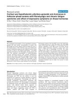
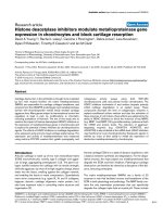
Relaxin (R) administration increases serum R concentrations, which is enhanced by β-estradiol (E) and attenuated by progesterone (P)Relaxin (R) administration increases serum R concentrations, which is enhanced by β-estradiol (E) and attenuated by progesterone (P). Systemic R
concentrations were assayed in serum from sham-operated controls (SC), ovariectomized normal controls (NC), and rabbits treated with E, R, E + R
(ER), P, P + R (PR), and E + P + R (EPR) groups on day 2 and day 6, respectively, using an enzyme-linked immunosorbent assay as described. (a)
Histograms of the median with inter-quartile value showed that there was no statistically significant difference in the basal levels of R on day 2
between any of the groups. Additionally, the SC, NC, E, and P groups showed no statistically significant differences in R concentrations between
day 2 and day 6. R administration resulted in a significant increase of systemic R concentration on day 6 in R, ER, PR, and EPR groups compared
with the corresponding basal levels of R on day 2. (b) The fold change in R was determined by normalizing the day-6 concentration to the corre-
sponding day-2 concentration of R for each animal, and the median with inter-quartile value was plotted for each group of animals. The fold increase
in R was significantly greater in R (35-fold), ER (46-fold), PR (13-fold), and EPR (32-fold) groups compared with that in SC, NC, E, and P groups. No
statistically significant difference of R ratio was found between any of the R-treated groups, with the exception of ER versus PR groups (p < 0.01).
Data were collected from a minimum of six rabbits in each group and expressed as median with inter-quartile value. (* < 0.05; ** < 0.01; *** < 0.001;
**** < 0.0001; a, versus SC; b, versus NC; c, versus E; d, versus P; e, versus PR.)
Arthritis Research & Therapy Vol 8 No 4 Hashem et al.
Page 4 of 9
(page number not for citation purposes)
absorbance was determined at 550 nm with a microtiter plate
reader. The collagen concentration (µg/ml) was determined
against a collagen standard curve, and the tissue collagen
content was standardized to the total disc dry weight.
Determination of serum relaxin levels
Systemic relaxin concentration in serum was measured using
a commercially available enzyme-linked immunosorbent assay
kit (American Laboratory Products Company, Windham, NH,
USA) according to the manufacturer's instructions. Briefly,
100 µl of samples or standards (0 to 250 pg/ml) was pipetted
in duplicate into wells coated with antibody to relaxin and was
incubated at 4°C for 12 hours. After further washes, the bioti-
nylated anti-relaxin antibody was added and incubated for 2
hours at 4°C. After the wells were washed again, horseradish
peroxidase-streptavidin was added, followed by incubation at
4°C for 1 hour. After washing, the color was developed using
tetramethylbenzidine, the reaction was stopped with 4.5 N sul-
furic acid, and the OD value was determined at 450 nm on a
plate reader.
Statistical analysis
GAG and collagen concentrations of the TMJ disc, knee
meniscus, and articular cartilage in each rabbit were calcu-
lated as a mean value from bilateral samples. Data from six to
eight rabbits in each group were expressed as mean ± stand-
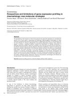
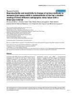
Figure 2
β-estradiol (E) or relaxin (R) treatment causes loss of GAGs from specific fibrocartilages while progesterone (P) inhibits this effectβ-estradiol (E) or relaxin (R) treatment causes loss of GAGs from specific fibrocartilages while progesterone (P) inhibits this effect. Temporomandib-
ular joint (TMJ) discs, pubic symphysis, knee meniscus, and articular cartilage were retrieved from control and hormone-treated rabbits, and total
GAG content was determined by dimethylmethylene blue assay and normalized to the total dry weight for each sample. Histograms of the mean (±
standard deviation) GAG concentration in sham-operated controls (SC), ovariectomized normal controls (NC), and rabbits treated with E, R, ER, P,
P + R (PR), and E + P + R (EPR) groups were plotted. Ovariectomy had minimal effect on total GAGs present in any of these four tissues. E, R, and
ER produced a significant reduction of GAGs in TMJ disc (a) and pubic symphysis (b) relative to SC and NC groups. P alone contributed to the
maintenance of GAGs in all these tissues or prevented E-, R-, or ER-mediated loss of GAGs from both TMJ disc and pubic symphysis. None of these
hormone treatments caused a significant change of GAGs in knee articular cartilage (c) and meniscus (d). Data were collected from a minimum of
six rabbits in each group. (* < 0.05; ** < 0.01; *** < 0.001; **** < 0.0001; a, versus SC; b, versus NC; c, versus P; d, versus PR; e, versus EPR.)
ANOVA, analysis of variance; NS, not significant.
Available online />Page 5 of 9
(page number not for citation purposes)
ard deviation. Statistical analysis was performed using single-
factorial analysis of variance, and inter-group differences were
determined using Fisher's multiple comparison tests, with p <
0.05 being considered statistically significant. Systemic
relaxin concentration of relaxin in each group was expressed
as median with inter-quartile value and was analyzed by the
non-parametric Kruskal-Wallis test, with a p < 0.05 being con-
sidered statistically significant.
Results
Relaxin administration increases systemic relaxin
concentration, which is accentuated by β-estradiol
priming and attenuated by progesterone treatment
On day 2, low to undetectable levels of endogenous relaxin
were noted in all groups of rabbits (Figure 1a). Similarly, con-
trol rabbits implanted with osmotic pumps containing saline
demonstrated low to non-detectable levels of relaxin on day 6.
In contrast, all four groups of rabbits implanted with osmotic
pumps containing relaxin showed substantial increases in sys-
temic relaxin concentration on day 6. The median concentra-
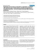
Figure 3
β-estradiol (E), relaxin (R), and progesterone (P) treatment have differential effects on the loss of collagen in fibrocartilagesβ-estradiol (E), relaxin (R), and progesterone (P) treatment have differential effects on the loss of collagen in fibrocartilages. Temporomandibular joint
(TMJ) discs, pubic symphysis, knee meniscus, and articular cartilage were retrieved from control and hormone-treated rabbits, and the total collagen
content was determined by the Sircol assay and normalized to the total dry weight for each sample. Histograms of the mean (± standard deviation)
collagen concentration in sham-operated controls (SC), ovariectomized normal controls (NC), and rabbits treated with E, R, E + R (ER), P, P + R
(PR), and E + P + R (EPR) groups were plotted. Ovariectomy resulted in a significant reduction of collagen content in TMJ disc (a) and increase col-
lage in the pubic symphysis (b) but had no effect on collagen in knee articular cartilage (c) and meniscus (d). Administration of E, R, and ER main-
tained the reduction of collagen content in TMJ disc produced by ovariectomy and caused a significant reduction in collagen in pubic symphysis
relative to NC and in knee articular cartilage relative to SC and NC. In contrast, progesterone antagonized the effects of E, R, and ER on collagen
content in TMJ disc, pubic symphysis, and the knee articular cartilage. Collagen content in knee meniscus was not affected by treatment with any of
these hormones. Data were collected from a minimum of six rabbits in each group. (* < 0.05; ** < 0.01; *** < 0.001; **** < 0.0001; a, versus SC; b,
versus NC; c, versus P; d, versus PR; e, versus EPR; NS, not significant.)
Arthritis Research & Therapy Vol 8 No 4 Hashem et al.
Page 6 of 9
(page number not for citation purposes)
tion was 38 pg/ml for rabbits that received relaxin alone, 58
pg/ml for those administered β-estradiol and relaxin, 37 pg/ml
for rabbits receiving progesterone + relaxin, and 44 pg/ml for
those administered progesterone + β-estradiol + relaxin. All
these groups showed significantly greater systemic relaxin lev-
els on day 6 than the corresponding basal relaxin levels on day
2.
The changes in systemic relaxin were further assessed by nor-
malizing relaxin concentrations on day 6 to the corresponding
basal level of relaxin on day 2 within each group (Figure 1b).
Overall, there was a 30-fold increase in median levels of sys-
temic relaxin in all four groups receiving relaxin, which is signif-
icantly higher than the changes in sham-operated and
ovariectomized normal controls, and in rabbits receiving either
β-estradiol or progesterone alone. The median fold increase in
systemic relaxin concentration ranged from a low of 13-fold in
the progesterone + relaxin group, followed by 32-fold in the β-
estradiol + progesterone + relaxin group, 35-fold in the relaxin
group, to a high of 46-fold in β-estradiol + relaxin group. The
fold increase in systemic relaxin levels between all but one pair
of the four groups receiving relaxin was not statistically signif-
icant. Interestingly, administration of relaxin to β-estradiol-
primed rabbits produced a significantly greater (p < 0.01) fold
increase in systemic relaxin concentrations as compared with
the fold increase observed when relaxin was administrated to
progesterone-primed rabbits.
β-estradiol, relaxin, and β-estradiol + relaxin, but not
progesterone, selectively causes the loss of GAGs from
specific fibrocartilages
The sham-operated rabbits demonstrated varied basal con-
centrations of GAGs in each tissue type; these concentrations
were highest in the TMJ disc and knee cartilage, followed by
the pubic symphysis, and then the knee meniscus (Figure 2a–
d). Ovariectomy had minimal effects on the total GAGs
present in any of the four tissues. Exposure of the rabbits to
various hormone treatments resulted in responses that varied
among tissue types. β-estradiol or relaxin alone or β-estradiol
+ relaxin produced a statistically significant reduction of
approximately 50% in TMJ disc GAG concentrations relative
to sham-operated and ovariectomized control rabbits (Figure
2a). These hormones also contributed to a statistically signifi-
cant reduction of GAGs in pubic symphyseal fibrocartilage,
which at a 30% decrease was less marked than for the TMJ
disc (Figure 2b). None of the hormone treatments caused any
significant changes in GAG content of the knee articular carti-
lage (Figure 2c) or knee meniscus (Figure 2d).
Treatment with progesterone alone contributed to the mainte-
nance of GAG concentrations in the TMJ disc and pubic sym-
physis fibrocartilages at levels found in sham-operated and
ovariectomized control rabbits. Furthermore, administration of
progesterone prevented the relaxin-mediated or β-estradiol +
relaxin-mediated loss of GAGs from these tissues. Also, the
combined administration of all three hormones maintained lev-
els of GAG similar to that in sham-operated rabbits, suggest-
ing that these hormones together produced a near physiologic
effect on the joint tissues.
Together, these findings demonstrate varied responses of dif-
ferent fibrocartilaginous and hyaline cartilaginous tissues to
relaxin, β-estradiol, and progesterone. We also show that pro-
gesterone has an important effect in minimizing the degrada-
tive effects of β-estradiol and relaxin on TMJ disc and pubic
symphyseal GAG content.
β-estradiol, relaxin, and progesterone have differential
effects on the loss of collagen in fibrocartilaginous
tissues
The TMJ disc in ovariectomized controls showed a significant
loss of collagen relative to sham-operated controls (Figure 3a).
This decrease in TMJ disc collagen after ovariectomies was
maintained when β-estradiol, relaxin, or β-estradiol + relaxin
was administered to the rabbits. In contrast, administration of
progesterone alone or in combination with relaxin or β-estra-
diol + relaxin maintained collagen concentrations in the TMJ
disc at levels found in sham-operated controls and at signifi-
cantly greater levels than in ovariectomized controls. This
implies that the decrease in progesterone after ovariectomy
leads to collagen loss from the TMJ disc and that administra-
tion of exogenous progesterone protects these tissues from
the loss of collagen.
Changes in collagen concentrations in the pubic symphysis
after ovariectomies with or without administration of various
hormones were very similar to those reported previously [12],
validating the use of this tissue as an appropriate positive con-
trol for our studies. Ovariectomized controls had significantly
higher concentrations of collagen than did sham-operated
controls (Figure 3b). Administration of β-estradiol caused a
significant decrease in the pubic symphyseal collagen relative
to ovariectomized controls but not relative to sham-operated
controls. Additionally, relaxin or β-estradiol + relaxin produced
a significant reduction in collagen relative to both sham-oper-
ated and ovariectomized control rabbits. When relaxin was
administered to β-estradiol-primed animals, the loss of colla-
gen was accentuated relative to those exposed to β-estradiol
or relaxin alone as evidenced by the level of significance
between these groups and control groups. Administration of
progesterone alone, progesterone + relaxin, or β-estradiol +
progesterone + relaxin restored the collagen concentrations
to, or slightly above, those found in sham-operated rabbits.
Rabbits receiving progesterone alone had significantly lower
concentrations of pubic symphyseal collagen than did ovariec-
tomized controls or those administered β-estradiol + proges-
terone + relaxin. Finally, pubic symphyseal collagen
concentrations in rabbits receiving either progesterone +
relaxin or β-estradiol + progesterone + relaxin were similar to
those of ovariectomized controls.
Available online />Page 7 of 9
(page number not for citation purposes)
Treatments of rabbits with any one of the three hormones or
any combination of these hormones produced some loss of
articular cartilage collagen relative to that in ovariectomized
controls (Figure 3c). Additionally, administration of β-estradiol
or β-estradiol + relaxin resulted in significantly lower collagen
concentrations relative to sham-operated controls or rabbits
receiving progesterone alone or progesterone + relaxin.
Finally, as with GAGs, the knee meniscus did not show any
changes in collagen concentrations either after ovariectomy or
with the administration of any of the hormones alone or in var-
ious combinations (Figure 3d).
Discussion
Here, we show for the first time, the contribution of relaxin and
also of β-estradiol to the degradation of TMJ fibrocartilage and
(to a lesser extent) of knee articular cartilage in vivo, the lack
of potentiation of these responses to relaxin by β-estradiol, and
progesterone's prevention of matrix loss mediated by β-estra-
diol and/or relaxin. These findings suggest that, by modulating
the remodeling of the ECM of cartilage, these hormones may
play an important regulatory role in the normal and pathologic
metabolism of cartilaginous tissues. In the latter scenario, it is
conceivable that individuals with abnormal absolute or relative
levels of one or more of these hormones or their receptors
might incur progressive loss of matrix macromolecules, lead-
ing to joint disorders characterized by the degeneration of spe-
cific cartilages or fibrocartilages [27]. Such potential
hormone-mediated changes in the composition of the ECM
can significantly impact the ability of joints to sustain and dis-
tribute mechanical loading and can also substantially affect the
normal function and survival of cells in tissues [28-30].
There is a striking female preponderance for many types of
joint diseases in general and for TMJ diseases in particular.
TMJ disorder is an umbrella term describing a group of clinical
signs and symptoms involving the masticatory musculature,
the TMJ, and associated structures such as the fibrocartilagi-
nous disc [31]. TMJ disorders are distinguished from similar
diseases of other joints by one specific epidemiologic differ-
ence, namely that, unlike many similar diseases of other joints
that afflict postmenopausal women, TMJ diseases are
observed primarily in women of reproductive age. On the basis
of our findings, it is plausible that specific reproductive hor-
mones target this highly fibrocartilaginous joint for degradative
activity by upregulating particular MMPs that contribute to the
loss of collagen and GAGs [5,21]. In keeping with this postu-
late, our studies show that the responses of TMJ disc to
relaxin, β-estradiol, and progesterone are more similar to those
observed in the pubic symphysis than to those seen in the
knee meniscus or articular cartilage, suggesting that the
observed effects of these hormones are specific to cell type
and tissue type. Additionally, the link between the relaxin- and
β-estradiol-mediated induction of MMPs and the loss of colla-
gen and GAGs in the TMJ disc by these hormones has been
demonstrated previously in our tissue explant studies. How-
ever, the in vivo induction of MMPs by these hormones and the
association between MMP induction and matrix loss remain to
be established.
The reasons for the observed differences in responsiveness of
the TMJ disc fibrocartilage, knee meniscus fibrocartilage, artic-
ular cartilage, and pubic symphysis to the hormones are not
known. However, our recent work on identifying and quantify-
ing estrogen receptor-α and -β and relaxin receptors LGR7
(leucine-rich repeat-containing, G protein-coupled receptor 7)
and LGR8 may provide some insights into one potential rea-
son for the observed differences. These findings show varied
expression of these receptors between these tissues, support-
ing the conclusion that the more robust responses of the pubic
symphysis and TMJ disc to relaxin and β-estradiol are possibly
related to the presence of higher levels of receptors in these
tissues than in the knee meniscus [32]. The findings of the cur-
rent study also raise the possibility that the distinct composi-
tion, organization, and biomechanical characteristics in
different subtypes of cartilages [22] may influence the ability
of systemic hormones to access each of the tissues, thereby
influencing the net amount of matrix loss. These concepts
need further study.
The similarities of our findings on the hormone-mediated
changes in the collagen content of the pubic symphysis with
those of previous investigators [12,13] suggest that our model
has several commonalities to animal models used previously
and attest to its relevance for the purposes of this study. Sam-
uel and colleagues [12], for example, showed that relaxin
causes 64% (± 4%) and 68% (± 6%) decreases, respectively,
in pubic symphysis collagen in unprimed and β-estradiol-
primed ovariectomized non-pregnant rats. This compares with
relaxin contributing to collagen loss of approximately 60% in
unprimed and 80% in β-estradiol-primed rabbit pubic symph-
ysis in the present study. Also, in agreement with our observa-
tions on the pubic symphysis, previous findings show that
progesterone treatment rescues the collagen loss mediated
by relaxin in estrogen-primed animals [12,16]. Together, these
observations indicate that the pubic symphysis served as an
appropriate positive control for our experiments. In addition to
confirming these previous observations on relaxin's contribu-
tion to collagen loss in the pubic symphysis, we also show new
findings that β-estradiol and relaxin cause a slight, but statisti-
cally significant, loss of GAGs from this tissue.
Whereas a few previous studies have demonstrated other
non-reproductive target sites, including the in vivo matrix turn-
over of lung alveolar tissues [33] and the in vitro modulation of
tissue degradation in dermal fibroblasts [34] as well as fibro-
cartilaginous cells or tissues from joints [5,21], our findings
show for the first time that relaxin alters matrix composition of
specific cartilages from non-reproductive sites in vivo. As with
findings on reproductive tissues [9,12,16,17,22], we had pre-
Arthritis Research & Therapy Vol 8 No 4 Hashem et al.
Page 8 of 9
(page number not for citation purposes)
viously demonstrated that the effect of relaxin on MMP induc-
tion is potentiated by β-estradiol in isolated fibrocartilaginous
cells from the TMJ [5]. In contrast, TMJ fibrocartilaginous
explants [21] and the current in vivo findings show no poten-
tiation by β-estradiol of relaxin's induction of MMPs or loss of
collagen and GAGs. These differences in responses between
cultured cells and tissue explants can likely be attributed to the
differences in the behavior of isolated cells from cells in their
natural matrix environment within the context of the explants
and in the intact animal.
Our results also show a significant increase in systemic relaxin
levels in all groups administered relaxin, with median concen-
trations ranging from a low of 37 pg/ml for rabbits receiving
progesterone + relaxin to a high of 58 pg/ml for those admin-
istered β-estradiol + relaxin. This range of concentrations of
relaxin in all groups of animals administered this hormone
alone or in combination with other hormones is similar to that
found systemically in cycling women [35-37]. Interestingly, our
findings showed that β-estradiol priming enhances systemic
relaxin concentrations, whereas priming with progesterone
tended to diminish the serum levels of relaxin. These effects of
hormone priming on systemic relaxin levels were reflected in
the statistically greater fold increase of relaxin in β-estradiol +
relaxin versus progesterone + relaxin groups (p < 0.01) (Fig-
ure 1a). This finding corresponds to some degree with the
decreases in collagen and GAGs mediated by relaxin and β-
estradiol + relaxin, but not by progesterone, progesterone +
relaxin, or β-estradiol + progesterone + relaxin in the more
responsive of the tissues studied, namely the TMJ disc and
pubic symphysis. These results suggest that administration of
relaxin using an implanted subcutaneous osmotic pump con-
tributes to the increases in systemic relaxin concentration in
which β-estradiol and progesterone may act as regulators of
systemic relaxin concentration. However, further studies are
required to clarify the mechanism by which β-estradiol, relaxin,
and progesterone interact with each other to modulate sys-
temic relaxin levels and subsequently maintain or disturb the
homeostasis of the ECM in fibrocartilage.
Conclusion
Our study demonstrates a cross-talk between relaxin, β-estra-
diol, and progesterone that may affect systemic relaxin con-
centrations and ultimately the net matrix remodeling activity of
these hormones alone or in various combinations in target tis-
sues in vivo. We also show specificity in the responses of var-
ious cartilages and fibrocartilages to modulation of matrix loss
by relaxin and β-estradiol. Excessive degradation of collagen
and GAGs by relaxin and/or β-estradiol may perturb the home-
ostasis of ECM in these joints and eventually contribute to the
degenerative disease of the target joint.
Competing interests
The authors declare that they have no competing interests.
Authors' contributions
GH performed animal procedures, including hormone injec-
tions and implantation of osmotic pumps, performed collagen
and GAG assays, and compiled the data. QZ assisted GH in
the above animal procedures and performed the relaxin
enzyme-linked immunosorbent assays. JC performed all initial
studies to establish the model and also helped to establish the
experimental procedures for sample collection and assays. TH
performed the surgeries to retrieve all the tissues and blood
from the animals. WW assisted SK in data analysis and con-
tributed to the interpretation of the data and preparation of the
manuscript. SK conceived, designed, and supervised the
study, performed the statistical analysis, and contributed to the
interpretation of the data and the revision of the manuscript. All
authors have read and approved the final manuscript.
Acknowledgements
This study was supported by grants DE11993 and DE00458 from the
National Institutes of Health and by a University of California at San Fran-
cisco Academic Senate Shared Equipment Grant to SK.
References
1. Buckwalter JA, Mankin HJ, Grodzinsky AJ: Articular cartilage and
osteoarthritis. Instructional Course Lectures 2005, 54:465-480.
2. Bramono DS, Richmond JC, Weitzel PP, Kaplan DL, Altman GH:
Matrix metalloproteinases and their clinical applications in
orthopaedics. Clin Orthop Relat Res 2004, 428:272-285.
3. Ushiyama T, Inoue K, Nishioka J: Expression of estrogen recep-
tor related protein (p29) and estradiol binding in human
arthritic synovium. J Rheumatol 1995, 22:421-426.
4. Khalkhali-Ellis Z, Seftor EA, Nieva DR, Handa RJ, Price RH Jr, Kir-
schmann DA, Baragi VM, Sharma RV, Bhalla RC, Moore TL, Hen-
drix MJ: Estrogen and progesterone regulation of human
fibroblast-like synoviocytes function in vitro: implications in
rheumatoid arthritis. J Rheumatol 2000, 27:1622-1631.
5. Kapila S, Xie YQ: Targeted induction of collagenase and
stromelysin by relaxin in unprimed and β-estradiol-primed
diarthrodial joint fibrocartilaginous cells but not in
synoviocytes. Lab Invest 1998, 78:925-938.
6. Warren MP, Fried JL: Temporomandibular disorders and hor-
mones in women. Cells Tissues Organs 2001, 169:187-192.
7. Carlsson GE, LeResche L: Epidemiology of temporomandibular
disorders. In Progress in Pain Research and Management: Tem-
poromandibular Disorders and Related Pain Conditions Edited
by: Sessle BJ, Bryant PS, Dionne RA. Seattle: IASP Press;
1995:211-226.
8. Sheffield LG, Anderson RR: Effect of estradiol and relaxin on
collagen and non-collagen protein synthesis by mammary
fibroblasts. Life Sci 1984, 35:2199-2203.
9. Too CK, Kong JK, Greenwood FC, Bryant-Greenwood GD: The
effect of oestrogen and relaxin on uterine and cervical
enzymes: Collagenase, proteoglycanase and beta-glycuroni-
dase. Acta Endocrinology (Copenh) 1986, 111:394-403.
10. Mushayandebvu TI, Rajabi MR: Relaxin stimulates interstitial
collagenase activity in cultured uterine cervical cells from non-
pregnant and pregnant but not immature guinea pigs; estra-
diol-17 beta restores relaxin's effect in immature cervical cells.
Biol Reprod 1995, 53:1030-1037.
11. Hwang JJ, Lin SW, Teng CH, Ke FC, Lee MT: Relaxin modulates
the ovulatory process and increases secretion of different
gelatinases from granulosa and theca-interstitial cells in rats.
Biol Reprod 1996, 55:1276-1283.
12. Samuel CS, Butkus A, Coghlan JP, Bateman JF: The effect of
relaxin on collagen metabolism in the nonpregnant rat pubic
symphysis: the influence of estrogen and progesterone in reg-
ulating relaxin activity. Endocrinology 1996, 137:3884-3890.
13. Samuel CS, Coghlan JP, Bateman JF: Effects of relaxin, preg-
nancy and parturition on collagen metabolism in the rat pubic
symphysis. J Endocrinol 1998, 159:117-125.
Available online />Page 9 of 9
(page number not for citation purposes)
14. Goldsmith LT, Crob HS, Scherer KJ, Surve A, Steinetz BG, Weiss
G: Placental control of ovarian immunoreactive relaxin secre-
tion in the pregnant rat. Endocrinology 1981, 109:584-592.
15. Sherwood OD: Relaxin. In The Physiology of Reproduction
Edited by: Knobil E, Neill J. New York: Raven Press;
1994:861-1009.
16. O'Day-Bowman MB, Winn RJ, Dzuik PJ, Lindley ER, Sherwood
OD: Hormonal control of the cervix in pregnant gilts. III.
Relaxin's influence on cervical biochemical properties in ova-
riectomized hormone-treated gilts. Endocrinology 1991,
129:1967-1976.
17. Sherwood OD, Downing SJ, Guico LM: The physiological effects
of relaxin during pregnancy: studies in rats and pigs. Oxf Rev
Reprod Biol 1993, 15:143-149.
18. Cheah SH, Sherwood OD: Effect of preparturient 17 beta-estra-
diol and relaxin on parturition and pup survival in the rat. Endo-
crinology 1988, 122:1958-1963.
19. Palejwala S, Stein DE, Weiss G, Monia BP, Tortoriello D, Gold-
smith LT: Relaxin positively regulates matrix metalloproteinase
expression in human lower uterine segment fibroblasts using
a tyrosine kinase signaling pathway. Endocrinology 2001,
142:3405-3413.
20. Lenhart JA, Ryan PL, Ohleth KM, Palmer SS, Bagnell CA: Relaxin
increases secretion of matrix metalloproteinase-2 and matrix
metalloproteinase-9 during uterine and cervical growth and
remodeling in the pig. Endocrinology 2001, 142:3941-3949.
21. Naqvi T, Duong T, Hashem G, Shiga M, Zhang Q, Kapila S:
Relaxin's induction of metalloproteinases is associated with
the loss of collagen and glycosaminoglycans in synovial joint
fibrocartilaginous explants. Arthritis Res Ther 2004, 7:R1-R11.
22. Hall JA, Anthony RV: Influence of ovarian steroids on relaxin-
induced distensibility and compositional changes in the por-
cine cervix. Biol Reprod 1993, 48:1348-1353.
23. Chen B, Wen Y, Yu X, Polan ML: Elastin metabolism in pelvic
tissues: is it modulated by reproductive hormones? Am J
Obstet Gynecol 2005, 192:1605-1613.
24. Abubaker AO, Hebda PC, Gunsolley JN: Effects of sex hor-
mones on protein and collagen content of the temporoman-
dibular joint disc of the rat. J Oral Maxillofac Surg 1996,
54:721-727.
25. Yasuoka T, Nakasbima M, Okuda T, Tatematsu N: Effect of estro-
gen replacement on tempromandibular joint remodeling in
ovariectomized rats. J Oral Maxillofac Surg 2000, 58:189-196.
26. Unemori EN, Beck LS, Lee WP, Xu Y, Siegel M, Keller G, Liggitt
HD, Bauer EA, Amento EP: Human relaxin decreases collagen
accumulation in vivo in two rodent models of fibrosis. J Invest
Dermatol 1993, 101:280-285.
27. Kapila S: Does the relaxin, estrogen and matrix metalloprotei-
nase axis contribute to degradation of TMJ fibrocartilage? J
Musculoskelet Neuronal Interact 2003, 3:401-405.
28. Knudson CB, Knudson W: Hyaluronan and CD44: modulators
of chondrocyte metabolism [Review]. Clin Orthop Relat Res
2004:S152-162.
29. Kalya S, Rosenthal AK: Extracellular matrix changes regulate
calcium crystal formation in articular cartilage. Curr Opin
Rheumatol 2005, 17:325-329.
30. Hecht JT, Hayes E, Haynes R, Cole WG: COMP mutations,
chondrocyte function and cartilage matrix. Matrix Biol 2005,
23:525-533.
31. Haskin CL, Milam SB, Cameron IL: Pathogenesis of degenera-
tive joint disease in the human temporomandibular joint. Crit
Rev Oral Biol Med 1995, 6:248-277.
32. Wang W, Hayami T, Chen C, Kapila S: Relaxin and estrogen
receptor expression in TMJ and meniscus fibrocartilages
[abstract]. J Den Res 2006, 85(spec iss A):0021.
33. Unemori EN, Pickford LB, Salles AL, Piercy CE, Grove BH, Erikson
ME, Amento EP: Relaxin induces an extracellular matrix-
degrading phenotype in human lung fibroblasts in vitro and
inhibits lung fibrosis in a murine model in vivo. J Clin Invest
1996, 98:2739-2745.
34. Unemori EN, Amento EP: Relaxin modulates synthesis and
secretion of procollagenase and collagen by human dermal
fibroblasts. J Biol Chem 1990, 265:10681-10685.
35. Eddie LW, Bell RJ, Lester A, Geier M, Bennet G, Johnston PD, Niall
HD: Radioimmunoassay of relaxin in pregnancy with an ana-
logue of human relaxin. Lancet 1986, 1:1344-1346.
36. Bell RJ, Eddie LW, Lester AR, Wood EC, Johnston PD, Niall HD:
Relaxin in human pregnancy serum measured with a homolo-
gous RIA. Obstet Gynecol 1987, 69:585-589.
37. Stewart DR, Celniker AC, Taylor CA, Cragun JR, Overstreet J, Las-
ley B: Relaxin in the peri-implantation period. J Clin Endocrinol
Metab 1990, 70:1771-1773.