Báo cáo y học: " Diagnostic value of anti-human citrullinated fibrinogen ELISA and comparison with four other anti-citrullinated protein assays" potx
Bạn đang xem bản rút gọn của tài liệu. Xem và tải ngay bản đầy đủ của tài liệu tại đây (228.37 KB, 7 trang )
Open Access
Available online />Page 1 of 7
(page number not for citation purposes)
Vol 8 No 4
Research article
Diagnostic value of anti-human citrullinated fibrinogen ELISA and
comparison with four other anti-citrullinated protein assays
Bert Vander Cruyssen
1
*, Tineke Cantaert
1
*, Leonor Nogueira
2
, Cyril Clavel
2
, Leen De Rycke
1
,
Amélie Dendoven
1
, Mireille Sebag
2
, Dieter Deforce
3
, Christian Vincent
2
, Dirk Elewaut
1
, Guy Serre
2
and Filip De Keyser
1
1
Department of Rheumatology, Ghent University Hospital, Ghent, Belgium
2
UMR 5165 'Laboratory of Epidermis Differentiation and Rheumatoid Autoimmunity', CNRS – Toulouse III University, Toulouse, France
3
Laboratory of Pharmaceutical Biotechnology, Ghent University, Ghent, Belgium
* Contributed equally
Corresponding author: Bert Vander Cruyssen,
Received: 28 Mar 2006 Revisions requested: 1 Jun 2006 Revisions received: 6 Jul 2006 Accepted: 13 Jul 2006 Published: 19 Jul 2006
Arthritis Research & Therapy 2006, 8:R122 (doi:10.1186/ar2011)
This article is online at: />© 2006 Vander Cruyssen et al.; licensee BioMed Central Ltd.
This is an open access article distributed under the terms of the Creative Commons Attribution License ( />),
which permits unrestricted use, distribution, and reproduction in any medium, provided the original work is properly cited.
Abstract
We studied the diagnostic performance of the anti-human
citrullinated fibrinogen antibody (AhFibA) ELISA for rheumatoid
arthritis (RA) in a consecutive cohort (population 1) and
evaluated the agreement between the AhFibA ELISA and four
other assays for anti-citrullinated protein/peptide antibodies
(ACPAs) as well as rheumatoid factor in patients with
longstanding RA (population 2). Population 1 consisted of 1024
patients with rheumatic symptoms; serum samples from these
patients were sent to our laboratory for ACPA testing within the
context of a diagnostic investigation for RA. Ninety-two of these
patients were classified as having RA according to the American
College of Rheumatology criteria and 463 were classified as
non-RA patients. Population 2 consisted of 180 patients with
longstanding RA and was used to assess agreement and
correlations between five ACPA assays: anti-cyclic citrullinated
peptide (CCP)1 and anti-CCP2 antibodies were detected using
a commercially available ELISA, AhFibA using ELISA, and anti-
PepA and anti-PepB antibodies using line immunoassay.
Applying previously proposed cut-offs for AhFibA, we obtained
a sensitivity of 60.9% and a specificity of 98.7% in population 1.
Receiver operating characteristic curve analysis could not
detect a significant difference in diagnostic performance
between the AhFibA ELISA and anti-CCP2 assay. Performing a
hierarchical nearest neighborhood cluster analysis of the five
different ACPA assays in population 2, we identified two
clusters: a cluster of anti-pepA, anti-pepB and anti-CCP1, and a
cluster of AhFibA and anti-CCP2. In conclusion, we found that
AhFibA and anti-CCP2 antibodies had similar diagnostic
performance. However, disagreement between ACPA tests may
occur.
Introduction
Clinical indicators of rheumatoid arthritis (RA) are pain and
swelling of the proximal interphalangeal and metacarpophalan-
geal joints. Larger joints such as knee, elbow and ankle joints
may also be affected. Synovial inflammation and joint destruc-
tion together with the extra-articular manifestations of the dis-
ease are responsible for a severe decline in the RA patient's
quality of life. It is important to identify RA early. Joint erosions,
which are irreversible, occur early in the disease process and
intervention with aggressive therapy is most successful if it is
applied early in the disease course [1]. A sensitive and specific
serological test is needed for application in this window in the
disease course, when often not all clinical manifestations are
apparent.
Several autoantibody systems have been described in this
autoimmune disease [2]. The presence of the rheumatoid fac-
tor (RF), directed against the Fc part of an IgG molecule, is
one of the American College of Rheumatology (ACR) criteria
for RA [3]. This antibody is present in about 65–75% of RA
ACPA = anti-citrullinated protein/peptide antibody; ACR = American College of Rheumatology; AhFibA = anti-human fibrinogen (auto)antibodies;
CCP = cyclic citrullinated peptide; ELISA = enzyme-linked immunosorbent assay; HRP = horseradish peroxidase; OD = optical density; PBS = phos-
phate-buffered saline; RA = rheumatoid arthritis; RF = rheumatoid factor; ROC = receiver operating curve.
Arthritis Research & Therapy Vol 8 No 4 Vander Cruyssen et al.
Page 2 of 7
(page number not for citation purposes)
patients. However, because it is also found in patients with
other autoimmune diseases or infectious diseases, and even in
the healthy elderly, it has limited specificity. The presence of
anti-citrullinated protein/peptide antibodies (ACPAs), on the
other hand, is significantly more specific for RA. ACPAs are
directed against various proteins that have one trait in com-
mon; some of their arginines have been converted to citrulline
by post-translational modification, catalyzed by peptidy-
larginine deiminase enzymes [4,5].
Depending on the substrate, various assays for detection of
ACPAs have been developed. Human buccal mucosa cells
and rat oesophagus provide the antigenic substrate for anti-
perinuclear factor and anti-keratin antibodies [6,7]. The diffi-
culty in standardizing these natural substrates, together with
arbitrary interpretation of the indirect immunofluorescence pat-
tern, has hampered the widespread use of these tests.
Because it was shown that both anti-perinuclear factor and
anti-keratin antibodies reacted against citrullinated filaggrin
related proteins [8], the latter was used for detection of
ACPAs in immunoblot assays and in an ELISA, resulting in an
assay with 52% sensitivity at a specificity of 95% in a cohort
of patients with established disease [9,10]. An ELISA using a
cyclic citrullinated peptide (CCP) derived from filaggrin was
commercialized (anti-CCP1) [11]. Numerous studies were
reported in which sensitivities ranged from 41% (with a corre-
sponding specificity of 97.8% [12]) to 68% (with a corre-
sponding specificity of 98% [11]) in established RA. The line
immunoassay format was used with two filaggrin based pep-
tides (pepA and pepB), obtained from the results of epitope
mapping as well as molecular modelling and computational
chemistry [13]. The sensitivity of this assay was 63.6% for
pepA and 54.2 % for pepB at a specificity of 98.5% in estab-
lished RA [14]. With the development of the second genera-
tion anti-CCP2 ELISA, sensitivities ranging from 65% [15] to
80% [16] at a high level of specificity have been reported in
established RA. Recently, the presence of citrullinated fibrin in
the synovial membrane of RA patients [17] and the use of cit-
rullinated fibrinogen to assay the serum antibodies to deimi-
nated fibrinogen (anti-human citrullinated fibrinogen antibody
[AhFibA]) was described [18,19].
The aim of the present study was to assess the diagnostic per-
formance of the AhFibA ELISA for RA in a consecutive popu-
lation of patients of whom serum was sent to our laboratory for
RA serological testing. We also studied the agreement
between five different ACPA assays and RF in a cohort of
patients with established RA.
Materials and methods
Study population 1
Study population 1 was established to evaluate the diagnostic
performance of the AhFibA assay and to compare the diagnos-
tic performance with an anti-CCP2 antibody assay by means
of receiver operator characteristic (ROC) curve analysis. This
cohort consisted of 1024 patients with rheumatic symptoms,
from whom serum samples were consecutively sent to our lab-
oratory for ACPA determination within the context of a diag-
nostic investigation. Patients were diagnosed by a clinician by
reviewing of files and the patients were classified in accord-
ance with the ACR classification criteria for RA [3]. Eighty-one
patients were lost to follow up. We thus diagnosed 92 individ-
uals as having RA, and all of these patients met the ACR crite-
ria for RA. In 463 patients the diagnosis of RA could be
excluded. A further 388 patients had undifferentiated arthritis
and were further withdrawn from the analysis. The most fre-
quent diseases diagnosed in the non-RA patients were oste-
oarthritis (31%), soft tissue mechanical complaints 20%
(including peri-arthritis scapulohumeralis and tendinopathies),
spondyloarthropathy (13%), systemic lupus erythematosus
(9%), vasculitis (6%), polymyalgia rheumatica (5%), other con-
nective tissue diseases (including scleroderma and Sjögren's
syndrome; 2%), adult patients with juvenile idiopathyic arthritis
(1%), psoriatic arthritis (5%), crystal arthritis (3%) and other
diseases including infections, malignancies and neurological
disorders (5%).
Of the RA patients, 65.2% were female, the median age was
55 years (range 22–85 years) and the median disease dura-
tion was 3 years (range 0–40 years). In the non-RA patients,
66.2% were female and the median age was 51 years (range
11–83 years). Of the RA patients 64% were receiving dis-
ease-modifying antirheumatic drug therapy, predominantly
methotrexate (n = 34), sulphasalazine (n = 11) and lefluno-
mide (n = 5). Combination therapies were administered to six
patients. None of the patients were being treated with anti-
tumour necrosis factor therapy at the time of sampling. Corti-
costeroids were being received by 30% of patients.
Study population 2
Study population 2 consisted of 180 consecutive RA patients
with longstanding disease of at least 4 years (median disease
duration 9 years; range 4–39 years) [14]. In this cohort we
compared the AhFibA assay with four other ACPA assays. All
patients were treated with classic disease-modifying antirheu-
matic drug therapy (methotrexate, gold salts, or sulphasala-
zine). None of the patients received leflunomide, anti-tumour
necrosis factor therapy, or other biologicals. Concomitant cor-
ticosteroids were used in one-third of the patients.
Rheumatoid factor assay
RF was determined using the latex fixation test. A suspension
of uniform polystyrene particles sensitized in glycine buffer
with heat-altered human IgG (Difco Laboratories, Detroit, MI,
USA) was diluted 1/20 and incubated with progressive dilu-
tions of human sera in microtitre wells. The reagents were
mixed and incubated at 37°C for 2 hours. The plates were then
shaken gently and inspected for observable agglutination. The
dilution titre present in the last well showing agglutination was
recorded.
Available online />Page 3 of 7
(page number not for citation purposes)
Detection of anti-pepA and anti-pepB antibodies by line
immunoassay
Anti-pepA and anti-pepB Abs were detected by a research
line immunoassay containing two citrulline-containing pep-
tides, as described previously (INNO-LIA™ RA [for research
use only]; Innogenetics, Ghent, Belgium) [13,14]. The cut-off
defined for anti-pepA and anti-pepB antibodies corresponds
with a specificity of 100% and 99.3% and a sensitivity of
63.6% and 54.2%, respectively [14].
Detection of anti-CCP1 and anti-CCP2 antibodies by
ELISA
Anti-CCP1 and anti-CCP2 antibodies were detected using a
commercially available ELISA containing synthetic CCPs
(Immunoscan RA, mark 1 and mark 2; Eurodiagnostica, Arn-
hem, The Netherlands). The ELISA was performed in accord-
ance with the manufacturer's instructions. Briefly, serum
samples were diluted 1/50 with dilution buffer and incubated
for 1 hour at 37°C. After removing the liquid and washing three
times with rinsing buffer, the conjugate solution (peroxidase
conjugated anti-human IgG antibodies) was added into each
well and incubated for 1 hour at 37°C. After three washing
steps with rinsing buffer, the substrate solution (tetramethyl
benzidine) was added and incubated for 30 min at room tem-
perature. The stop solution (sulfuric acid; 0.5 mol/l) was added
and the absorbance values were read immediately at 450 nm.
A 98.5% specificity cut-off has previously been set at 42 U/ml
(sensitivity 75.4%) for the anti-CCP2 assay [14] and a 98%
specific cut-off has been set at 92 U/ml for the anti-CCP1
assay [11].
Detection of AhFibA antibodies by ELISA
The AhFibA-ELISA was developed previously [18,20]. Briefly,
plasminogen depleted human fibrinogen (Calbiochem, Meu-
don, France) was further affinity purified on a protein G column
(HiTrap protein G; Amersham Biosciences, Orsay, France).
Deimination was performed for 2 hours at 37°C with 7 units
rabbit skeletal muscle peptidylarginine deiminase per milli-
gram fibrinogen (Sigma, Lyon, France) in deimination buffer
(0.1 mol/l Tris-Hcl [pH 7.4], 10 mmol/l CaCl
2
, 5 mmol/l DTT).
Microtitration plates (MaxiSorp, Nunc, Denmark) were coated
overnight with human deiminated fibrinogen (5 μg/ml) diluted
in phosphate-buffered saline (PBS; pH 7.4). The plates were
blocked with PBS containing 2% bovine serum albumin. A vol-
ume of 100 μl sera, diluted to 1:50 in 2 mol/l NaCl PBS, was
applied and plates were incubated for 1 hour. After washing,
plates were incubated with horseradish peroxidase labelled
goat anti-human IgG antibodies (γ-chain specific) for 1 hour
and washed again. All incubations and washing steps were
performed at 4°C. Bound antibodies were detected with
ortho-phenylene diamine dihydrochloride (Sigma, St. Louis,
MO, USA). The reaction was stopped with 50 μm of 3 mol/l
sulphuric acid.
Plates were read using a Multiskan plate reader (Thermo Lab-
system, Cergy-Pontoise, France). Serum samples were tested
twice and results were averaged. A serum was considered
positive for AhFibA above a previously defined cut-off corre-
sponding with the 98.5% specificity level (optical densitiy
(OD) ≥ 0.12 nm) [18].
Statistical analysis
ROC curve analyses were performed. Differences between
areas under the curve were evaluated, as proposed by Hanley
[21]. Agreement between dichotomized variables was meas-
ured by the (weighted) κ statistic. Proportions of matched-pair
data were compared by means of the McNemar test. A hierar-
chical nearest neighbourhood cluster analysis of variables was
performed based on squared Euclidian distances. All analyses
were performed using the commercial available statistical
package SPSS 11.0 (SPSS Institute Inc., Chicago, Il, USA).
Results
Diagnostic performance of the AhFibA assay for RA and
comparison with the anti-CCP2 and RF assay in
population 1
The previously proposed AhFibA cut-off of OD ≥ 0.12 corre-
sponded to a specificity level of 98.5% [18]. When we applied
this cut-off to population 1, we obtained a sensitivity of 60.9%
and a specificity of 98.7% for the AhFibA assay. Seven non-
RA patients tested false positive for AhFibA: one patient with
systemic lupus erythematosus, one with psoriatic arthritis, one
with polymyositis, one with polymyalgia rheumatica and three
with osteoarthritis.
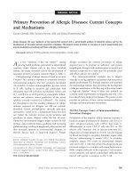
The ROC curve comparing the diagnostic performance of
AhFibA ELISA, anti-CCP2 assay, and RF in population 1 is
shown in Figure 1a, with detail of the curve in the high specif-
icity region shown in Figure 1b. There were no significant dif-
ferences in the area under the ROC curve analyses of the
AhFibA assay compared with the anti-CCP2 assay (0.824 ver-
sus 0.854; P = NS) [21]. The sensitivities at cut-offs defining
comparable specificity levels were similar for the AhFibA
ELISA and the anti-CCP2 assay, but they were significantly
higher than the sensitivities of the RF test (Table 1). Applying
the McNemar test, there were no significant differences
between the two ACPA tests after dichotomization at the cut-
offs presented in Table 1.
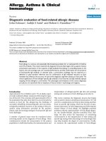
Agreement between the AhFibA assay and anti-CCP2 in
population 1
In Figure 2 the results of the anti-CCP2 assay are plotted
against the results of the AhFibA assay. Table 2 shows the
cross-tabulation of the results of the AhFibA and anti-CCP2
ELISAs after dichotomization at the 98.5 % specificity level
both for the RA and the non-RA patients. The κ statistic, as a
measure of agreement between AhFibA and anti-CCP2
ELISA, calculated on the global population, was 0.845. After
splitting the population into RA and non-RA patients, we
Arthritis Research & Therapy Vol 8 No 4 Vander Cruyssen et al.
Page 4 of 7
(page number not for citation purposes)
obtained a κ of 0.765 for the RA patients and κ of 0.420 for
the non-RA patients. Hence, agreement of both assays is
especially impaired in non-RA patients; only 11 non-RA
patients exhibited any ACPA reactivity, of which only three
were positive for both AhFibA and anti-CCP2 ELISAs. Those
three patients had the following diagnoses: osteoarthritis, pso-
riatic arthritis and polymyositis. We calculated that the specif-
icity in case of double ACPA positivity is 99.4%, with a
sensitivity of 58.7%.
Agreement between the five ACPA assays and RF in
population 2
In this cohort of longstanding RA patients, the agreement
between the AhFibA assay and the anti-CCP2 assay corre-
sponded with the agreement observed in the RA patients of
population 1. After dichotomization at a >98% specificity level,
as defined in Materials and methods (see above), we calcu-
lated the sensitivities listed in Table 3; sensitivities, especially
for the AhFibA assay, observed in population 2 were higher
than those in population 1. The results of the κ statistic as a
measure of agreement between dichotomized tests are listed
in Table 4, confirming the moderate agreement between the
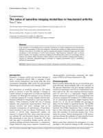
different ACPA tests. A hierarchical nearest neighborhood
cluster analysis of variables was performed with the results of
RF and five ACPA assays: anti-CCP1, anti-CCP2, anti-pepA,
anti-pepB and AhFibA (Figure 3).
Figure 1
ROC curve analyses of the RF, AhFibA and anti-CCP2 assay in population 1ROC curve analyses of the RF, AhFibA and anti-CCP2 assay in population 1. (a) The whole curve is shown, and (b) with a focus on detail of the
ROC curve at the 98% specificity level for the AhFibA and anti-CCP2 assay in population 1. AhFibA, anti-human fibrinogen (auto)antibodies; CCP,
cyclic citrullinated peptide; RF, rheumatoid factor; ROC, receiver operating curve.
Table 1
Sensitivities and specificities of AhFibA and anti-CCP2 assay in the two populations
Cut-off Population 1 Population 2
Specificity Sensitivity Sensitivity
AhFibA antibody (OD) 0.027 0.945 0.685 0.800
0.11 0.985 0.609 0.711
0.12 0.987 0.609 0.700
0.19 0.990 0.598 0.678
anti-CCP2 antibody (U/ml) 14.5 0.950 0.728 0.744
37 0.985 0.674 0.680
42 0.985 0.641 0.650
135 0.990 0.565 0.551
RF 160 0.948 0.391 0.556
640 0.985 0.163 0.244
1280 0.993 0.087 0.122
AhFibA, anti-human fibrinogen (auto)antibodies; CCP, cyclic citrullinated peptide; OD, optical density; RF, rheumatoid factor.
Available online />Page 5 of 7
(page number not for citation purposes)
This analysis identified the ACPAs apart from the RF. Within
ACPAs, we identified different clusters: a cluster of pepA,
pepB and anti-CCP1, and a cluster of AhFibA and anti-CCP2.
Discussion
In the present study, we describe the diagnostic performance
of an assay, based on the detection of AhFibA. We compared
the diagnostic value of the AhFibA ELISA and the anti-CCP2
ELISA, and conclude that both assays perform equally well,
which is reflected by similar ROC curves and similar sensitivi-
ties and specificities. There were some non-significant differ-
ences in sensitivities of the AhFibA and anti-CCP2 assay
between populations 1 and 2.
In contrast to the comparable diagnostic performance of the
AhFibA and anti-CCP2 antibodies, the agreement between
the two assays in population 1 was only moderate, and was
especially impaired in the non-RA patients. Indeed, at the
98.5% specificity level only 11 non-RA patients exhibited any
ACPA reactivity, of which only three were positive for both
AhFibA and anti-CCP2 antibodies (Table 2). Double ACPA
positivity thus resulted in a specificity of 99.4% with a sensitiv-
ity of 58.7%.
In population 2, we also evaluated the agreement between the
AhFibA ELISA and four other ACPA tests. This confirmed the
moderate agreement between the different ACPA assays.
Agreement between the different ACPA assays may be impor-
tant for the implementation of prediction models. Different pre-
diction models for diagnosis of (persistent) erosive disease
have been described by means of different ACPA assays
[22,23]. Taking into account the similarities between the differ-
ent ACPA tests, we performed a cluster analysis. Separated
from RF, we found a clustering of anti-pepA, anti-pepB and
anti-CCP1 assays on one side and anti-CCP2 and AhFibA
assays on the other. The clustering of RF at a long distance
from ACPAs illustrates the different nature of the antibody sys-
tems [24]. Two different explanations can be hypothesized to
account for the two clusters within the ACPA tests. First, the
anti-pepA, anti-pepB and anti-CCP1 assay use a citrullinated
epitope derived from filaggrin. Filaggrin is not the natural
autoantigen for ACPA because it is only expressed in epider-
mis. The substrate of the anti-CCP2 ELISA comprises cyclic
peptides selected from libraries containing citrullinated
peptides screened with RA sera; these peptides could have a
lower degree of homology with filaggrin [25]. The second
potential explanation is that both the AhFibA and the anti-
CCP2 ELISA use multiple citrullinated epitopes for the detec-
tion of ACPAs. Because it was demonstrated that individual
Figure 2
Scatter plot: AhFibA assay versus anti-CCP2 assay in population 1Scatter plot: AhFibA assay versus anti-CCP2 assay in population 1. Ab,
antibody; AhFibA = anti-human fibrinogen (auto)antibodies; CCP =
cyclic citrullinated peptide.
Table 2
Agreement between AhFibA assay and anti-CCP2 assay at the
98.5% specificity level in population 1
Anti-CCP2 Total κ
Neg Pos
Non-RA AhFibA Neg 452 4 456 0.420 0.845
Pos 4 3 7
Total 456 7 463
RA AhFibA Neg 28 8 36 0.765
Pos 2 54 56
Total 30 62 92
AhFibA, anti-human fibrinogen (auto)antibodies; CCP, cyclic
citrullinated peptide; Neg, negative; Pos, positive; RA, rheumatoid
arthritis.
Table 3
Sensitivities of the different ACPA assays in population 2 after
dichotomization at specificity level ≥ 98%
Sensitivity Cut-off (at
98%
specificity)
Ref.
AhFibA 70% 0.12 OD [18]
Anti-CCP1 51% 92 U/ml [11]
Anti-CCP2 65% 42 U/ml [14]
Anti-PepA 57% 1 [13,14]
Anti-PepB 55% 1 [13,14]
ACPA, anti-citrullinated protein/peptide antibody; AhFibA, anti-
human fibrinogen (auto)antibodies; CCP, cyclic citrullinated peptide;
OD, optical density.
Arthritis Research & Therapy Vol 8 No 4 Vander Cruyssen et al.
Page 6 of 7
(page number not for citation purposes)
RA patients reacted with different citrullinated epitopes [5],
the sensitivity of an ACPA test is expected to increase when
more than one citrullinated epitope is used.
Increasing the sensitivity at a high specificity level for ACPA
detection appears difficult to achieve. Further characterization
of the synovial citrullinated proteins apart from fibrinogen may
provide new substrates for detection of ACPAs, which might
increase the sensitivity and specificity of the future ACPA
assays [26]. However, it can be hypothesized that there may
be a limit to the sensitivity of ACPA assays for RA. It could be
argued that there are two subpopulations within RA patients
[27,28]: a population with ACPAs can be detected, which has
an increased prevalence of the HLA shared epitope and with
a worse functional and radiological outcome; and a population
without ACPAs but with reactivities against several human car-
tilage gp39 peptides and type II collagen, with no increased
prevalence of the HLA shared epitope and with a better radio-
logical and functional prognosis. Also, ACPA positivity, if
observed in non-RA patients, can preferentially be observed in
patients who carry the HLA shared epitope, suggesting an
important association between ACPA and the HLA shared
epitope [29].
Conclusion
Detection of autoantibodies against human citrullinated fibrin-
ogen performs as well as the anti-CCP2 ELISA, because it has
similar diagnostic characteristics. Despite the similar diagnos-
tic characteristics of the different ACPA tests, we found that
the agreement between the different available assays is only
moderate, especially in non-RA patients.
Competing interests
The authors declare that they have no competing interests.
Authors' contributions
BVC and TC drafted the manuscript. BVC performed the sta-
tistical analysis. BVC, TC, LDR and AD constructed the data-
sets. BVC, TC, DD, DE, GS and FDK participated in the study
design. LN, CC, MS, CV and GS participated in the develop-
ment of the AhFibA ELISA. All authors read and approved the
final manuscript.
Acknowledgements
The authors wish to thank Innogenetics, Ghent, Belgium for the delivery
of the INNO-LIA™ RA kits.
Grant supports: Bert Vander Cruyssen was supported by a concerted
action grant GOA 2001/12051501 of the Ghent University, Belgium;
Tineke Cantaert was supported by a research grant from the 'Bijzonder
Onderzoeksfonds', Ghent University (B/04608); and Leen De Rycke is
Table 4
Value of κ statistic between the different ACPA assays in population 2
κ
AhFibA Anti-CCP1 Anti-CCP2 Anti-PepA Anti-PepB
AhFibA -
Anti-CCP1 0.552 -
Anti-CCP2 0.710 0.609 -
Anti-PepA 0.540 0.766 0.605 -
Anti-PepB 0.618 0.811 0.679 0.842 -
Values for κ statistic were calculated after dichotomization with previously defined >98% specific cut-offs [5,14,18]. ACPA, anti-citrullinated
protein/peptide antibody; AhFibA, anti-human fibrinogen (auto)antibodies; CCP, cyclic citrullinated peptide.
Figure 3
Dendrogram of the cluster analysis of the different ACPA assays in population 2Dendrogram of the cluster analysis of the different ACPA assays in population 2. ACPA, anti-citrullinated protein/peptide antibody; AhFibA, anti-
human fibrinogen (auto)antibodies; CCP, cyclic citrullinated peptide; RF, rheumatoid factor.
Available online />Page 7 of 7
(page number not for citation purposes)
supported by a grant from the 'Vlaams instituut voor de bevordering van
het wetenschappelijk-technologisch onderzoek in de industrie' (IWT/
SB/11127). This work was supported by a grant of the 'Association
pour la Recherche sur la Polyarthrite' and of the 'Fondation de l'Avenir
pour la Recherche medicale appliquée'.
References
1. Grigor C, Capell H, Stirling A, McMahon AD, Lock P, Vallance R,
Kincaid W, Porter D: Effect of a treatment strategy of tight con-
trol for rheumatoid arthritis (the TICORA study): a single-blind
randomised controlled trial. Lancet 2004, 364:263-269.
2. van Boekel MA, Vossenaar ER, van den Hoogen FH, van Venrooij
WJ: Autoantibody systems in rheumatoid arthritis: specificity,
sensitivity and diagnostic value. Arthritis Res 2002, 4:87-93.
3. Arnett FC, Edworthy SM, Bloch DA, McShane DJ, Fries JF, Cooper
NS, Healey LA, Kaplan SR, Liang MH, Luthra HS, et al.: The Amer-
ican Rheumatism Association 1987 revised criteria for the
classification of rheumatoid arthritis. Arthritis Rheum 1988,
31:315-324.
4. Girbal-Neuhauser E, Durieux JJ, Arnaud M, Dalbon P, Sebbag M,
Vincent C, Simon M, Senshu T, Masson-Bessiere C, Jolivet-Rey-
naud C, et al.: The epitopes targeted by the rheumatoid arthri-
tis-associated antifilaggrin autoantibodies are
posttranslationally generated on various sites of (pro)filaggrin
by deimination of arginine residues. J Immunol 1999,
162:585-594.
5. Schellekens GA, de Jong BA, van den Hoogen FH, van de Putte
LB, van Venrooij WJ: Citrulline is an essential constituent of
antigenic determinants recognized by rheumatoid arthritis-
specific autoantibodies. J Clin Invest 1998, 101:273-281.
6. Young BJ, Mallya RK, Leslie RD, Clark CJ, Hamblin TJ: Anti-kera-
tin antibodies in rheumatoid arthritis. Br Med J 1979, 2:97-99.
7. Nienhuis RL, Mandema E: A new serum factor in patients with
rheumatoid arthritis: the antiperinuclear factor. Ann Rheum
Dis 1964, 23:302-305.
8. Sebbag M, Simon M, Vincent C, Masson-Bessiere C, Girbal E,
Durieux JJ, Serre G: The antiperinuclear factor and the so-called
antikeratin antibodies are the same rheumatoid arthritis-spe-
cific autoantibodies. J Clin Invest 1995, 95:2672-2679.
9. Nogueira L, Sebbag M, Vincent C, Arnaud M, Fournie B, Cantagrel
A, Jolivet M, Serre G: Performance of two ELISAs for antifilag-
grin autoantibodies, using either affinity purified or deiminated
recombinant human filaggrin, in the diagnosis of rheumatoid
arthritis. Ann Rheum Dis 2001, 60:882-887.
10. Vincent C, Nogueira L, Sebbag M, Chapuy-Regaud S, Arnaud M,
Letourneur O, Rolland D, Fournie B, Cantagrel A, Jolivet M, et al.:
Detection of antibodies to deiminated recombinant rat filag-
grin by enzyme-linked immunosorbent assay: a highly effec-
tive test for the diagnosis of rheumatoid arthritis. Arthritis
Rheum 2002, 46:2051-2058.
11. Schellekens GA, Visser H, de Jong BA, van den Hoogen FH,
Hazes JM, Breedveld FC, van Venrooij WJ: The diagnostic prop-
erties of rheumatoid arthritis antibodies recognizing a cyclic
citrullinated peptide. Arthritis Rheum 2000, 43:155-163.
12. Bizzaro N, Mazzanti G, Tonutti E, Villalta D, Tozzoli R: Diagnostic
accuracy of the anti-citrulline antibody assay for rheumatoid
arthritis. Clin Chem 2001, 47:1089-1093.
13. Union A, Meheus L, Humbel RL, Conrad K, Steiner G, Moereels H,
Pottel H, Serre G, De Keyser F: Identification of citrullinated
rheumatoid arthritis-specific epitopes in natural filaggrin rele-
vant for antifilaggrin autoantibody detection by line
immunoassay. Arthritis Rheum 2002, 46:1185-1195.
14. De Rycke L, Peene I, Hoffman IE, Kruithof E, Union A, Meheus L,
Lebeer K, Wyns B, Vincent C, Mielants H, et al.: Rheumatoid fac-
tor and anticitrullinated protein antibodies in rheumatoid
arthritis: diagnostic value, associations with radiological pro-
gression rate, and extra-articular manifestations. Ann Rheum
Dis 2004, 63:1587-1593.
15. Dubucquoi S, Solau-Gervais E, Lefranc D, Marguerie L, Sibilia J,
Goetz J, Dutoit V, Fauchais AL, Hachulla E, Flipo , et al.: Evalua-
tion of anti-citrullinated filaggrin antibodies as hallmarks for
the diagnosis of rheumatic diseases. Ann Rheum Dis 2004,
63:415-419.
16. Pinheiro GC, Scheinberg MA, Aparecida da Silva M, Maciel S:
Anti-cyclic citrullinated peptide antibodies in advanced rheu-
matoid arthritis. Ann Intern Med 2003, 139:234-235.
17. Masson-Bessiere C, Sebbag M, Girbal-Neuhauser E, Nogueira L,
Vincent C, Senshu T, Serre G: The major synovial targets of the
rheumatoid arthritis-specific antifilaggrin autoantibodies are
deiminated forms of the alpha- and beta-chains of fibrin. J
Immunol 2001,
166:4177-4184.
18. Nogueira , Chapuy-Regaud S, Constantin A, Clavel C, Sebbag M,
Cantagrel A, Vincent C, Serre G: Autoantibodies to deiminated
fibrinogen are the most efficient serological criterion for the
diagnosis of rheumatoid arthritis. Arthritis Res 2002, 4 (Suppl
1):90.
19. Nielen MM, van der Horst AR, van Schaardenburg D, van der
Horst-Bruinsma IE, van de Stadt RJ, Aarden L, Dijkmans BA,
Hamann D: Antibodies to citrullinated human fibrinogen (ACF)
have diagnostic and prognostic value in early arthritis. Ann
Rheum Dis 2005, 64:1199-1204.
20. Chapuy-Regaud S, Nogueira L, Clavel C, Sebbag M, Vincent C,
Serre G: IgG subclass distribution of the rheumatoid arthritis-
specific autoantibodies to citrullinated fibrin. Clin Exp Immunol
2005, 139:542-550.
21. Hanley JA, McNeil BJ: A method of comparing the areas under
receiver operating characteristic curves derived from the
same cases. Radiology 1983, 148:839-843.
22. Visser H, le Cessie S, Vos K, Breedveld FC, Hazes JM: How to
diagnose rheumatoid arthritis early: a prediction model for
persistent (erosive) arthritis. Arthritis Rheum 2002,
46:357-365.
23. Nell VP, Machold KP, Stamm TA, Eberl G, Heinzl H, Uffmann M,
Smolen JS, Steiner G: Autoantibody profiling as early diagnos-
tic and prognostic tool for rheumatoid arthritis. Ann Rheum Dis
2005, 64:1731-1736.
24. Vander Cruyssen B, Peene I, Cantaert T, Hoffman IE, De Rycke L,
Veys EM, De Keyser F: Anti-citrullinated protein/peptide anti-
bodies (ACPA) in rheumatoid arthritis: specificity and relation
with rheumatoid factor. Autoimmun Rev 2005, 4:468-474.
25. Vossenaar ER, Van Venrooij WJ: Anti-CCP antibody, a highly
specific marker for (early) rheumatoid arthritis. Clin Appl
Immunol Rev 2004, 4:239-262.
26. Chapuy-Regaud S, Sebbag M, Baeten D, Clavel C, Foulquier C,
De Keyser F, Serre G: Fibrin deimination in synovial tissue is
not specific for rheumatoid arthritis but commonly occurs dur-
ing synovitides. J Immunol 2005, 174:5057-5064.
27. Hueber W, Kidd BA, Tomooka BH, Lee BJ, Bruce B, Fries JF,
Sonderstrup G, Monach P, Drijfhout JW, van Venrooij WJ, et al.:
Antigen microarray profiling of autoantibodies in rheumatoid
arthritis. Arthritis Rheum 2005, 52:2645-2655.
28. Huizinga TW, Amos CI, van der Helm-van Mil AH, Chen W, van
Gaalen FA, Jawaheer D, Schreuder GM, Wener M, Breedveld FC,
Ahmad N, et al.: Refining the complex rheumatoid arthritis phe-
notype based on specificity of the HLA-DRB1 shared epitope
for antibodies to citrullinated proteins. Arthritis Rheum 2005,
52:3433-3458.
29. van der Helm-van Mil AH, Verpoort KN, Breedveld FC, Huizinga
TW, Toes RE, de Vries RR: The HLA-DRB1 shared epitope alle-
les are primarily a risk factor for anti-cyclic citrullinated pep-
tide antibodies and are not an independent risk factor for
development of rheumatoid arthritis. Arthritis Rheum 2006,
54:1117-1121.