Báo cáo khoa học: "SemiWhole brain radiotherapy with a conformational external beam radiation boost for lung cancer patients with 1-3 brain metastasis: a multi institutional study" pdf
Bạn đang xem bản rút gọn của tài liệu. Xem và tải ngay bản đầy đủ của tài liệu tại đây (301.95 KB, 8 trang )
RESEARCH Open Access
Whole brain radiotherapy with a conformational
external beam radiation boost for lung cancer
patients with 1-3 brain metastasis: a multi
institutional study
Nathalie Casanova
1
, Zohra Mazouni
2
, Sabine Bieri
3
, Christophe Combescure
4
, Alessia Pica
2
, Damien C Weber
1,5*
Abstract
Background: To determine the outcome of patients with brain metastasis (BM) from lung cancer treated with an
external beam radiotherapy boost (RTB) after whole brain radiotherapy (WBRT ).
Methods: A total of 53 BM patients with lung cancer were treated sequentially with WBRT and RTB between 1996
and 2008 according to our institution al protocol. Mean age was 58.8 years. The median KPS was 90. Median
recursive partitioning analysis (RPA) and graded prognostic assessment (GPA) grouping were 2 and 2.5,
respectively. Surgery was performed on 38 (71%) patients. The median number of BM was 1 (range, 1-3). Median
WBRT and RTB combined dose was 39 Gy (range, 37.5 - 54). Median follow-up was 12.0 months.
Results: During the period of follow-up, 37 (70%) patients died. The median overall survival (OS) was 14.5 months.
Only 13 patients failed in the brain. The majority of patients (n = 29) failed distantly. The 1-year OS, -local control,
extracranial failure rates were 61.2%, 75.2% and 60.8%, respectively. On univariate analysis, improved OS was found
to be significantly associated with total dose (≤ 39 Gy vs. > 39 Gy; p < 0.01), age < 65 (p < 0.01), absence of
extracranial metastasis (p < 0.01), GPA ≥ 2.5 (p = 0.01), KPS ≥ 90 (p = 0.01), and RPA < 2 (p = 0.04). On multivariate
analysis, total dose (p < 0.01) and the absence of extracr anial metastasis (p = 0.03) retained statistical significance.
Conclusions: The majority of lung cancer patients treate d with WBRT and RTB progressed extracranially. There
might be a subgroup of younger patients with good performance status and no extracranial disease who may
benefit from dose escalation after WBRT to the metastatic site.
Background
Brain metastases (BMs) occur in up to 40% of al l adult
cancer patients[1], and are themostfrequenttypeof
brain malignancy. They represent usually a late event
during the course of the malignancy. Up to 200,000 new
cases per year are newly diagnosed in North America[2].
The incidence of BM may have increased, possibly as a
paradoxical result of the effectiveness of anti-cancer
drugs that do not cross the blood-brain barrier, but acts
effectively on the primary tumour and/or extracranial
metastases[ 3]. Alternatively, improved diagnostic strate-
gies[4] or clonal selection[5] could also explain the
observed increase of BM incidence. As such, BMs repre-
sent a major complication of cancer patient’ s
survivorship.
Most BMs originate from the lun g (40-50%), breast
(15-25%), melanoma (5-20%) or kidney (5-10%)[1]. Even
after whole brain radiotherapy (WBRT), the prognosis
of BM patients is poor, with a reported median overall
survival (OS) of 2.5 to > 6.0 months [6-8] and may be
somewhat overestimated by the patient and referring
physician alike[9].
WBRT, when compared to best supportive care only,
increases significantly OS. WBRT results, more often
than not, in a worthwhile, albeit temporary, improve-
ment in the patient’s medical condition. In a multi-
centric prospective phase III trial, the 3-months
* Correspondence:
1
Radiation Oncology, Geneva University Hospital, 6 rue Gabriel le Perret
Gentil, CH-1211 Geneva, Switzerland
Casanova et al. Radiation Oncology 2010, 5:13
/>© 2010 Casanova et al; licensee BioMed Central Ltd. This is an Open Access article distributed under the terms of the Creative
Commons Attribution License ( 0), whic h permits unrestricte d use, distribution, and
reproduction in any medium, provided the original work is properly cited.
radiological response rate, assessed by central review,
was 70% after WBRT[10]. Nevertheless, the prognosis of
these BM patients remains dismal, as they fail locally in
substantial number cases. In the RTOG 9508 trial, the
observed 1-year local failure rate was approximately 30%
[10]. In another phase III study, the 1-year brai n failure
ratewasashighas100%[11].Assuch,decreasingthe
local tumour failure rate after WBRT is desirable in BM
patients. It has been recently shown that brain recur-
rencehadamajorimpactonthepatient’sneuro-cogni-
tive function[12] and thus quality of life (QoL)[13].
For multiple BMs, several retrospective [14-17] and
prospective[18,19] historical studies have assessed the
influence of do se on outcome but none of these studies
have shown a survival advantage for high doses. Two
prospective randomized trials have however shown that
adjuvant radiosurgery increased significantly the brain
control rate in patients with a limited number of BMs
[10,11].
In this Swiss multicenter retrospective study we
assessed the outcome and pattern of failures in lung
cancer patient pre senting 1 to 3 BM treated sequentially
with WBRT and external beam radiotherapy boost
(RTB).
Methods
Patients
Cases were identified in the radiation oncology depart-
men ts of Geneva University Hospital (HUG), Sion Can-
tonal Hospital (CHCVS) and the University Hospital of
Lausanne (CHUV) databases. All three institutions
shared a common therapeuti c proto col for BM patients.
The inclusion criteria for this retrospective analysis
were: 1) patients with 1 - 3 brain metastasis; 2) KPS ≥
50; 3) age ≤ 80 years; 4) No previous radiotherapy to
the brain; 5) WBRT and 6) conformational boost using
external beam RT. No histopathology of the brain lesion
was required b ut a pathological diagnosis of cancer for
the primary tumour was necessary. Eighty three of such
patients were identified. Only patients with a primary
lung cancer tumour were retained for this analysis. As
such , a cohort of 53 patients is the basis of the analysis,
treated between May 1996 and November 2008 in the
three institutions. The patient’ s charact eristics are
detailed in Table 1. No significant patient characteristics’
differences were observed when stratified by centers,
except for dose and lung cancer type (Table 1). Sixteen
(30%) and 37 (70%) patients presented with and wit hout
extracranial disease, respectively. KPS ranged from 50 to
100 (median, 90). All patients were classified prospec-
tively using the KPS performance and RPA prognostic
[20] scales in the institutional databases and retrospec-
tively using the GPA prognostic scale[21] for the pur-
pose of this study.
Treatment
Surgery w as performed i n 38 (72%) patients (gross total
excision, n = 36; partial excision, n = 2; Table 1). WBRT
was administered using megavoltage photons with two
lateral fields. Median dose of WBRT was 25 Gy (range,
25 - 45). The WB RT dose per fraction ranged from 1.8
to 3 Gy (median, 3). After WBRT, a boost t o the meta-
static site was administered with external beam radio-
therapy. Stereotactic radiotherapy was not delivered for
RTB. Virtual simulation was used for RTB planning,
with a median margin of 10 mm (range, 10 - 25) around
the metastasis/metastases, in all patients. Median boost
dose was 9 Gy (range, 7.5 - 18). The RTB dose per frac-
tion ranged from 1.8 to 3 Gy (median, 3). The median
total dose administered to the metastatic sites was 39
Gy (range, 34.5 - 54).
Table 1 Patient characteristics (n = 53)
Variable CHUV Number
(%)
HUG
CHCVS p*
Age (years) 0.68
Median 57 61 55
Range 48 - 73 41 - 76 25 - 78
Gender 0.99
Female 6 (46) 7 (26) 5 (39)
Male 7 (54) 20 (74) 8 (61)
GPA 0.50
Median 3.0 2.5 2.5
Range 2 - 4 1 - 4 1 - 4
RPA 0.28
1 5 (38) 8 (30) 6 (46)
2 8 (62) 12 (44) 5 (39)
3 0 (0) 7 (26) 2 (15.4)
Lung cancer, type 0.03
Adenocarcinoma 9 (69) 17 (63) 6 (46)
SCC 1 (23) 4 (22) 7 (54)
Neuro-endocrine 3 (8) 6 (15) 0 (0)
Number of metastasis 0.88
1 12 (92) 22 (82) 11 (85)
2 - 3 1 (8) 5 (18) 2 (15)
Brain metastasis 0.13
Synchronous 21 (78) 6 (46) 8 (62)
Metachronous 6 (22) 7 (54) 5 (38)
Brain surgery
(metastatectomy)
0.44
Yes 11 (85) 19 (70) 8 (62)
No 2 (15) 8 (30) 5 (38)
Dose (Gy) < 0.01
≤ 39 0 26 3
>39 13 1 10
* Fisher test, except for age and GPA (Kruskal-Wallis test)
Casanova et al. Radiation Oncology 2010, 5:13
/>Page 2 of 8
Follow-up evaluation
Follow-up was obtained by office visit in the authors
(SB, AP and DCW) clinics, correspondence with the
referring physician or by direct telephone contact with
patients. Serial brain imaging studies (MRI or contrast-
enhanced CT) were requested usually before or after the
cli nica l follow-up, or if the patient presented with clini-
cal progressive disease (PD). All side effects seen after
90 days from the end of radiotherapy were considered
late adverse events. These were classified according to
the National Cancer Institute Common Terminology
Criteria for Adverse Events (CTCAE), ver. 3.0 grading
system .
Statistical analysis
Local control (LC), extracranial failure (ECF), progres-
sion-free survival (PFS) and overall survival (OS) rates at
1 year were calculated from the date of WBRT using
Kaplan Meier estimates. Recorded events were the
absence of local failure at the metastatic brain site and
PD at non-CNS sites for LC and ECF, respectively, or
death, local, brain or extra cranial failure or death for
PFS and death (all causes of death included) for OS. PD
was defined as any increase in tumour size or recurrent
tumour either at the metastati c brain site, in the brain or
extracra nially . The associatio n between the facto rs and
themortalityandtherelapsewasexploredbyunivariate
and multivariate survival analyses. In the univariate survi-
val analysis, the survival curves were assessed by using
the Kaplan-Meier’s est imator and compared with the log
rank’s test. In the multivariate analysis, a Cox regression
model was used and the hazard ratios are reported with
the 95% confidence intervals. The variables with a p-
value less than 0.10 were introduced in the Cox model,
and a selection procedure was performed. We checked
that the selected variables were the same by either for-
ward or backward procedure. Only the final model was
reported. Statistical te sts were based on a two-sided sig-
nificance level, and a p valueof0.05orlesswasconsid-
ered statistically significant. The statistical analysis was
performed on the Statistical Package for Social Sciences
system (SPSS, Ver.17.0, SPSS Inc., Chicago, IL).
Results
After a median follow-up of 12.0 months (range, 3.0 -
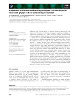
56.0), 37 (70%) patients died. The median OS was 14.5
± 1.3 months. The 6 month- and 1-year actuarial OS
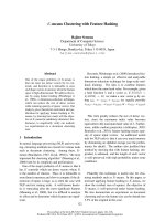
rates w ere 80.9% and 61.2%, respectively (Fig. 1). Cause
of death was PD in a majority of patients (n =33;
89.2%).Amongthese33PDpatients,25and8diedof
extracranial and brain progression, respectively. Three
(8.1%) patient died of bronchopneumonia. Postoperative
death for second Head & Neck cancer was observed in
(2.7%) another patient.
Overall, 38 disease progression were observed. The
median time to disease progression was 7.3 ± 1.1
months. The 6 month- and 1-year PFS rates were 62.9%
and 26.7%, respectively. The majority of patients with
PD presented with extra cranial PD. Eighteen (47.4%)
patients failed extracranially as the sole side of PD, 14
(36.8%) failed in the brain only and 6 (15.8%) progressed
at the metastatic brain site only.
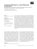
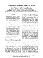
Overall, local failure was observed in (24.5%) 13
patients (Fig. 2). The median time to local failure was
48.9 ± 11.5 months. The 6 month- and 1-year local con-
trol rates were 98.1% and 75.2%, respectively. Local fail-
ure only was observed in 6 patients and another 7
patients presented local brain failure with concomitant
distant brain failure.
Distant brain failure was observed in 14 (26.4%)
patients. The median time to distant brain failure only
was 48.9 ± 25.1 months. The 6 month- and 1-year brain
failure rates were 10.8% and 28.2%, respectively. Brain
failure only was observed in 7 patients and another 7
patients presented local brain failure with concomitant
distant brain failure.
Extra cranial failure was observed in 29 (54.7%)
patients. Median time to extra cranial failure was 10.4 ±
1.1 months. The 6 month- and 1-year local control rates
were 29.5% and 60.8%, respectively. Extra cranial failure
onlywasobservedin18patients,6and3patientspre-
sented with extra cranial failure/local brain failure/dis-
tant brain failure and extra cranial failure/distant brain
failure, respectively. Extra cranial failure and local brain
failure only was observed in another 2 patients.
Late radiation-induced toxicity was minimal: alopecia
(gradeCTCAE1,15andgradeCTCAE2,3patients)
was observed in 18 (33.9%) patients. No patient pre-
sented with gross neuro-cognitive dysfunction. Asthenia
grade C TCAE grade 1 and 2 was observed in 1 1
patients, respectively. No patient presented with asthenia
CTCAE grade 3.
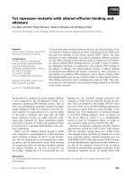
On univariate analysis (Table 2), improved OS was
found to be significantly associated with total dose (≤ 39
Gy vs. > 39 Gy; p < 0.01; Fig. 3), age < 65 (p < 0. 01),
absence of extracrani al metastasis (p < 0.01), GPA ≥ 2.5
(p = 0.01), KPS ≥ 90 (p = 0.01), and RPA = 2 (p = 0.02).
Gender was not found to be associated with survival but
there was a trend for statistica l significance of improved
OS in patients female vs. patients male (p = 0.07; Table
2). Likewise, there was a statistical trend toward signifi-
canceforsurgery(p=0.07;Table2)andcenter(p=
0.07; Table 2). The number of brain metastasis (p =
0.49; Table 2), histology (p = 0.58; Table 2) and syn-
chronous vs. metachronou s (p = 0.71) were however not
found to be associated significantly with survival. On
multivariate analysi s, only total dose (hazard ratio [HR],
3.55; 95% confide nce interval [95 %CI], 1.65 - 7.64; p <
Casanova et al. Radiation Oncology 2010, 5:13
/>Page 3 of 8
0.01) and the absence of extracranial metastasis (HR
2.29; 95%CI, 1.10 - 4.73; p = 0.03) retained statistical
significance.
Improved PFS was found to be significantly associated
with age < 65 (p < 0.01), total dose (≤ 39 Gy vs.>39
Gy; p < 0.01), absence of extracranial metastasis (p <
0.01), RPA < 2 (p = 0.01), GPA ≥ 2.5 (p = 0.01), T stage
(p = 0.02), metachronous vs.synchronousBM(p=
0.03), N stage (p = 0.05), KPS ≥ 90 (p = 0.05) and center
(p = 0.05). On multivariate analysis, total dose (HR 3.63;
95% CI 1.60 - 8 .24; p < 0.01), T stage (HR 3.02; 95% CI
1.32 - 6.89; p < 0.01), and the absence of extracranial
metastasis (HR 5.79; 95% CI 2.52 - 13.32; p < 0.01)
retained statistical significance.
Discussion
Tothebestofourknowledge,thepresentstudyisthe
largest series ever published on WBRT with RTB in the
treatment of lung cancer patients with BM. The
observed progression diseasepatternwasmainlyextra-
cranially, with 3 patients out of 4 with disease progres-
sion deceasing from systemic disease. As such, the
estimated LC rate was remarkable, with a 1-year LC rate
of more than 75% (Fig. 2)
The significant influence of total dose on duration of
survival in this cohort of patients with metastatic lung
cancer was the main finding of this analysis (Fig. 3). The
addition of a RTB to WBRT appeared to substantially
increase the median OS to approximately 15 months
(Fig. 1), which compares favourably with those of o ther
series of radiosurgery (SRS), with[22] or without surgery
[10,11,23] or concomitant targeted agent[24]. A survival
advantage of SRS to WBRT in patients with multiple
BMs was not observed in the RTOG 9805 study rando-
mising 333 patients with 1 to 4 BM[10]. The mean OS
was 6.5 and 5.7 months (p = 0.13) in the WBRT alone
and combined modality arms, respectively. Patients with
single BM treated with adjuvant SRS had however a sig-
nificant better survival (4.9 vs.6.5months;p=0.04)
than those who were not allocated boost treatment.
Likewise, a smaller prospective trial randomising 27
patients with 1 - 4 BM to WBRT ± SRS did not show a
signif ican t increase in survival (7.5 vs.11.0months,p=
0.22)[11].
The influence of RTB (15 Gy in 8 fractions) was also
assessed in 50 BM patients treated with 30 - 40 Gy
WBRT[25]. The mean OS of these patients was 4.6
months,comparedto3.8monthsforthose(n =114)
receiving WBRT alone. Hoskin et al. concluded that no
advantage of high dose adjuvant radiation treatment
could be foreseen using external beam radiotherapy.
Approximately 60% of patients with a single BM
received RTB in this study on the basis of stable disease
and good general condition. Possible explanations for
this discrepant finding include imbalances between the
two cohorts with respect to known and unknown base-
line prognostic factors (no prognostication was possi ble
for the Royal Marsden Hospital study) or imbalances in
the use of second and third-line therapies, as the major-
ity of patients (60% - 75%) died of metastatic disease
Figure 1 Overall survival in 53 lung cancer patients treated with WBRT and RTB.
Casanova et al. Radiation Oncology 2010, 5:13
/>Page 4 of 8
outside the brain in both studies. Our results are how-
ever in line with the retrospective analysis of 201
patients with 1 - 2 BMs[1]. All patients were RPA 1 or
2 and they underwent resection of the metastasis and
WBRT with (n = 102) or without (n = 99) a RTB. The
median OS was 18 and 9.5 months (p < 0.001) for the
former and latter group, respectiv ely. On multivariate
analysis, RTB, extent of surgical resection and interval
from the tumour diagnosis and RT were found to be
statistically significant. Interestingly, the median OS
observed in our study, constituted of a majority (>70%)
of patients undergoing surgery, is identical (14.5
months) to the one reported by the German group. The
addition of a RTB was also associated with improved
loca l tumour and brain control[1]. Noteworthy, increas-
ing the dose to the surgical bed with 10 - 15 Gy RTB
after WBRT did not modify the patient outcome in a
recent match-pair analysis with patient treated with
WBRT and radiosurgery[26].
The present study evaluated 11 prognostic factors for
OS and PFS. A n administered dose of > 39 Gy was
associated with a significant increase in OS and PFS
(Table 2). Interestingly, the parameter center was asso-
ciated with a significant improvement in patient out-
come in univariate analysis (Table 2). One center did
always administer sequentially 36 Gy with WBRT and
18 Gy with RTB (Table 1). As dose was a significant
prognosticator, this factor did not retain significance in
the multivariate analysis. Assuming a a-b ratio of 10 for
lung cancer, the 54 Gy (delivered in 2 Gy per fraction)
and 39 Gy (delivered in 3 Gy per fraction) will corre-
spond to a biological eff ective dose (BED) of 65 and 51
Gy
10
, respectively. The magnitude of the >25% increase
in BED might be expected to result in an increase in LC
for BM patients treated with the former dose schedule.
This strategy will however consequentially translate in
an increase of the overall treatment time that could be
detrimental for poor prognosis patients w ith a limited
OS. The other significant prognostic factor for OS and
PFS was the absence of extra cranial disease, which is a
recognized prognosticator for BM patients undergoing
RT[20].
Figure 2 Local control in 53 lung cancer patients treated with WBRT and RTB.
Casanova et al. Radiation Oncology 2010, 5:13
/>Page 5 of 8
We could not assess the long term neuro-cognitive
effect of this RTB strategy, as only one center prospec-
tively performed Mini Mental Status Examination in all
BM patients. The patients treated in this center had
however the lower survival rate, so we had unfortunately
insufficient baseline and follow-up data to adequately
assess neuro-cognition. We were however unaware of
any such toxicity in patients who were followed in our
respective clinics. The observed >75% of LC could possi-
bly resu lt in an increase of neuro-cognitive function for
our patients treat ed with WBRT and RTB. Regine et al.
reported on the neuro-cognitive outcome of 445 BM
patients treated in the RTOG 91-04 phase III study[27].
Control of BM had a significant impact on neuro-cogn i-
tion as measured by the Mini-Mental Status Examina-
tion. Likewise, Meyers et al. reported on another phase
III trial assessing the efficacy of gadolinium motexafin
[12]. Patien ts with BM from lung cancer presented with
an increase of fine motor and visual motor scanning
function if they had a partial response on brain MRI. All
patient with PD had a decline of neuro-cognitive
function.
It is appropriate to acknowledge that, in a retrospec-
tive analysis spanning more than 12 years, the apparent
striking impact of total dose on outcome might be at
least partially reflect c onfounding factors. RTB was
delivered only to patient s with a go od prognosis and, as
such, this treatment policy should not be delivered
indiscriminately to all BM patients. The majority of
patient underwent surgical resection, but 15% of the
cohort did not benefit from surgery. The patients trea-
ted in one center delivering high dose RT did present a
more favourable prognostic profile, although not signifi-
cantly so (Table 1). It should be noted however that
there was no difference in age, number of B M or per-
centage of operated patients (Table 1). We were thus
unable to identify other factors that might adequately
explain the observed effect. There was another limita-
tion to our study. The small sample size of 53 patients
and i ts consequential statistical power limits the overall
conclusions of this study. We have chosen to perform
however a multivariate analysis, as the ratio of observa-
tions to prognostic factors was appropriate[28]. Further
research regarding RT dose-outcome relationships is
justified in the framework of modern technique delivery.
Conclusions
This analysis of the outcome of 53 lung cancer patients
with BM treated with WBRT and RTB reveals an
increase in OS and PFS for patients treated with higher
39 Gy
> 39 Gy
Figure 3 Overall survival (OS) by RT dose group for 53 BM patients with lung cancer.
Casanova et al. Radiation Oncology 2010, 5:13
/>Page 6 of 8
radiation doses. Only one-quarter of the studied cohort
presented with local failure. The majority of patients
presented with extra cranial progression. There might
be a subgroup of younger patients with good perfor-
mance status and no extracranial disease who may bene-
fit from non-stereotactic dose escalation after WBRT t o
the metastatic site.
Abbreviations
BM: brain metastasis; RTB: radiotherapy boost; WBRT: whole brain radiation
therapy; QoL: quality of life; MRI: magnetic resonance imagery; CT: computed
tomography; PD: progressive disease; CTCAE: Common Terminology Criteria
for Adverse Events; LC: local control; ECF: extracranial failure; OS: overall
survival; PFS: progression-free survival; KPS: Karnofsky performance status;
RPA: recursive partitioning analysis; GPA: graded prognostic assessment; SCC:
Squamous cell carcinoma; BED: biologic effective dose.
Author details
1
Radiation Oncology, Geneva University Hospital, 6 rue Gabriel le Perret
Gentil, CH-1211 Geneva, Switzerland.
2
Radiation Oncology, Centre Hospitalier
Universitaire Vaudois, Rue du Bugnon 21, CH-1001 Lausanne, Switzerland.
3
Radiation Oncology, Sion Cantonal Hospital, Av. du Grand-Champsec 80,
CH-1950 Sion, Switzerland.
4
Clinical Epidemiology Unit, Geneva University
Hospital, 6 rue Gabrielle Perret Gentil, CH-1211 Geneva, Switzerland.
5
University of Geneva, 1 rue Michel Servet, CH-1205 Geneva, Switzerland.
Table 2 Summary of univariate anlaysis for OS and PFS
median
OS
(months)
p*
(HR [95%])
median
PFS
(months)
p*
Age, years
< 65 15.9 <0.01 9.3 <0.01
≥ 65 7.4 (3.75 [1.51-9.31]) 3.8 (3.10 [1.44-6.69])
Total dose, Gy
≤ 39 8.2 <0.01 3.9 <0.01
> 39 23.3 (3.84 [1.83-8.03]) 11.7 (0.29 [0.14-0.59])
GPA
≥ 2.5 15.9 0.01 9.3 0.01
< 2.5 7.4 (2.42 [1.18-4.93]) 3.8 (2.46 [1.23-4.92])
Extracranial metastasis
Yes 7.6 <0.01 3.8 <0.01
No 16.9 (2.71 [1.35-5.44]) 9.3 (2.93 [1.48-5.79])
KPS
≥ 90 14.7 0.01 5.1 0.05
< 90 7.6 (2.35 [1.19-4.63]) 9.3 (2.26 [0.98-5.22])
RPA
1 14.7 0.02 8.3 0.01
2-3 7.6 (2.46 [1.10-5.50]) 3.8 (2.72 [1.25-5.89])
Gender
Female 16.4 0.07 7.4 0.58
Male 12.6 (1.94 [0.93-4.03]) 6.2 (1.21 [0.61-2.41])
Center
CHUV 26.4 0.07 33.3 0.05
HUG 10.1 (2.76 [1.12-6.80]) 4.1 (3.23 [1.21-8.65])
CHCVS 16.4 9.0
Surgery
Yes 7.5 0.07 3.8 0.05
No 15.9 (1.90 [0.94-3.82]) 9.3 (0.51 [0.25-1.01])
Number of brain metastasis
1 16.4 0.49 7.4 0.61
2-3 14.3 (0.72 [0.28-1.86]) 6.6 (0.78 [0.30-2.02])
Type of primary ling cancer
SCC 14.5 0.58 6.2 0.40
AdenoCa 14.7 (1.61 [0.64-4.02]) 9.3 (1.67 [0.70-3.99])
Neuroendocrine 12.6 (1.61 [0.64-4.02]) 6.5
*log-rank
Casanova et al. Radiation Oncology 2010, 5:13
/>Page 7 of 8
Authors’ contributions
DCW was responsible for the primary concept and the design of the study;
DCW, NC, ZM and SB performed the data capture and analysis. NC and
DCW drafted the manuscript; DCW and CC performed the statistical analysis;
DCW, NC, ZM and SB reviewed patient data; AP, SB, CC and MZ revised the
manuscript.
All authors have read and approved the final manuscript.
Competing interests
The authors declare that they have no competing interests.
Received: 22 December 2009
Accepted: 18 February 2010 Published: 18 February 2010
References
1. Rades D, Pluemer A, Veninga T, Dunst J, Schild SE: A boost in addition to
whole-brain radiotherapy improves patient outcome after resection of 1
or 2 brain metastases in recursive partitioning analysis class 1 and 2
patients. Cancer 2007, 110:1551-1559.
2. Johnson JD, Young B: Demographics of brain metastasis. Neurosurgery
clinics of North America 1996, 7:337-344.
3. Gercovich FG, Luna MA, Gottlieb JA: Increased incidence of cerebral
metastases in sarcoma patients with prolonged survival from
chemotherapy. Report of cases of leiomysarcoma and chondrosarcoma.
Cancer 1975, 36:1843-1851.
4. Ryberg M, Nielsen D, Osterlind K, Andersen PK, Skovsgaard T,
Dombernowsky P: Predictors of central nervous system metastasis in
patients with metastatic breast cancer. A competing risk analysis of 579
patients treated with epirubicin-based chemotherapy. Breast cancer
research and treatment 2005, 91:217-225.
5. Duchnowska R, Szczylik C: Central nervous system metastases in breast
cancer patients administered trastuzumab. Cancer treatment reviews 2005,
31:312-318.
6. Sundstrom JT, Minn H, Lertola KK, Nordman E: Prognosis of patients
treated for intracranial metastases with whole-brain irradiation. Annals of
medicine 1998, 30:296-299.
7. Zimm S, Wampler GL, Stablein D, Hazra T, Young HF: Intracerebral
metastases in solid-tumor patients: natural history and results of
treatment. Cancer 1981, 48:384-394.
8. Lagerwaard FJ, Levendag PC, Nowak PJ, Eijkenboom WM, Hanssens PE,
Schmitz PI: Identification of prognostic factors in patients with brain
metastases: a review of 1292 patients. International journal of radiation
oncology biology, physics 1999, 43:795-803.
9. Barnes EA, Chow E, Tsao MN, Bradley NM, Doyle M, Li K, Lam K, Danjoux C:
Physician Expectations of Treatment Outcomes for Patients with Brain
Metastases Referred for Whole Brain Radiotherapy. International journal of
radiation oncology, biology, physics 2010, 76:187-192.
10. Andrews DW, Scott CB, Sperduto PW, Flanders AE, Gaspar LE, Schell MC,
Werner-Wasik M, Demas W, Ryu J, Bahary JP, Souhami L, Rotman M,
Mehta MP, Curran WJ Jr: Whole brain radiation therapy with or without
stereotactic radiosurgery boost for patients with one to three brain
metastases: phase III results of the RTOG 9508 randomised trial. Lancet
2004, 363:1665-1672.
11. Kondziolka D, Patel A, Lunsford LD, Kassam A, Flickinger JC: Stereotactic
radiosurgery plus whole brain radiotherapy versus radiotherapy alone
for patients with multiple brain metastases. International journal of
radiation oncology, biology, physics 1999, 45:427-434.
12. Meyers CA, Smith JA, Bezjak A, Mehta MP, Liebmann J, Illidge T, Kunkler I,
Caudrelier JM, Eisenberg PD, Meerwaldt J, Siemers R, Carrie C, Gaspar LE,
Curran W, Phan SC, Miller RA, Renschler MF: Neurocognitive function and
progression in patients with brain metastases treated with whole-brain
radiation and motexafin gadolinium: results of a randomized phase III
trial. J Clin Oncol 2004, 22:157-165.
13. Li J, Bentzen SM, Li J, Renschler M, Mehta MP: Relationship between
neurocognitive function and quality of life after whole-brain
radiotherapy in patients with brain metastasis. International journal of
radiation oncology, biology, physics 2008, 71:64-70.
14. Sham JS, Lau WH, Tung Y: Radiotherapy of brain metastases from
carcinoma of the bronchus. Clinical radiology 1989, 40:193-194.
15. Egawa S, Tukiyama I, Akine Y, Kajiura Y, Yanagawa S, Watai K, Nomura K:
Radiotherapy of brain metastases. International journal of radiation
oncology, biology, physics 1986, 12:1621-1625.
16. D’Elia F, Bonucci I, Biti GP, Pirtoli L: Different fractionation schedules in
radiation treatment of cerebral metastases. Acta radiologica 1986,
25:181-184.
17. Chatani M, Teshima T, Hata K, Inoue T, Suzuki T: Whole brain irradiation
for metastases from lung carcinoma. A clinical investigation. Acta
radiologica 1985, 24:311-314.
18. Harwood AR, Simson WJ: Radiation therapy of cerebral metastases: a
randomized prospective clinical trial. International journal of radiation
oncology, biology, physics 1977, 2:1091-1094.
19. Borgelt B, Gelber R, Kramer S, Brady LW, Chang CH, Davis LW, Perez CA,
Hendrickson FR: The palliation of brain metastases: final results of the
first two studies by the Radiation Therapy Oncology Group. International
journal of radiation oncology, biology, physics 1980, 6:1-9.
20. Gaspar L, Scott C, Rotman M, Asbell S, Phillips T, Wasserman T,
McKenna WG, Byhardt R: Recursive partitioning analysis (RPA) of
prognostic factors in three Radiation Therapy Oncology Group (RTOG)
brain metastases trials. International journal of radiation oncology, biology,
physics 1997, 37:745-751.
21. Sperduto PW, Berkey B, Gaspar LE, Mehta M, Curran W: A New Prognostic
Index and Comparison to Three Other Indices for Patients With Brain
Metastases: An Analysis of 1,960 Patients in the RTOG Database.
International journal of radiation oncology, biology, physics 2008, 70:510-514.
22. Roberge D, Petrecca K, El Refae M, Souhami L: Whole-brain radiotherapy
and tumor bed radiosurgery following resection of solitary brain
metastases. Journal of neuro-oncology 2009.
23. Kim YS, Kondziolka D, Flickinger JC, Lunsford LD: Stereotactic radiosurgery
for patients with nonsmall cell lung carcinoma metastatic to the brain.
Cancer 1997, 80:2075-2083.
24. Ma S, Xu Y, Deng Q, Yu X: Treatment of brain metastasis from non-small
cell lung cancer with whole brain radiotherapy and Gefitinib in a
Chinese population. Lung cancer (Amsterdam, Netherlands) 2009,
65:198-203.
25. Hoskin PJ, Crow J, Ford HT: The influence of extent and local
management on the outcome of radiotherapy for brain metastases.
International journal of radiation oncology, biology, physics 1990, 19:111-115.
26. Rades D, Kueter JD, Pluemer A, Veninga T, Schild SE: A matched-pair
analysis comparing whole-brain radiotherapy plus stereotactic
radiosurgery versus surgery plus whole-brain radiotherapy and a boost
to the metastatic site for one or two brain metastases. International
journal of radiation oncology, biology, physics 2009, 73
:1077-1081.
27. Regine WF, Scott C, Murray K, Curran W: Neurocognitive outcome in brain
metastases patients treated with accelerated-fractionation vs.
accelerated-hyperfractionated radiotherapy: an analysis from Radiation
Therapy Oncology Group Study 91-04. International journal of radiation
oncology, biology, physics 2001, 51:711-717.
28. Peduzzi P, Concato J, Kemper E, Holford TR, Feinstein AR: A simulation
study of the number of events per variable in logistic regression
analysis. Journal of clinical epidemiology 1996, 49:1373-1379.
doi:10.1186/1748-717X-5-13
Cite this article as: Casanova et al.: Whole brain radiotherapy with a
conformational external beam radiation boost for lung cancer patients
with 1-3 brain metastasis: a multi institutional study. Radiation Oncology
2010 5:13.
Casanova et al. Radiation Oncology 2010, 5:13
/>Page 8 of 8