Báo cáo khoa học: "Choline PET based dose-painting in prostate cancer - Modelling of dose effects" potx
Bạn đang xem bản rút gọn của tài liệu. Xem và tải ngay bản đầy đủ của tài liệu tại đây (297.49 KB, 9 trang )
RESEARC H Open Access
Choline PET based dose-painting in prostate
cancer - Modelling of dose effects
Maximilian Niyazi
1
, Peter Bartenstein
2
, Claus Belka
1
, Ute Ganswindt
1*
Abstract
Background: Several randomized trials have documented the value of radiation dose escalation in patients with
prostate cancer, especially in patients with in termediate risk profile. Up to now dose escalation is usually applied to
the whole prostate. IMRT and related techniques currently allow for dose escalation in sub-volumes of the organ.
However, the sensitivity of the imaging modality and the fact that small islands of cancer are often dispersed
within the whole organ may limit these approaches with regard to a clear clinical benefit. In order to assess
potential effects of a dose escalation in certain sub-volumes based on choline PET imaging a mathematical dose-
response model was developed .
Methods: Based on different assumptions for a/b, g50, sensitivity and specificity of choline PET, the in fluence of
the whole prostate and simultaneous integrated boost (SIB) dose on tumor control probability (TCP) was
calculated. Based on the given heterogeneity of all potential variables certain representative permutations of the
parameters were chosen and, subsequently, the influence on TCP was assessed.
Results: Using schedules with 74 Gy within the whole prostate and a SIB dose of 90 Gy the TCP increase ranged
from 23.1% (high detection rate of choline PET, low whole prostate dose, high g50/ASTRO definition for tumor
control) to 1.4% TCP gain (low sensitivity of PET, high whole prostate dose, CN + 2 definition for tumor contr ol) or
even 0% in selected cases. The corresponding initial TCP values without integrated boost ranged from 67.3 % to
100%. According to a large data set of intermediate-risk prostate cancer patients the resulting TCP gains ranged
from 22.2% to 10.1% (ASTRO definition) or from 13.2% to 6.0% (CN + 2 definition).
Discussion: Although a simplified mathematical mode l was employed, the presented model allows for an
estimation in how far given schedules are relevant for clinical practi ce. However, the benefit of a SIB based on
choline PET seems less than intuitively expected. Only under the assumption of high detection rates and low initia l
TCP values the TCP gain has been shown to be relevant.
Conclusions: Based on the employed assumptions, specific dose escalation to choline PET positive areas within
the prostate may increase the local control rates. Due to the lack of exact PET sensitivity and prostate a/b
parameter, no firm conclusions can be made. Small variations may completely abrogate the clinical benefit of a SIB
based on choline PET imaging.
Introduction
Several randomized trials have documented a clear dose-
response relationship for prostate cancer. Although not
employing modern IMRT techniques t he M. D. Ander-
son phase III dose escalation trial was the first rando-
mized trial to prove 78 Gy vs. 70 Gy. It resulted in
better biochemical control for the higher radiation dose
in patients with inter mediate-risk features [1]. Other
groups obtained similar results [2-6]. This interpretation
is corroborated by population based approaches showing
that only doses ≥ 72 Gy are associated with adequate
tumor control [7,8].
The implementation of IMRT into clinical practice o f
prostate cancer radiation treatment enables the physi-
cian to increase the doses in focal areas of the gland,
which is in contrast to the central d ogma in radiation
oncology to strive for a homogeneous dose to the target
volume [9]. How ever, this approach might have two
* Correspondence:
1
Department of Radiation Oncology, Ludwig-Maximilians-University
München, Marchioninistr. 15, 81377 München, Germany
Niyazi et al. Radiation Oncology 2010, 5:23
/>© 2010 Niyazi et al; licensee BioMed Central Ltd. This is an Open Access article distributed under the terms of the Creative Commons
Attribution License ( s/by/2.0), w hich permits unrestricted use, distribution, and reproduction in
any medium, provide d the original work is properly cited.
advantages: Firstly the dose escalation is limited to a
minor part of the target volume and thus, the probabil -
ityofsideeffectsshouldbelowered [10]. Secondly the
biological efficacy may be incr eased by the use of higher
doses per fraction.
The first who addressed this issue were Pickett, Xia
and colleagues [11,12], later on further studies were
conducted [13,14], also in case of high-risk prostate can-
cer [15]. Li et al. reported a new IMRT simultaneous
integrated boost (SIB) strategy that irradiates prostate
via hypo-fractionation while irradiating pelvic nodes
with the conventiona l fractionat ion. Compared to the
conventional two-phase treatment, the proposed SIB
technique offers potential advantages, including bett er
sparing of critical structures leading to less inconti-
nence, rectal bleeding, irritative symptoms [16-20] or
urethral toxicity [21], more efficient delivery, shorter
treatment duration, and better biological efficacy [22].
Fonteyne et al. reported that addition of an IMRT SIB
to an intra-prostatic lesion (defined by magnetic reso-
nance imaging) did not increase the severity or inci-
dence of acute toxicity [23]. Furthermore new
techniques like volumetric modulated arcs, helical
tomotherapy or IMPT additionally showed improve-
ments in conformal avoidance relative to fixed beam
IMRT [24,25].
Despite the technical advances in radiotherapy the
optimal treatment for prostate cancer strongly depends
on the accuracy of tumor characterization and staging.
Positron emission tomography (PET) is an exquisitely
sensitive molecular imaging technique using positron-
emitting radioisotopes coupled to specific ligands [26].
Different PET tracers, including [
11
C] choline, [
18
F]
choline and [
11
C] acetate, have been described for the
detection of prostate cancer. However, larger trials are
still needed to establish their final c linical value con-
cerning the primary detection and the staging of pros-
tate cancer [27].
In principle, signal-genera tion is based on an incre ased
choline metabolism in prostate cancer leading to a n
increased up-take in tumor tissue compared to that of
benign tissue [28]. However, benign prosta te hyperplasia
and inflammatory changes may also lead to increased
uptake thereby lowering the specificity of the PET signal.
A precise volumetric assessment of PET signals is of
rising importance for radiotherapy (RT) planning [29].
The use of choline PET/CT data to detect tumor spots
within the prostate has been analyzed and first c linical
experiences in lymph node-positive patients were
reported [30]. In t his regard, Ciernik et al. investigated
the utility of F-18-choline PET signals to serve as a tar-
get for semi-automatic segm entation for forward t reat-
ment planning of prostate cancer. F-18-choline PET and
CT scans of ten patients with histologically proven
prostate cancer without extra-capsular tumor extension
were acquired using a combined PET/CT scanner. Plan-
ning target volumes (PTV’s) derived from CT and F-18-
choline PET yielded comparable results. 3D-conformal
planning with CT or F-18-choline PET resulted in com-
parable doses to the rectal wall. Choline PET signals of
the prostate provided adequate spatial information to be
used for standardized PET-based target volume defini-
tion [31].
As PET allows for detection of small lesions within
the prostate and modern IMRT techniques can b e used
for integrated focal boosting, it is evident to use PET
info rmation in order to escalate the dose within defined
tumor spots also called biologically guided radiotherapy
[32]. This type of selective dose-escalation has already
been implemented successfully using spectroscopic MRI
data [23,33,34]. Although doing so may be intuitively
reasonable, the true effect of such procedures is strongly
influenced by a multitude of facto rs. We therefore
attempted to develop a method to estimate the increas e
of local tumor control using an IMRT SIB to choline
PET positive hotspots within the gland. The computa-
tions were done in a putative intermediate-risk collective
reflecting the fact that these patients will have the most
benefit by any dose escalation approach.
Methods
The best currently available dataset for dose-response
relationships in prostate cancer was derived from a
study of 235 low-risk and 382 intermediate-risk patients
treated between 1987 and 1998 with external beam RT
alone at the M. D. Anderson Cancer Center [35].
Local control (biochemical no evidence of disease) was
def ined in two different ways; Firstly, ASTRO definition
was employed: Time to PSA failure is defined as the end
of RT to the mid-point between the PSA nadir and the
first PSA rise [35]. Secondly, the Ho uston definition
defines biochemical failure as PSA rise of ≥ 2ng/ml
above the current nadir PSA (CN + 2) [ 36-38]. In both
settings detectable local, nodal and distant relapses a s
well as initiation of hormonal treatment are scored as
failures.
In order to develop a mathematical TCP model for
prostate cancer, we firstly assumed the prostate to be a
geom etrical structure subdivided into a fixed number of
voxels (defining their v olume as v
i
= 1 ). Voxels includ-
ing tumor cells are called tumorlets.
N is defined as the number of clonogenic cells within
the tumor, V as the volume of the target volume and n
i
is defined as the densit y of tumor cells wit hin a tumor-
let. We furthermore ass umed that all tumorlets have the
same density of clonogenic cells. In order to achieve this
in practice one has to define the voxels as sufficiently
small.
Niyazi et al. Radiation Oncology 2010, 5:23
/>Page 2 of 9
The tumor control probability (T CP) is modelled a s a
Poisson distribution [39]. In such a geometrical setting
it is defined as:
TCP e
nSF
i
ii
SF
i
is the surviving fraction within the single sub-
volume with the running index i (ranging from 1 to m
=V/v
i
). Using the well-known linea r-quadratic model
the surviving fraction can be calculated as:
SF e
d
j
nd
j
1
/
with d
j
as single dose (usually 1.8 or 2 Gy), n as the
number of fractions and a, b as the parameters from
the linear-quadratic model which refer to the radio-sen-
sitivity of the tumor cells (a represents lethal lesions
made by one-track action and b accounts for lethal
lesions m ade by two-track action, [40]). In this formula
the tumor doubling time is not considered.
Relevant a/b ratios can be obtained from both in
vitro experiments and clinical fractionation studies and
give the dose where linear and quadratic effect are
equal according to total cell kill [41] whereas in vitro
data do not necessarily predict the radio-sensitivity of
tissues in clinical radiotherapy. There is a wide varia-
tion of a/b values for prostat e cancer in the literature
with the exact value of a/b being still unknown
[41-51].
Thus, the following calculations were based on the
values determined by Fowler et al. (a/b =1.5Gy,a =
0.04 Gy
-1
) [43], Wang et al. (a/b =3.1Gy,a =0.15
Gy
-1
[49,52]) and Valdagni et al. (a/b = 8.3 Gy [46,48]).
Another relevant parameter to describe the TCP is the
slope of the killing curve (g50) which relates to the
number of clonogens within the tumor in the following
way [53]:
50
2
2
2
ln
ln
N
ln
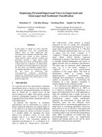
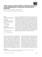
Cheung et al. calculated a g50 value of 2.2 [1.1-3.2,
95% CI] and TCD50 = 67.5 Gy [65.5-69.5 Gy, 95% CI]
(ASTRO definition) or g50 = 1.4 [0.2-2.5, 95% CI] and
TCD50 = 57.8 Gy [49.8-65.9 Gy, 95% CI] (CN + 2 defi-
nition) for intermediate-risk patients [35]. The corre-
sponding TCP curves are shown in Figure 1.
Those voxels not containing a clonogenic cell (pure
prostate tissue) do not contribute to the overall TCP as
the corresponding factor equals 1.
Summarizing all these equations, and after some alge-
braic manipulations keeping in mind that v
i
=1,one
obtains:
TCP TCP e
SIB conv
SF N
/
ΔSF denotes the difference between boosted and con-
ventional surviving f raction (conventional means with-
out boost, but 3D-conformal RT or IMRT technique).
This expression has to be corrected due to the limited
sensi tivity in detecting all clonogenic cells. The sensitiv-
ityvaluesforcholinePETrangefrom81%(foraSUV
of 2.65) [54] down to 73% [28,55] or 64% [56] (Addi-
tional file 1 offers the possibility to specify different
parameters for intermediate-risk prostate cancer to cal-
culate the effect of an IMRT SIB).
This is a simplified picture of reality as the sensitivity
of detecting tumor cells within the prostate is depen-
dent on the size or more precise intensity of the enhan-
cing tumor lesion. Par tial volume effects c an severely
affect images both qualitatively and quantitatively: For
any hot lesion of a small size and embedded in a colder
background, this effect spreads out the signal. It typi-
cally occurs whenever the tumor size is less than 3
times the full width at half maximum (FWHM) of the
rec onstructed image resolution. The maximum value in
the hot tumor then will be lower than the actual maxi-
mum value. A small tumor will look larger but less
aggressive than it actually is [57]. The model assumes
the detection rate for t he sake of simplicity size-inde-
pendent and constant, the aforementioned sensitivities
from the literature are taken as best guesses for the
detection rate.
The model used for our ca lculation is based on a
number of additional assumptions. Thus, several
Figure 1 Tumor control probability curves for both definitions
of local control derived by data of Cheung et al. (RT of the
whole prostate).
Niyazi et al. Radiation Oncology 2010, 5:23
/>Page 3 of 9
shortcomings have to be taken into account wh en inter-
preting the data:
1) The assumption of a homogeneous density of clo-
nogenic tumor-cells is notobvious.Theremaybe
islands within the prostate with a higher clonogenic
density. However, this is no strict contradiction to
our assumption as the sub-voxels may be scaled
down until only empty voxels and voxels with a
small but uniform number of clonogenic cells
remain left.
2) The given model is incapable of reflecting biologi-
cal sub-volume effects adequately: For e xample, one
may assume that hypoxic areas within high-density
tumor foci may cause a locally enhanced radio-resis-
tance. Since all values used for our calculation are
basedonwholeorganTCPs,thegivenmodel
ignores issues of focally increased resistance.
3) Biologically, a complex feedback between the
tumor and surrounding normal tissue exists. For
example, the release of certain cytokines after radia-
tion damage may influence the surrounding tumor
tissue and vice versa. Again the given model is not
able to integrate the putative interaction of adjacent
clonogenic tumor and stroma cells.
4) It is assumed that all clonogenic cells within the
tumor have a uniform radiosensitivity.
All these effects may be in place but do not seem to
have much influence in practice. One prominent exam-
ple is the comparison between primary and salvage
radiotherapy.
After prostatectomy with positive surgical margins
adjuvant radiotherapy improves disease-free survival
rates and thus it is discussed as a new standard of adju-
vant treatment in selected cases [58]; in cases of local
relapse, salvage radiotherapy is the only potentially cura-
tive treatment approach [59]. The doses being necessary
to control microscop ic tumor seem to be higher than
initially expected and to be similar to those for
macroscopic tumor within the setting of a primary treat-
ment [60].
Results
The relevant parameters fed into our model in order to
calculate the increase in w hole organ TCP are: Sensitiv-
ity of choline PET, a, a/b, g50, whole prostate dose, SIB
dose and dose per fraction.
In order to present the calculations different represen-
tative scenarios have been tested:
1. High sensitivity of choline PET, low whole prostate
dose, high g50 (ASTRO consensus), Fowler’s a/b
This parameter set was chosen to calculate a putativ e
maximum TCP increase: Choline PET sensitivity was set
to 81% and 74 Gy were chosen as homogeneous pros-
tate dose. a/b was set to 1.5 Gy (a =0.04Gy
-1
), g50
was chosen according to Cheung’sdatawiththe
ASTRO definition. As shown in Figure 1 this parameter
set leads to a higher steepness of the TCP curve. The
results are shown in Table 1. The TCP in this setting
with homogeneous dose of 74 G y within the prostate
was 67.3% and was improved by 23.1% up to 9 0.4%
using a SIB.
2. High sensitivity of choline PET, low whole prostate
dose, low g50 (CN + 2 definition), Fowler’s a/b
In contrast, one may assume a parameter set with
slightly less optimal conditions for a SIB. Table 2 sum-
marizes the results when assuming a higher detection
rate for PET (81%), a low homogeneous whole prostate
dose(74Gy),aSIBdoseof90Gyandradio-sensitivity
parameters as described by Fowler et al. (a/b =1.5Gy,
a =0.04Gy
-1
)andg50 taken again from Cheun g’s data
but this time according to the CN + 2 definition. The
calculated TCP without SIB was 96.0% which leaves
only an increase of 2.9% with a SIB.
This result is basically driven by a high initial control
probability. In reality the initial clinical control probabil-
ity is lower [35].
Table 1 TCP-increase for high sensitivity of choline PET, low whole prostate dose, high g50(ASTRO consensus) and
Fowler’s a/b
a [Gy
-1
] a/b [Gy] g50 Det. rate PET [%] Dose [Gy] SIB [Gy] Single dose [Gy] TCP
conv
[%] TCP Increase [%]
0.04 1.5 2.2 81 74 90 2 67.3 23.1
Calculation of the increase in TCP with whole prostate dose of 74 Gy after boosting choline PET positive regions within the prostate up to 90 Gy. a and a/b are
estimated from Fowler’s data and g50 from Cheung’s data (ASTRO definition). For choline PET a high sensitivity was used.
Table 2 TCP-increase for high sensitivity of choline PET, low whole prostate dose, low g50(CN + 2 definition) and
Fowler’s a/b
a [Gy
-1
] a/b [Gy] g50 Det. rate PET [%] Dose [Gy] SIB [Gy] Single dose [Gy] TCP
conv
[%] TCP Increase [%]
0.04 1.5 1.4 81 74 90 2 96.0 2.9
Calculation of the TCP-increase after boosting choline PET positive regions within the prostate up to 90 Gy. a/b is estimated from Fowler’s data and g50 from
Cheung’s data (CN + 2 definition). For choline PET a high sensitivity w as used.
Niyazi et al. Radiation Oncology 2010, 5:23
/>Page 4 of 9
3. Low sensitivity of choline PET, high whole prostate
dose, low g50 (CN + 2 definition), Fowler’s a/b
A “worst case” scenario is considered where a low sensi-
tivity of PET is presumed, the homogeneous whole
prostate dose is hig h (see Table 3, 78 Gy along the dose
concept of the M. D. Anderson trial [1]), a/b is low and
g50 is less steep than the corresponding ASTRO value.
Based on these assumptions the gain of a SIB is low,
as the initial TCP is again very high (97.0%) and as the
remaining SIB effect is smal l (1.4%). Again, this result is
in contrast to clinical reality reflected in the Cheung
data [35].
4. High sensitivity of choline PET, low whole prostate
dose, g50 arbitrary, Wang’s a/b
Using a/b and a values originally obtained by Wang et
al. one obtains independently of g50 or the whole organ
dose a TCP of 100% which leaves no benefit f or a SIB
(Table 4). This result is probably due to the fact that
the respective g50 as well as a/b p arameters were
derived from independent clinical trials.
5. Different sensitivities of choline PET, low whole
prostate dose, different a/b values, calculated a, high g50
(ASTRO definition)
In order to circumvent the problem of overestimating
the initial TCP one can try to reproduce the M. D.
Anderson data (Cheung et al.) employing different a/b
values (Fowler, Wang, Valdagni) and fitting an optimal
a value to finally achieve a realistic co ncordance
between observed TCD50 and calculated TCD50 value.
InTable5theASTROconsensuswasusedforthe
definition of tumor control, leading to a steeper TCP
curve (see Figure 1). Using a low whole prostate dose
(74 Gy), the baseline tumor control was 68.7%. In this
setting the SIB mediated TCP increase was strongly
dependent on the sensitivity of the choline PET. Assum-
ing a sensitivity rate of 81%, the TCP was increased by
22.2%, for 64% the increase was lowered to 17.0%.
Using higher a/b values automatically resulted in a
lower TCP gain. This differenceisbasedonthefact
that in the given model a was optimized with fixed g50
and a/b, resulting in different TCP curves.
Table 3 TCP-increase for low sensitivity of choline PET, high whole prostate dose, low g50(CN + 2 definition) and
Fowler’s a/b
a [Gy
-1
] a/b [Gy] g50 Det. rate PET [%] Dose [Gy] SIB [Gy] Single dose [Gy] TCP
conv
[%] TCP Increase [%]
0.04 1.5 1.4 64 78 90 2 97.0 1.4
Calculation of the TCP-increase after boosting choline PET positive regions within the prostate up to 90 Gy; the whole organ dose was set to 78 Gy. a/b is
estimated from Fowler’s data and g50 from Cheung’s data (CN + 2 definition). For choline PET a low sensitivity was used.
Table 4 TCP-increase for high sensitivity of choline PET, low whole prostate dose, g50arbitrary and Wang’s a/b
a [Gy
-1
] a/b [Gy] g50 Det. rate PET [%] Dose [Gy] SIB [Gy] Single dose [Gy] TCP
conv
[%] TCP Increase [%]
0.15 3.1 1.4 81 74 90 2 100 0
0.15 3.1 2.2 81 74 90 2 100 0
Calculation of the TCP-increase after boosting choline PET positive regions within the prostate up to 90 Gy. a/b is estimated from Wang’s data and g50 arbitrary.
For choline PET a high sensitivity was assumed.
Table 5 TCP-increase for different sensitivities of choline PET, low whole prostate dose, different a/b values, calculated
a and high g50 (ASTRO definition)
a [Gy
-1
] a/b [Gy] g50 Det. rate PET [%] Dose [Gy] SIB [Gy] Single dose [Gy] TCP
conv
[%] TCP Increase [%]
0.04 1.5 2.2 81 74 90 2 68.7 22.2
0.04 1.5 2.2 64 74 90 2 68.7 17.0
0.06 3.1 2.2 81 74 90 2 68.7 21.4
0.06 3.1 2.2 64 74 90 2 68.7 16.4
0.08 8.3 2.2 81 74 90 2 68.7 20.1
0.08 8.3 2.2 64 74 90 2 68.7 15.4
Calculation of the TCP-increase after boosting choline PET positive regions within the prostate up to 90 Gy. a/b was set to either Fowler’s/Wang’s or Valdagni’s
value, a was analytically determined in order to achieve agreement between calculated TCD50 and TCD50 obtained by Cheung et al. g50 was again taken from
Cheung’s data (ASTRO definition).
Niyazi et al. Radiation Oncology 2010, 5:23
/>Page 5 of 9
6. Different sensitivities of choline PET, high whole
prostate dose, different a/b values, calculated a, high g50
(ASTRO definition)
Compared to Table 5 in Table 6 a higher whole prostate
dose (78 Gy) was used. The initial TCP could be
improved to 77.2%. The increase in TCP by the given
SIB dos e was lower ranging from 14.9% (high PET sen-
sitivity, low a/ b) to 10.1% (low detection rate, high a/b).
7. Different sensitivities of PET, low whole prostate dose,
different a/b values, calculated a, low g50 (CN + 2
definition)
InTable7theCN+2consensuswasusedtodefine
tumor control, leading to less steep TCP curves (see Fig-
ure1).Again,alowwholeprostatedosewasused;the
baseline tumor control then was calculated to be 80.0%.
Similarly to the previous scenario, the TCP-increase by
a given SIB was also strongly rela ted to the assumed
sensitivity of the choline PET. Using a sensitivity of 81%
the T CP was increased by 13.2% compared to 22.2% in
the same setting employing the ASTRO definition. In
contrast, for 64% sensitivity the increase was only 10.3%.
Replacing the given a/b by higher values resulted in
lower TCP gains. The lowest increase for TCP was seen
for Valdagni’s a/b with a low choline PET sensitivity:
9.0%.
8. Different sensitivities of PET, high whole prostate dose,
different a/b values, calculated a, low g50 (CN + 2
definition)
Compared to Table 7 in Table 8 a higher whole prostate
dose was used (78 Gy). The initial TCP could be
improved to 80%. The increase in TCP was low er as it
ranged from 9.1% (high detectio n rate of PET, low a/b)
to 6.0% (low detection rate, high a/b).
Discussion
Using a simplified mathematical model allowed us to
determine the increase in TCP after an IMRT SIB based
on choline PET positive intra-prostatic lesions. The
model has been based on several fundamental assump-
tions including uniform clonogenic cell density, no
interaction between adjacent tumor cells, no sub-volume
effects and a uniform radio-sensitivity of all tumor cells.
Furthermore the model does not consider population
differences o r time factors [61]. This model is substan-
tiated by the fact that doses being needed to control
microscopic tumor in an adjuvant/salvage setting seem
to be almost as high as those used in primary therapy
for macroscopic tumors [60].
It was shown that a SIB mediated increase of the
given TCP is strongly dependent on the sensitivity of
the choline PET, the g50-value with special em phasis on
Table 6 TCP-increase for different sensitivities of choline PET, high whole prostate dose, different a/b values,
calculated a and high g50(ASTRO definition)
a [Gy
-1
] a/b [Gy] g50 Det. rate PET [%] Dose [Gy] SIB [Gy] Single dose [Gy] TCP
conv
[%] TCP Increase [%]
0.04 1.5 2.2 81 78 90 2 77.2 14.9
0.04 1.5 2.2 64 78 90 2 77.2 11.5
0.06 3.1 2.2 81 78 90 2 77.2 14.1
0.06 3.1 2.2 64 78 90 2 77.2 11.0
0.08 8.3 2.2 81 78 90 2 77.2 13.1
0.08 8.3 2.2 64 78 90 2 77.2 10.1
Calculation of the TCP-increase after boosting choline PET positive regions within the prostate up to 90 Gy, higher homogeneous whole prostate dose. a/b was
analytically determined in order to achieve agreement between calculated TCD50 and TCD50 obtained by Cheung et al. g50 was again taken from Cheung’s data
(ASTRO definition).
Table 7 TCP-increase for different sensitivities of PET, low whole prostate dose, different a/b values, calculated a and
low g50(CN + 2 definition)
a [Gy
-1
] a/b [Gy] g50 Det. rate PET [%] Dose [Gy] SIB [Gy] Single dose [Gy] TCP
conv
[%] TCP Increase
[%]
0.03 1.5 1.4 81 74 90 2 80 13.2
0.03 1.5 1.4 64 74 90 2 80 10.3
0.04 3.1 1.4 81 74 90 2 80 12.6
0.04 3.1 1.4 64 74 90 2 80 9.8
0.06 8.3 1.4 81 74 90 2 80 11.6
0.06 8.3 1.4 64 74 90 2 80 9.0
Calculation of the TCP-increase after boosting choline PET positive regions within the prostate up to 90 Gy. a/b was set to either Fowler’s/Wang’s or Valdagni’s
value, a was analytically determined in order to achieve agreement between calculated TCD50 and TCD50 obtained by Cheung et al. g50 was again taken from
Cheung’s data (CN + 2 definition).
Niyazi et al. Radiation Oncology 2010, 5:23
/>Page 6 of 9
thedefinitionoftumorcontrol,thedoseusedforthe
treatment of the whole organ and the a/b values.
We observed a high variat ion between the outcomes
based on different initial assumptions. A critical limita-
tion is the fact that there is no chance to derive a/b and
a values for the calculation of dose-response relation-
ships from the trial by Cheung et al. (the best data avail-
able to date) since a single fixed fractionation schedule
was applied [35].
In keeping with this several inconsistencies occurred
(Table 5, Table 6, Table 7 and Table 8): the calculated
a values did not fit their counterpart in literature except
for Fowler’s data where the deviation was small. In this
case the dependence on g50 and the detection rate of
choline PET became more important.
On the one hand, g50 depends on the failure defini-
tion and data are different with longer follow-up data,
and at present the confidence interval is still wide as g50
= 2.2 [1.1-3.2, 95% CI] (ASTRO definition) or g50 = 1.4
[0.2-2.5, 95% CI] (CN + 2 definition).
On the other hand, a study from Farsad et al . demon-
stratedthatC-11-cholinePET/CT has a relatively high
rate of false-negative results on a sextant basis. In addi-
tion it has been clearly shown that non-malignant pro-
static disorders may induce an increased
11
C-choline
uptake [62]. Our model calculations are not dependent
on specificity as the irradiation of non-infiltrated voxels
does not influence TCP but this will lead to unessen-
tially big SIB target volumes.
Taken togethe r, the relatively high efficacy rates of an
IMRT based SIB are potentially overestimating the real
benefit (Table 5, Table 6, Table 7 and Table 8, bet ween
7.1% and 22.2%). Patient setup errors as well as intra-
fractionmotionoftheprostatewerenotconsidered
throughout the whole estimation process which could
potentially hamper the results in a negative way [63-67].
Another important factor influencing tumor control was
neglected in the model: the risk of regional, i.e. pelvic
nodal and/or systemic failure. This may be a potential
source of limiting the effectiveness of this approach as it
was assumed that local control entails biochemical con-
trol; in this regard a sing le cancer cell outside the pros-
tate could violate this assumption and diminish tumor
control.
Despite all of our considerations our model data are
not in contrast to data provided by Kim et al. [68]
claiming that selective boosting is more effective than
homogeneous dose escalation as sparing of normal tis-
sue is easier to achieve.
Furthermore, risk-adaptive optimization increases the
therapeutic ratio as compared to conventional selective
boosting IMRT. In another paper Kim et al. derive simi-
lar results, but mention the importance of the underly-
ing imaging modality and consecutively their sensitivity
in detecting occult tumor cells [69].
Utilizing an IMRT boost is an elegant technique but
one has to mention another classical but suitable
method: With brachytherapy the doses to the organs at
risk are lower or similar to IMRT-only. Dose escalation
for prostate tumors may also be easily achieved by bra-
chytherapy alone [70].
Conclusions
Regarding treatment planning in radiotherapy, choline
PETmayoffersomeadvantagesintermsofstaging,
tumor delineation and the description of biological pro-
cesses. However, a TCP-increase related to any IMRT
SIB on choline PET positive regions has to be consid-
ered as realistically low.
Additional file 1: This file contains a sheet where parameters like
choline PET sensitivity/specificity, a, a/b, g50, TCD50, dose, SIB dose and
single dose can be specified and a sheet carrying out all necessary
calculation steps.
Click here for file
[ />S1.XLS ]
Abbreviations
RT: radiotherapy; IMRT: intensity-modulated radiotherapy; SIB: simultaneous
integrated boost; PTV: Planning target volume; LQ: linear-quadratic; TCP:
Table 8 TCP-increase for different sensitivities of PET, high whole prostate dose, different a/b values, calculated a and
low g50(CN + 2 definition)
a [Gy
-1
] a/b [Gy] g50 Det. rate PET [%] Dose [Gy] SIB [Gy] Single dose [Gy] TCP
conv
[%] TCP Increase
[%]
0.03 1.5 1.4 81 78 90 2 84.5 9.1
0.03 1.5 1.4 64 78 90 2 84.5 7.1
0.04 3.1 1.4 81 78 90 2 84.5 8.5
0.04 3.1 1.4 64 78 90 2 84.5 6.6
0.06 8.3 1.4 81 78 90 2 84.5 7.7
0.06 8.3 1.4 64 78 90 2 84.5 6.0
Calculation of the TCP-increase after boosting choline PET positive regions within the prostate up to 90 Gy, low homogeneous dose 78 Gy. a/b was set to either
Fowler’s/Wang’s or Valdagni’s result, a was analytically determined in order to achieve agreement between calculated TCD50 and TCD50 obtained by Cheung et
al. g50 was again taken from Cheung’s data (CN + 2 definition).
Niyazi et al. Radiation Oncology 2010, 5:23
/>Page 7 of 9
tumor control probability; TCD50: tumor control dose 50%; PET: positron
emission tomography; SUV: standardized uptake value; CI: confidence
interval; SF: surviving fraction; EBRT: external beam radiotherapy; PSA:
prostate-specific antigen; ASTRO: American Society for Therapeutic Radiology
and Oncology; CN: current nadir; FWHM: full width at half maximum.
Author details
1
Department of Radiation Oncology, Ludwig-Maximilians-University
München, Marchioninistr. 15, 81377 München, Germany.
2
Department of
Nuclear Medicine, Ludwig-Maximilians-University München, Marchioninistr.
15, 81377 München, Germany.
Authors’ contributions
MN developed the underlying mathematical model and wrote the
manuscript. PB participated in the preparation of the manuscript. UG and CB
provided the idea and participated in the conception as well as the
preparation of the manuscript. All authors read and approved the final
manuscript.
Competing interests
The authors declare that they have no competing interests.
Received: 19 January 2010 Accepted: 18 March 2010
Published: 18 March 2010
References
1. Pollack A, Zagars GK, Starkschall G, Antolak JA, Lee JJ, Huang E, von
Eschenbach AC, Kuban DA, Rosen I: Prostate cancer radiation dose
response: Results of the M. D. Anderson phase III randomized trial.
International Journal of Radiation Oncology Biology Physics 2002,
53(5):1097-1105.
2. Zelefsky MJ, Leibel SA, Gaudin PB, Kutcher GJ, Fleshner NE, Venkatramen ES,
Reuter VE, Fair WR, Ling CC, Fuks Z: Dose escalation with three-
dimensional conformal radiation therapy affects the outcome in
prostate cancer. International Journal of Radiation Oncology Biology Physics
1998, 41(3):491-500.
3. Hanks GE, Hanlon AL, Epstein B, Horwitz EM: Dose response in prostate
cancer with 8-12 years’ follow-up. Elsevier Science Inc 2002, 427-435.
4. Bey P, Carrie C, Beckendorf V, Ginestet C, Aletti P, Madelis G, Luporsi E,
Pommier P, Cowen D, Gonzague-Casabianca L, et al: Dose escalation with
3D-CRT in prostate cancer: French study of dose escalation with conformal
3D radiotherapy in prostate cancer - Preliminary results. International
Journal of Radiation Oncology Biology Physics 2000, 48(2):513-517.
5. Boersma LJ, Brink van den M, Bruce AM, Shouman T, Gras L, te Velde A,
Lebesque JV: Estimation of the incidence of late bladder and rectum
complications after high-dose (70-78 Gy) conformal radiotherapy for
prostate cancer, using dose-volume histograms. International Journal of
Radiation Oncology Biology Physics 1998, 41(1):83-92.
6. Forman JD, Duclos M, Shamsa F, Porter AT, Orton C: Hyperfractionated
conformal radiotherapy in locally advanced prostate cancer: Results of a
dose escalation study. International Journal of Radiation Oncology Biology
Physics 1996, 34(3):655-662.
7. Welz S, Nyazi M, Belka C, Ganswindt U: Surgery vs. radiotherapy in
localized prostate cancer. Which is best? Radiation Oncology 2008, 3.
8. Ganswindt U, Paulsen F, Anastasiadis AG, Stenzl A, Bamberg M, Belka C: 70
Gy or more: which dose for which prostate cancer? J Cancer Res Clin
Oncol 2005, 131(7):407-419.
9. Tanderup K, Olsen DR, Grau C: Dose painting: Art or science? Radiotherapy
and Oncology 2006, 79(3):245-248.
10. Al-Mamgani A, Heemsbergen WD, Peeters STH, Lebesque JV: ROLE OF
INTENSITY-MODULATED RADIOTHERAPY IN REDUCING TOXICITY IN DOSE
ESCALATION FOR LOCALIZED PROSTATE CANCER. International Journal of
Radiation Oncology Biology Physics 2009, 73(3):685-691.
11. Pickett B, Vigneault E, Kurhanewicz J, Verhey L, Roach M: Static field
intensity modulation to treat a dominant intra-prostatic lesion to 90 Gy
compared to seven field 3-dimensional radiotherapy. Int J Radiat Oncol
Biol Phys 1999, 44(4):921-929.
12. Xia P, Pickett B, Vigneault E, Verhey LJ, Roach M: Forward or inversely
planned segmental multileaf collimator IMRT and sequential
tomotherapy to treat multiple dominant intraprostatic lesions of
prostate cancer to 90 Gy. Int J Radiat Oncol Biol Phys 2001, 51(1):244-254.
13. Dogan N, Wu Y, Hagan MP: Simultaneous-integrated boost (SIB) IMRT for
treatment of indermediate-risk prostate cancer with nodal irradiation.
International Journal of Radiation Oncology Biology Physics 2006,
66(3):2805.
14. Singh AK, Guion P, Sears-Crouse N, Ullman K, Smith S, Albert PS,
Fichtinger G, Choyke PL, Xu S, Kruecker J, et al: Simultaneous integrated
boost of biopsy proven, MRI defined dominant intra-prostatic lesions to
95 Gray with IMRT: early results of a phase I NCI study. Radiation
Oncology 2007, 2.
15. Li X, Wang JZ, Jursinic P, Lawton CA: IMRT simultaneous integrated boost
for high-risk prostate cancer. International Journal of Radiation Oncology
Biology Physics 2004, 60(1):2486.
16. Pinkawa M, Piroth MD, Fischedick K, Nussen S, Klotz J, Holy R, Eble MJ: Self-
assessed bowel toxicity after external beam radiotherapy for prostate
cancer–predictive factors on irritative symptoms, incontinence and
rectal bleeding. Radiat Oncol 2009, 4:36.
17. Onal C, Topkan E, Efe E, Yavuz M, Sonmez S, Yavuz A: Comparison of rectal
volume definition techniques and their influence on rectal toxicity in
patients with prostate cancer treated with 3D conformal radiotherapy: a
dose-volume analysis. Radiat Oncol 2009, 4:14.
18. Martin JM, Bayley A, Bristow R, Chung P, Gospodarowicz M, Menard C,
Milosevic M, Rosewall T, Warde PR, Catton CN: Image guided dose
escalated prostate radiotherapy: still room to improve. Radiat Oncol 2009,
4:50.
19. Guckenberger M, Baier K, Richter A, Vordermark D, Flentje M: Does
intensity modulated radiation therapy (IMRT) prevent additional toxicity
of treating the pelvic lymph nodes compared to treatment of the
prostate only? Radiat Oncol 2008, 3:3.
20. Ghadjar P, Vock J, Vetterli D, Manser P, Bigler R, Tille J, Madlung A,
Behrensmeier F, Mini R, Aebersold DM: Acute and late toxicity in prostate
cancer patients treated by dose escalated intensity modulated radiation
therapy and organ tracking. Radiat Oncol 2008, 3:35.
21. Ghadjar P, Matzinger O, Isaak B, Behrensmeier F, Stroux A, Rentsch CA,
Thalmann GN, Aebersold DM: Association of urethral toxicity with dose
exposure in combined high-dose-rate brachytherapy and intensity-
modulated radiation therapy in intermediate- and high-risk prostate
cancer. Radiotherapy and Oncology 2009, 91(2):237-242.
22. Li XA, Wang JZ, Jursinic PA, Lawton CA, Wang D: Dosimetric advantages
of IMRT simultaneous integrated boost for high-risk prostate cancer.
International Journal of Radiation Oncology Biology Physics 2005,
61(4):1251-1257.
23. Fonteyne V, Villeirs G, Speleers B, De Neve W, De Wagter C, Lumen N, De
Meerleer G: Intensity-modulated radiotherapy as primary therapy for
prostate cancer: Report on acute toxicity after dose escalation with
simultaneous integrated boost to intraprostatic lesion. International
Journal of Radiation Oncology Biology Physics 2008, 72(3):799-807.
24. Weber DC, Wang H, Cozzi L, Dipasquale G, Khan HG, Ratib O, Rouzaud M,
Vees H, Zaidi H, Miralbell R: RapidArc, intensity modulated photon and
proton techniques for recurrent prostate cancer in previously irradiated
patients: a treatment planning comparison study. Radiat Oncol 2009, 4:34.
25. Yuen J, Rodrigues G, Trenka K, Coad T, Yartsev S, D’Souza D, Lock M,
Bauman G: Comparing two strategies of dynamic intensity modulated
radiation therapy (dIMRT) with 3-dimensional conformal radiation
therapy (3DCRT) in the hypofractionated treatment of high-risk prostate
cancer. Radiat Oncol 2008, 3:1.
26. Groves AM, Win T, Ben Haim S, Ell PJ: Non-[F-18]FDG PET in clinical
oncology. Lancet Oncology 2007, 8(9):822-830.
27. Picchio M, Crivellaro C, Giovacchini G, Gianolli L, Messa C: PET-CT for
treatment planning in prostate cancer. Q J Nucl Med Mol Imag 2009,
53(2):245-268.
28. Reske SN, Blumstein NM, Glatting G: [C-11]choline PET/CT imaging in
occult local relapse of prostate cancer after radical prostatectomy.
European Journal of Nuclear Medicine and Molecular Imaging 2008,
35(1):9-17.
29. Grosu AL, Piert M, Weber WA, Jeremic B, Picchio M, Schratzenstaller U,
Zimmermann FB, Schwaiger M, Molls M: Positron emission tomography
for radiation treatment planning. Strahlentherapie Und Onkologie 2005,
181(8):483-499.
30. Ganswindt U, Paulsen F, Alber M, Bares R, Bamberg M, Belka C: Intensity-
modulated radiotherapy (IMRT) for lymph node-positive patients in
prostate cancer under consideration of C-11-choline-PET data - first
clinical experiences. Strahlentherapie Und Onkologie 2006, 182:71-71.
Niyazi et al. Radiation Oncology 2010, 5:23
/>Page 8 of 9
31. Ciernik IF, Brown DW, Schmid D, Hany T, Egli P, Davis JB: 3D-Segmentation
of the F-18-choline PET signal for target volume definition in radiation
therapy of the prostate. Technology in Cancer Research & Treatment 2007,
6(1):23-30.
32. Stewart RD, Li XA: BGRT: Biologically guided radiation therapy - The
future is fast approaching!. Medical Physics 2007, 34:3739-3751.
33. Payne GS, Leach MO: Applications of magnetic resonance spectroscopy
in radiotherapy treatment planning. British Journal of Radiology 2006, 79:
S16-S26.
34. van Lin E, Futterer JJ, Heumink S, Vight Van Der LP, Hoffmann AL, Van
Kollenburg P, Huisman HJJ, Scheenen TWJ, Witjes JA, Leer JWK, et al: IMRT
boost dose planning on dominant intraprostatic lesions: Gold marker-
based three-dimensional fusion of CT with dynamic contrast-enhanced
and H-1-spectroscopic MRI. International Journal of Radiation Oncology
Biology Physics 2006, 65(1):291-303.
35. Cheung R, Tucker SL, Lee AK, De Crevoisier R, Dong L, Kamat A, Pisters L,
Kuban D: Dose-response characteristics of low- and intermediate-risk
prostate cancer treated with external beam radiotherapy. Elsevier
Science Inc 2005, 993-1002.
36. Thames H, Kuban D, Levy L, Horwitz EM, Kupelian P, Martinez A, Michalski J,
Pisansky T, Sandler H, Shipley W, et al: Comparison of alternative
biochemical failure definitions’ based on clinical outcome in 4839
prostate cancer patients treated by external beam radiotherapy
between 1986 and 1995. International Journal of Radiation Oncology
Biology Physics 2003, 57(4):929-943.
37. Kuban DA, Thames HD, Levy LB: Radiation for prostate cancer: use of
biochemical failure as an endpoint following radiotherapy. World Journal
of Urology 2003, 21(4):253-264.
38. Kuban DA, Thames HD, Levy LB, Horwitz EM, Kupelian PA, Martinez AA,
Michalski JM, Pisansky TM, Sandler HM, Shipley WU, et al: Failure definition-
dependent differences in outcome following radiation for localized
prostate cancer. Can one size fit all? International Journal of Radiation
Oncology Biology Physics 2003, 57(2 Supplement):S146-S147.
39. Munro TR, Gilbert CW: THE RELATION BETWEEN TUMOUR LETHAL DOSES
AND THE RADIOSENSITIVITY OF TUMOUR CELLS. British Journal of
Radiology 1961, 34(400):246-251.
40. O’Rourke SFC, McAneney H, Hillen T: Linear quadratic and tumour control
probability modelling in external beam radiotherapy. Journal of
Mathematical Biology 2009, 58(4-5):799-817.
41. Garcia LM, Wilkins DE, Raaphorst GP: alpha/beta ratio: A dose range
dependence study. International Journal of Radiation Oncology Biology
Physics 2007, 67(2):587-593.
42. Fowler JF, Ritter MA, Fenwick JD, Chappell RJ:
How low is the alpha/beta
ratio for prostate cancer? In regard to Wang et al., IJROBP 2003; 55: 194-
203. International Journal of Radiation Oncology Biology Physics 2003,
57(2):593-595.
43. Fowler J, Chappell R, Ritter M: Is alpha/beta for prostate tumors really
low? International Journal of Radiation Oncology Biology Physics 2001,
50(4):1021-1031.
44. D’Souza WD, Thames HD: Is the alpha/beta ratio for prostate cancer low?
International Journal of Radiation Oncology Biology Physics 2001, 51(1):1-3.
45. Chappell R, Fowler J, Ritter M: New data on the value of alpha/beta -
Evidence mounts that it is low. (vol 60, pg 2004). International Journal of
Radiation Oncology Biology Physics 1002, 61(2):635-635.
46. Bentzen SM, Ritter MA: The alpha/beta ratio for prostate cancer: What is
it, really? Radiotherapy and Oncology 2005, 76(1):1-3.
47. Kal HB, Van Gellekom MPR: How low is the alpha/beta ratio for prostate
cancer? International Journal of Radiation Oncology Biology Physics 2003,
57(4):1116-1121.
48. Valdagni R, Italia C, Montanaro P, Lanceni A, Lattuada P, Magnani T,
Fiorino C, Nahum A: Is the alpha-beta ratio of prostate cancer really low?
A prospective, non-randomized trial comparing standard and
hyperfractionated conformal radiation therapy. Radiotherapy and
Oncology 2005, 75(1):74-82.
49. Wang JZ, Guerrero M, Li XA: How low is the alpha/beta ratio for prostate
cancer? International Journal of Radiation Oncology Biology Physics 2003,
55(1):194-203.
50. Wang JZ, Li XA, Yu CX, DiBiase SJ: The low alpha/beta ratio for prostate
cancer: What does the clinical outcome of HDR brachytherapy tell.
International Journal of Radiation Oncology Biology Physics 2003,
57(4):1101-1108.
51. Williams SG, Taylor JMG, Liu N, Tra Y, Duchesne GM, Kestin LL, Martinez A,
Pratt GR, Sandler H: Use of individual fraction size data from 3756
patients to directly determine the alpha/beta ratio of prostate cancer.
International Journal of Radiation Oncology Biology Physics 2007, 68(1):24-33.
52. Li XA, Wang JZ, Stewart RD, Dibiase SJ, Wang D, Lawton CA: Designing
equivalent treatment regimens for prostate radiotherapy based on
equivalent uniform dose. British Journal of Radiology 2008, 81(961):59-68.
53. Tome WA, Fowler JF: On cold spots in tumor subvolumes. Medical Physics
2002, 29(7):1590-1598.
54. Reske SN, Blumstein NM, Glatting G: Advancement of PET and PET/CT in
prostate carcinoma. Urologe 2006, 45(6).
55. Krause BJ, Souvatzoglou M, Tuncel M, Herrmann K, Buck AK, Praus C,
Schuster T, Geinitz H, Treiber U, Schwaiger M: The detection rate of [C-11]
Choline-PET/CT depends on the serum PSA-value in patients with
biochemical recurrence of prostate cancer. European Journal of Nuclear
Medicine and Molecular Imaging
2008, 35(1):18-23.
56. Scattoni V, Picchio M, Suardi N, Messa C, Freschi M, Roscigno M, Da
Pozzo L, Bocciardi A, Rigatti P, Fazio F: Detection of lymph-node
metastases with integrated [C-11]choline PET/CT in patients with PSA
failure after radical retropubic prostatectomy: Results confirmed by open
pelvic-retroperitoneal lymphadenectomy. Eur Urol 2007, 52(2):423-429.
57. Soret M, Bacharach SL, Buvat I: Partial-volume effect in PET tumor
imaging. J Nucl Med 2007, 48(6):932-945.
58. Ganswindt U, Stenzl A, Bamberg M, Belka C: Adjuvant radiotherapy for
patients with locally advanced prostate cancer - A new standard? Eur
Urol 2008, 54(3):528-542.
59. Ganswindt U, Belka C: Radiotherapy in prostate cancer. Urologe 2008,
47(9):1245-1254.
60. King CR, Kapp DS: Radiotherapy after prostatectomy: Is the evidence for
dose escalation out there? International Journal of Radiation Oncology
Biology Physics 2008, 71(2):346-350.
61. Warkentin B, Stavrev P, Stavreva NA, Fallone BG: Limitations of a TCP
model incorporating population heterogeneity. Physics in Medicine and
Biology 2005, 50(15):3571-3588.
62. Farsad M, Schiavina R, Castellucci P, Nanni C, Corti B, Martorana G, Canini R,
Grigioni W, Boschi S, Marengo M, et al: Detection and localization of
prostate cancer: Correlation of C-11-choline PET/CT with histopathologic
step-section analysis. Journal of Nuclear Medicine 2005, 46(10):1642-1649.
63. Krengli M, Gaiano S, Mones E, Ballare A, Beldi D, Bolchini C, Loi G:
Reproducibility of patient setup by surface image registration system in
conformal radiotherapy of prostate cancer. Radiat Oncol 2009, 4:9.
64. Nyholm T, Nyberg M, Karlsson MG, Karlsson M: Systematisation of spatial
uncertainties for comparison between a MR and a CT-based
radiotherapy workflow for prostate treatments. Radiat Oncol 2009, 4:54.
65. Graf R, Wust P, Budach V, Boehmer D: Potentials of on-line repositioning
based on implanted fiducial markers and electronic portal imaging in
prostate cancer radiotherapy. Radiat Oncol 2009, 4:13.
66. Lips IM, Dehnad H, van Gils CH, Boeken Kruger AE, Heide van der UA, van
Vulpen M: High-dose intensity-modulated radiotherapy for prostate
cancer using daily fiducial marker-based position verification: acute and
late toxicity in 331 patients. Radiat Oncol 2008, 3:15.
67. Boda-Heggemann J, Kohler FM, Wertz H, Ehmann M, Hermann B,
Riesenacker N, Kupper B, Lohr F, Wenz F: Intrafraction motion of the
prostate during an IMRT session: a fiducial-based 3D measurement with
Cone-beam CT. Radiat Oncol 2008, 3:37.
68. Kim Y, Tome WA: Is it beneficial to selectively boost high-risk tumor
subvolumes? A comparison of selectively boosting high-risk tumor
subvolumes versus homogeneous dose escalation of the entire tumor
based on equivalent EUD plans. Acta Oncologica 2008, 47(5):906-916.
69. Kim Y, Tome WA: On the impact of functional imaging accuracy on
selective boosting IMRT. Physica Medica 2009, 25(1):12-24.
70. Pieters BR, Kamer van de JB, van Herten YR, van Wieringen N,
D’Olieslager GM, Heide van der UA, Koning CC: Comparison of biologically
equivalent dose-volume parameters for the treatment of prostate cancer
with concomitant boost IMRT versus IMRT combined with
brachytherapy. Radiother Oncol 2008, 88(1):46-52.
doi:10.1186/1748-717X-5-23
Cite this article as: Niyazi et al.: Choline PET based dose-painting in
prostate cancer - Modelling of dose effects. Radiation Oncology 2010
5:23.
Niyazi et al. Radiation Oncology 2010, 5:23
/>Page 9 of 9