Báo cáo y học: " What is MRI bone oedema in rheumatoid arthritis and why does it matte" ppt
Bạn đang xem bản rút gọn của tài liệu. Xem và tải ngay bản đầy đủ của tài liệu tại đây (2.19 MB, 5 trang )
Page 1 of 5
(page number not for citation purposes)
Available online />Abstract
MRI bone oedema occurs in various forms of inflammatory and non-
inflammatory arthritis and probably represents a cellular infiltrate
within bone. It is common in early rheumatoid arthritis and is
associated with erosive progression and poor functional outcome.
Histopathological studies suggest that a cellular infiltrate
comprising lymphocytes and osteoclasts may be detected in
subchondral bone and could mediate the development of erosions
from the marrow towards the joint surface. There is emerging
evidence from animal models that such an infiltrate corresponds
with MRI bone oedema, pointing towards the bone marrow as a site
for important pathology driving joint damage in rheumatoid arthritis.
In the mid-17th century, a Dutch apprentice to a textile
merchant, Anton van Leeuwenhoek, was the first to see and
describe bacteria, yeasts and the circulation of blood
corpuscles in capillaries using a new tool, the light micro-
scope [1]. The subsequent elucidation of the microbiological
basis of infectious disease can be traced back, in part, to his
pioneering work in imaging. A parallel exists between the
invention of the microscope and the development of magnetic
resonance imaging (MRI), which allows new ways to explore
biological systems. In rheumatoid arthritis (RA), MRI provides
information about synovitis and erosion in early disease [2,3]
when inflammatory and destructive articular change is
typically subradiographic. In addition, it has revealed
something new and unexpected; the appearance referred to
as bone oedema. This MRI finding has been reported in other
conditions, such as osteonecrosis [4], osteoarthritis [5], and
ankylosing spondylitis [6], and in the sports medicine setting
where it appears associated with mechanical stress [7].
However, in RA there is evidence to suggest that bone
oedema represents a pivotal change occurring within
subchondral bone that may be associated with early events in
disease pathogenesis, which have not previously been
accessible to any form of imaging.
Just as the existence of micro-organisms was not expected
prior to the invention of the microscope, so the presence and
importance of bone oedema could not have been predicted
using the other sonographic and radiographic imaging
techniques used to investigate RA. MRI is unique in that it
images protons, which are usually contained within water
molecules (hence ‘oedema’), these in turn frequently being
contained within cells [8]. Although ultrasound can be used to
image synovitis by detecting thickening of the synovial
membrane [9] and can reveal increased synovial blood flow
using Doppler imaging [10], cellular infiltration within bone
remains invisible. Radiography, while an excellent technique
for imaging cortical bone, also cannot detect subcortical
cellular infiltrates, which are not necessarily associated with
periarticular osteopenia [11]. Histology could be used to
examine subchondral bone but resection of this tissue is
almost never done in early RA and the primary focus for tissue
immunohistochemistry has been the accessible synovium.
There are currently no published studies comparing the
histopathology of subchondral bone in RA with MRI appear-
ances (specifically bone oedema) but these are underway.
Unfortunately, they are likely to include patients with long-
standing disease where erosive and secondary degenerative
change could complicate the picture. In ankylosing spondylitis,
such a study has recently been published, describing
preoperative bone oedema in three of eight ankylosing
spondylitis patients with longstanding disease who underwent
spinal surgery involving resection of zygapophyseal joints [12].
Concordance was observed between bone oedema and a
mononuclear inflammatory infiltrate in bone marrow, but only
when the latter was relatively intense, suggesting that the MRI
feature is only apparent above a certain threshold.
Until recently, it was necessary to go back to literature
published in the early 1980s for a description of the histology
Review
What is MRI bone oedema in rheumatoid arthritis and why does
it matter?
Fiona M McQueen
1
and Benedikt Ostendorf
2
1
Department of Molecular Medicine and Pathology, Faculty of Medicine and Health Sciences, University of Auckland, Park Rd, Auckland, New Zealand
2
Center for Rheumatology , Department of Endocrinology, Diabetology and Rheumatology, Heinrich-Heine University Dusseldorf, Dusseldorf, Germany
Corresponding author: Fiona M McQueen,
Published: 5 December 2006 Arthritis Research & Therapy 2006, 8:222 (doi:10.1186/ar2075)
This article is online at />© 2006 BioMed Central Ltd
MPH-SPECT = high-resolution multipinhole single-photon-emission computed tomography; MRI = magnetic resonance imaging; NFκB = nuclear
factor kappa B; RA = rheumatoid arthritis; TNF = tumor necrosis factor.
Page 2 of 5
(page number not for citation purposes)
Arthritis Research & Therapy Vol 8 No 6 McQueen and Ostendorf
of subchondral bone in RA. Barrie [13] in 1981 described
“diffuse osteitis” within subchondral bone in 35% of patients
undergoing metatarsal head resection. In the November
2005 issue of Arthritis and Rheumatism, Bugatti and
colleagues [14] published a similar immunohistochemical
study of RA subchondral bone (from specimens obtained at
the time of joint replacement), using contemporary
techniques. They found lymphoid aggregates on the sub-
chondral side of the joint in established RA, often associated
with osteoclasts within the bone marrow abutting the cortex.
They concluded that “an inflammatory lymphoid infiltrate … is
a characteristic feature of RA subchondral bone marrow…
raising the hypothesis that subchondral bone marrow
inflammation might develop independent of the propagation
of synovial tissue.”
The MRI finding of bone oedema has been an important
driver in refocusing interest towards the subchondral bone in
early RA. A cohort study published in 1998 [2] revealed bone
oedema to be present at the carpus in 64% of RA patients
within 6 months of disease onset and in 45% after 6 years
[15]. There was clear evidence at one and six years after
disease onset [15,16] that bone oedema was a pre-erosive
lesion. The bone oedema score at presentation and one year
later was correlated with radiographic erosion and joint space
narrowing scores six years later [15] and, interestingly, even
with function, as measured by the physical function
component of the short-form-36 score [17]. A later study also
showed a link between bone oedema scores and tendon
function at eight years in these patients [18]. Others have
also found bone oedema to be common in RA [19], and it
was described by Ostendorf and colleagues [20] at the
metatarsal heads within only two months of the onset of
symptoms. Tamai and colleagues [21] recently confirmed its
association with disease severity as indicated by inflammatory
markers such as C-reactive protein and interleukin-6 levels in
early RA. At the other end of the spectrum of disease
duration, we have recently described florid bone oedema, at
the site of intended surgery, in RA patients awaiting joint
replacement or fusion. These data suggested that bone
oedema may be especially associated with painful and
aggressive disease [22]. Taken together, these lines of
evidence suggest that the process we recognize as MRI
bone oedema is widespread and relatively common in early
and late disease and tied to the development of long term
joint damage. Before the advent of MRI, this process sited in
the subchondral bone was unsuspected and certainly not
accorded any significance in terms of disease pathogenesis.
New work is now emerging to link the entity of bone oedema
with current theories of the immunopathogenesis of RA.
Hirohata and colleagues [23], in a highly accessed article
published in Arthritis Research and Therapy in early 2006,
described a study of bone marrow cells aspirated from the
iliac crests of RA patients. CD34+ stem cells that were
abnormally sensitive to tumor necrosis factor (TNF)α [24]
were found to express high levels of the nuclear factor kappa
B (NFκB) transcription factor, contrasting with cells from
osteoarthritis patients where NFκB expression was normal
and TNF sensitivity not observed. These authors suggested
that a bone marrow stem cell abnormality could underlie RA
and proposed a disease model where such cells could, under
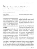
Figure 1
MRI scans from a 65 year old female rheumatoid arthritis patient with disease duration of one year. (a) Coronal T1 weighted image of the dominant
wrist with reduced signal indicating florid bone oedema involving the entire lunate bone (circle). (b) Equivalent image following the injection of
contrast (gadolinium diethylenetriamine pentaacetic acid (GdDPTA)) shows very bright signal within the lunate, suggesting the presence of
vascularized tissue (slice does not exactly correspond with pre-GdDPTA image). (c) Axial T2w image with bright signal confirming bone oedema at
the lunate.
Page 3 of 5
(page number not for citation purposes)
the influence of TNF, differentiate into fibroblast-like cells, and
travel to the synovial membrane where they might appear as
type B synoviocytes and promote synovitis [23]. Alternatively,
they could travel via the systemic circulation to the
subchondral bone marrow and initiate inflammatory and pre-
erosive changes from there, possibly including activation of
osteoclasts as described by Schwarz and colleagues [25].
Angiogenesis is known to accompany cellular proliferation in
rheumatoid synovial membrane via mediators such as
vascular endothelial growth factor and platelet derived growth
factor [26]. Ostendorf and colleagues [27] investigated
rheumatoid finger joints using miniarthroscopy and found that
macroscopic vascularization of the synovial membrane
correlated with histological features of angiogenesis and
clinical signs of disease activity. If the subchondral bone is
proposed as another site of cellular proliferation in RA, one
would also expect to find angiogenesis there. Interestingly,
there is a suggestion from MRI data that this may occur as
regions of bone oedema which are typically recognized as
areas of hyperintense signal on T2w images, also exhibit
increased signal after intravenous injection of gadolinium
diethylenetriamine pentaacetic acid (Gd-DTPA). This contrast
agent travels within blood vessels and causes hyperintensity
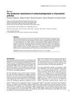
Available online />Figure 2
Potential role of bone marrow-derived stem cells in trafficking to the subchondral bone and synovial membrane in rheumatoid arthritis joints,
resulting in a subchondral cellular infiltrate (seen as bone oedema on MRI) followed by erosion. (a) CD34+ stem cells from bone marrow express
high levels of NFkB, which leads to unusual sensitivity to TNFα. (b) Stem cells differentiate into fibroblast-like cells and travel via the circulation to
synovial membrane to become type B synoviocytes - here they mediate formation of erosions via production of proinflammatory cytokines and
matrix metalloproteinases [23,25]. (c) Stem cells may also traffic to the subchondral bone marrow where they differentiate into mesenchymal cells.
These cells could then travel via bony canals from bone marrow to synovium [29] to excite an inflammatory response. (d) Alternatively, stem cells
could travel to subchondral bone and at this site could mediate an inflammatory response via T/B cell interactions associated with angiogenesis
[26] and osteoclast activation. This could lead to erosions originating from inside the bone, directed outwards towards joint surface [14]. (e)
Coronal T2 weighted MRI scan of the wrist in early rheumatoid arthritis reveals bone oedema at the bases of the 2nd and 3rd metacarpals and
adjacent regions of trapezoid and capitate carpal bones. Small intraosseous erosions are also apparent.
in vascular tissue [28]. An example from a patient with a
1 year history of RA is shown in Figure 1.
Finally, animal studies are emerging to clarify the role of the
bone marrow as a site of pathology in RA. Marinova-
Mutafchieva and colleagues [29] described an inflammatory
infiltrate in the subchondral bone of TNF-transgenic mice
where TNF-responsive mesenchymal cells were identified
within enlarged bony canals connecting bone marrow to
synovium. Most recently, Proulx and colleagues [30]
examined TNF-transgenic mice using a high-resolution 7
Tesla MRI scanner. They described the presence of bone
oedema and correlated this histologically with a highly cellular
infiltrate within the bone marrow. Another form of imaging,
high-resolution multipinhole single-photon-emission computed
tomography (MPH-SPECT), has revealed accelerated bone
turnover within the joints of interleukin-1 receptor antagonist
deficient mice [31]. In a single patient with early RA,
increased uptake in a central, interarticular distribution was
detected by MPH-SPECT when the MRI signal for bone
marrow on short tau inversion recovery (STIR) images was
normal, raising the possibility that even earlier changes in the
subchondral bone could be apparent using this sensitive,
high-resolution technique [32].
Figure 2 combines evidence from several imaging and
histological studies to suggest a disease model for RA,
where cells originating from bone marrow travel to the joint
and either mediate erosion from synovial membrane inwards
or from the subchondral bone outwards towards the joint
surface. This bone-marrow-centered model would be
consistent with the therapeutic success of drugs such as
rituximab [33], aimed at B cells, which may reside in the
synovium but originate from the bone marrow. It also predicts
that repopulation of the bone marrow with allotypically
different cells might effect remission of RA and this has been
described in recipients of allogeneic bone marrow transplants
performed in the 1980s [34]. It seems we are now on the
road to unraveling the mystery of what MRI bone oedema
actually means in RA. The implications are exciting and
suggest a new focus for understanding disease pathology
and influencing disease progression; moving away from the
synovium and towards the bone marrow.
Competing interests
The authors declare that they have no competing interests.
Acknowledgments
The authors wish to thank Professor P Conaghan for use of the MRI
scan in Figure 2.
References
1. The Collected Letters of Antoni van Leeuwenhoek 1701-1704.
Vol 14. ISBN: 9026514506. Lisse, The Netherlands: Swets and
Zeitlinger; 1996.
2. McQueen FM, Stewart N, Crabbe J, Robinson E, Yeoman S, Tan
PLJ, McLean L: Magnetic resonance imaging of the wrist in
early rheumatoid arthritis reveals a high prevalence of
erosion at four months after symptom onset. Ann Rheum Dis
1998, 57:350-356.
3. Klarlund M, Østergaard M, Jensen KE, Madsen JL, Skjødt H, the
TIRA group: Magnetic resonance imaging, radiography, and
scintigraphy of the finger joints: one year follow up of
patients with early arthritis. Ann Rheum Dis 2000, 59:521-528.
4. Lecouvet FE, van de Berg BC, Maldague BE, Lebon CJ, Jamart J,
Saleh M, Noel H, Malghem J: Early irreversible osteonecrosis
versus transient lesions of the femoral condyles: prognostic
value of subchondral bone and marrow changes on MR
imaging. Am J Roentgenol 1998, 170:71-77.
5. Carrino JA, Blum J, Parellada JA, Schweitzer ME, Morrison WB:
MRI of bone marrow edema-like signal in the pathogenesis of
subchondral cysts. Osteoarthritis Cartilage 2006, 14:1081-
1085.
6. Braun J, Baraliakos X, Golder W, Brandt J, Rudwaleit M, Listing J,
Bollow M, Sieper J, van der Heijde D: Magnetic resonance
imaging examinations of the spine in patients with ankylosing
spondylitis, before and after successful therapy with inflix-
imab: Evaluation of a new scoring system. Arthritis Rheum
2003, 48:1126-1136.
7. Hoy G, Wood T, Phillips N, Connell D: When physiology
becomes pathology: the role of magnetic resonance imaging
in evaluating bone marrow oedema in the humerus in elite
tennis players with an upper limb pain syndrome. Br J Sports
Med 2006, 40:710-713.
8. Peterfy CG: New developments in imaging in rheumatoid
arthritis. Curr Opin Rheumatol 2003, 15:288-295.
9. Grassi W, Cervini C: Ultrasonography in rheumatology: an
evolving technique. Ann Rheum Dis 1998, 57:268-271.
10. Szkudlarek M, Court-Payen, Strandberg C, Klarlund M, Klausen T,
Østergaard M: Power Doppler ultrasonography for assess-
ment of synovitis in the metacarpophalangeal joints of
patients with rheumatoid arthritis: a comparison with
dynamic magnetic resonance imaging. Arthritis Rheum 2001,
44:2018-2023.
11. Peterfy CG: MR imaging. Baillieres Clin Rheumatol 1996, 10:
635-678.
12. Appel H, Loddenkemper C, Grozdanowicz Z, Ebhardt H,
Dreimann M, Hempfing A, Stein H, Metz-Stavenhagen P, Rud-
waleit M, Sieper J: Correlation of histopathological findings
and magnetic resonance imaging (MRI) in the spine of
patients with ankylosing spondylitis. Arthritis Res Ther 2006,
8:R143.
13. Barrie HJ: Histologic changes in rheumatoid disease of the
metacarpal and metatarsal heads as seen in surgical mater-
ial. J Rheumatol 1981, 8:246-257.
14. Bugatti S, Caporali R, Manzo A, Vitolo B, Pitzalis C, Montecucco
C: Involvement of subchondral bone marrow in rheumatoid
arthritis. Lymphoid neogenesis and in situ relationship to
subchondral bone marrow osteoclast recruitment. Arthritis
Rheum 2005, 52:3448-3459.
15. McQueen FM, Benton N, Perry D, Crabbe J, Robinson E, Yeoman
S, McLean L, Stewart N: Bone oedema scored on magnetic
resonance scans of the dominant carpus at presentation pre-
dicts radiographic joint damage at the hands and feet six
years later in patients with rheumatoid arthritis. Arthritis
Rheum 2003, 48:1814-1827.
16. McQueen FM, Stewart N, Crabbe J, Robinson E, Yeoman S, Tan
PLJ, McLean L: Magnetic resonance imaging of the wrist in
early rheumatoid arthritis reveals progression of erosions
despite clinical improvement. Ann Rheum Dis 1999, 58:156-
163.
17. Benton N, Stewart N, Crabbe J, Robinson E, Yeoman S,
McQueen FM: MRI of the wrist in early rheumatoid arthritis
can be used to predict functional outcome at 6 years. Ann
Rheum Dis 2004, 63:555-561.
18. Zheng S, Yeoman, S, Robinson E, Crabbe J, Stewart N, Rouse J,
McQueen FM: Magnetic resonance imaging (MRI) bone
oedema predicts 8 year tendon function at the wrist but not
the requirement for orthopaedic surgery in rheumatoid arthri-
tis patients. Ann Rheum Dis 2006, 65:607-611.
19. Savnik A, Malmskov H, Thomsen HS, Graff LB, Nielsen H,
Danneskiold-Samsoe B, Boesen J, Bliddal H: MRI of the wrist
and finger joints in inflammatory joint diseases at 1-yr inter-
val: MRI features to predict bone erosions. Eur Radiol 2002,
12:1203-1210.
Arthritis Research & Therapy Vol 8 No 6 McQueen and Ostendorf
Page 4 of 5
(page number not for citation purposes)
20. Ostendorf B, Scherer A, Modder U, Schneider M: Diagnostic
value of magnetic resonance imaging of the forefeet in early
rheumatoid arthritis when findings on imaging of the
metacarpophalageal joints of the hands remain normal.
Arthritis Rheum 2004, 50:2094-2102.
21. Tamai M, Kawakami A, Takao S, Uetani M, Arima K, Tanaka F,
Fujikawa K, Aramaki T, Iwanaga N, Izumi Y, et al.: Bone marrow
oedema determined by MRI reflects severe disease status in
patients with early-stage rheumatoid arthritis. Ann Rheum Dis
2006, 65(Suppl II):629.
22. Gao A, Østergaard M, Robinson E, Dalbeth N, Doyle A, Shalley
G, McQueen F: Unexpected finding of frequent high grade
MRI bone oedema within the field of surgery in RA patients
awaiting joint replacement/fusion at the hands or feet. Arthri-
tis Rheum 2006, 54(Suppl):S625.
23. Hirohata S, Miura Y, Tmita T, Yoshikawa H, Ochi T, Chiorazzi N:
Enhanced expression of mRNA for nuclear factor kB1 (p50) in
CD34+ cells of the bone marrow in rheumatoid arthritis.
Arthritis Res Ther 2006, 8:R54.
24. Hirohata S, Yanagida T, Nagai T, Sawada T, Nakamura H,
Yoshino S, Tomita T, Ochi T: Induction of fibroblast-like cells
from CD34(+) progenitor cells of the bone marrow in
rheumatoid arthritis. J Leukoc Biol 2001, 70:413-421.
25. Schwarz EM, Looney RJ, Drissi MH, O’Keefe RJ, Boyce BF, Xing
L, Ritchlin CT: Autoimmunity and bone. Ann NY Acad Sci 2006,
1068:275-283.
26. Maruotti N, Cantatore FP, Crivellato E, Vacca A, Ribatti D: Angio-
genesis in rheumatoid arthritis. Review. Histol Histopathol
2006, 21:557-566.
27. Ostendorf B, Iking-Konert C, Bleck E, Dann P, Pauly Th, Friemann
J, Schneider M: Vascular imaging of rheumatoid synovium:
Macroscopic and microscopic analysis of synovial tissue
obtained by miniarthroscopy from finger joints of patients
with rheumatoid arthritis. Ann Rheum Dis 2006, 65(Suppl II):
66.
28. Stewart N, McQueen FM, Crabbe JC: MRI of the wrist: a pictor-
ial essay. Australas Radiol 2001, 45:268-273.
29. Marinova-Mutafchieva L, Williams RO, Funa K, Maini RN, Zvaifler
NJ: Inflammation is preceded by tumour necrosis factor-
dependent infiltration of mesenchymal cells in experimental
arthritis. Arthritis Rheum 2002, 46:507-513.
30. Proulx S, Kwok E, Shealy DJ, Ritchlin CT, Schwarz EM: Under-
standing bone marrow edema in arthritis: 3D-MRI and histol-
ogy analyses of TNF-Tg mice. Arthritis Rheum 2006, 54
(Suppl):S798-S799.
31. Ostendorf B, Scherer A, Wirrwar A, Hoppin JW, Lackas C,
Schramm NU, Cohnen M, Mödder U, van den Berg WB, Müller
HW, et al.: High-resolution multi-pinhole single photon emis-
sion computed tomography in imaging experimental and
human arthritis. Arthritis Rheum 2006, 54:1096-1104.
32. Ostendorf B, Scherer A, Wirrwar A, Hoppin JW, Schramm NU,
Cohnen M, Moedder U, Müller HW, Schneider M: Precise detec-
tion of bony changes in arthritis and osteoarthritis with high-
resolution scintigraphy. Ann Rheum Dis 2006, 65(Suppl II):
590.
33. Edwards JCW, Cambridge G: B-cell targeting in rheumatoid
arthritis and other autoimmune diseases. Nature 2006, 6:394-
403.
34. Lowenthal RM. Francis H, Gill DS: Twenty-year remission of
rheumatoid arthritis in 2 patients after allogeneic bone
marrow transplant. J Rheumatol 2006, 33:812-813.
Available online />Page 5 of 5
(page number not for citation purposes)