Báo cáo khoa học: "Association of telomerase activity with radio- and chemosensitivity of neuroblastomas" pdf
Bạn đang xem bản rút gọn của tài liệu. Xem và tải ngay bản đầy đủ của tài liệu tại đây (866.88 KB, 8 trang )
RESEARC H Open Access
Association of telomerase activity with radio- and
chemosensitivity of neuroblastomas
Simone Wesbuer
1†
, Claudia Lanvers-Kaminsky
2†
, Ines Duran-Seuberth
2
, Tobias Bölling
1
, Karl-Ludwig Schäfer
3
,
Yvonne Braun
3
, Normann Willich
1
, Burkhard Greve
1*
Abstract
Background: Telomerase activity compensates shortening of telomeres during cell division and enables cancer
cells to escape senescent processes. It is also supposed, that telomerase is associated with radio- and
chemoresistance. In the here described study we systematically investigated the influence of telomerase activity
(TA) and telomere length on the outcome of radio- and chemotherapy in neuroblastoma.
Methods: We studied the effects on dominant negative (DN) mutant, wild type (WT) of the telomerase catalytic
unit (hTERT) using neuroblastoma cell lines. The cells were irradiated with
60
Co and treated with doxorubicin,
etoposide, cisplatin and ifosfamide, respectively. Viability was determined by MTS/MTT-test and the GI
50
was
calculated. Telomere length was measured by southernblot analysis and TA by Trap-Assay.
Results: Compared to the hTERT expressing cells the dominant negative cells showed increased radiosensitivity
with decreased telomere length. Independent of telomere length, telomerase negative cells are significantly more
sensitive to irradiation. The effect of TA knock-down or overexpression on chemosensitivity were dependent on TA,
the anticancer drug, and the chemosensitivity of the maternal cell line.
Conclusions: Our results supported the concept of telomerase inhibition as an antiprolifera tive treatment
approach in neuroblastomas. Telomerase inhibition increases the outcome of radiotherapy while in combination
with chemotherapy the outcome depends on drug- and cell line and can be additive/synergistic or antagonistic.
High telomerase activity is one distinct cancer stem cell feature and the here described cellular constructs in
combination with stem cell markers like CD133, Aldehyddehydrogenase-1 (ALDH-1) or Side population (SP) may
help to investigate the impact of telomerase activity on cancer stem cell survival under therapy.
Background
Telomeres a re special structure s at the end of chromo-
somes, which comprise repetitive DNA-sequences
((TTAGGG)n) combined with distinct proteins. They
protect chromosomes from end-to-end fusions and from
loosing coding sequences during mitosis. They are 15-
20 kB in length and are shortened in the range of 20 to
200 basepairs with each cell cycle and by this preventing
loss of coding DNA-sequences and end to end fusion of
chromosomes during cell cycle. If telomere length
reaches a critical length, cells become senescent. Thus
telomeres serve as a mitotic clock and determine senes-
cence processes.
The telomeric se quence is a structural feature of all
cells but some have the potential to recover telomere
length by the activity of the enzyme telomerase, a ribo-
nucleoprotein-complex which elongates telomeric
sequences by its internal RNA-template and which is
expressed prefere ntially in germ cells, stem cells or acti-
vated lymphocytes. However, it is well known, that
more than 90% of all human malignant tumor entities
reactivate telomerase activity [1] and especially cancer
stem ce lls are reported to have the potential to r ecover
high telomerase activity [2, 3]. By rea ctivation, tumor
cells achieve the ability for unlimited proliferation dur-
ing carcinogenesis [4-6]. In this way, telomerase is
expected to be a promising target in malignant tumor
treatment and a prognostic marker in tumor progression
and therapeutic response [7].
* Correspondence:
† Contributed equally
1
Department of Radiotherapy -Radiooncology-, University Hospital Münster,
Albert-Schweitzer-Straße 33, D-48149 Münster
Wesbuer et al. Radiation Oncology 2010, 5:66
/>© 2010 Wesbuer et al; licensee BioMed Central Ltd. This is an Open Access article distributed under the terms of the Creative
Commons Attribution License ( which permits unrestricted use, distribution, and
reproduction in any medium, provided the original work is properly cited.
Current literature indicates a relationship between
cellular radiosensitivity and telomere length [8-10]. Goy-
tisolo et al. reported a clear synergistic effect of telomer-
ase inhibition, telomere shortening and radiation
response of normal tissue [11]. These findings were con-
firmed by Wong et al. investigating telomer e length and
radiosensitivity in knock-out mice [12]. Irradiation and
chemotherapy also seem to modulate telomerase activity
and human telo merase reverse transcriptase (hTERT)
gene expression in vitro and in xenograft-tumors in vivo
[13-16]. Inhibition of telomerase has a significant influ-
ence on cell death processes and was reported to
increase apoptosis probably by loss of chromosomal
T-loop protection [17]. Accordingly, it would be of high
interest to know whether the modulatio n of telomerase
activity has an impact on radio- and chemotherapy or
not especially in those tumors with high telomerase
expression and high radioresistance which both are also
distinctive freatures of cancer stem cells [2,18].
Therefore, we transformed different cell lines of a
tumor which was described to be radioresistant (Neuro-
blastoma) [19] with vectors which either lead to a stable
overexpression or to a complete downregulation of telo-
merase activity. These cells were used as models to
investigate the influence of telomerase activity as well as
telomere length on the outcome of chemo- and/or
radiotherapy.
Methods
Cell transformation
The neuroblastoma cell lines CHLA-90 and SK-N-SH
were transfected. CHLA-90 was kindly provided from
C.P. Reyn olds, Division of Hematology-Oncology, USC-
CHLA Institute for Pediatric Clinical Research,
Children’ s Hospital Los Angeles, Los Angeles, USA).
SK-N-SH was purchased from the American Tissue
Culture Collection, Promochem). All cell lines were of
polyclonal origin.
Cell culture
The cells were grown in RPMI1640 cell culture medium
supplemented with 10% fetal calf serum, 2 mmol/L
L-glutamine, penicillin and streptomycin. Cells were
passaged twice a week and used for drug t reatment and
irradiation after 20 to 22 population doublings. The
dominant negative SK-N-SH cells survive only a limited
number of doublings. For viability tests cells we re trans-
ferred onto 96 well plates with a density of 5,000 cells
per well. After 72 h cells were either irradiated with 1,
2, 5, 10, 20 Gy X-ray (Telekobalt Phillips, Hamburg,
Germany) or exposed to 2.5 × 10
-6
-2.5×10
-10
mol/L
doxorubicin (Adriblastin™,Pharmacia,Karlsruhe,Ger-
many), 1 × 10
-4
-1×10
-8
mol/L etoposide (Eto-GRY™,
Gry-Pharma, Kirchzarten, Germany), 1 × 10
-4
-1×10
-8
mol/L cisplatin (Platinex™ , Bristol-Myer Squibb,
München, Germany), 1 × 10
-4
-1×10
-8
mol/L
4-Hydroxy-pe roxy-ifosfamide (ASTA, Frankfurt, Ger-
many ). Cell viability was analysed after 24 h, 48 h, 72 h,
and 96 h using the MTS or MTT assay. Experiments
were carried out in quadruplate an d each experiment
was repeated independently three times. From each
MTS/MTT experiment aliquots of cells were frozen in
liquid nitrogen for telomere length and telomerase activ-
ity measurements.
MTS-Test
After treatment cell viability was determined after 24 h,
48 h, 72 h , and 96 h by the MTS or the MTT assay as
described previously [20].
TheMTTandMTSassaybaseonthesameprinciple.
Both rely on the formation of a purple formazan dye by
mitochondrial aldehyd dehydrogenases of viable cells.
The formazan dye formed from MTS is water soluble
and can be determined spectrophotometrically 3 h after
MTS addition at a wavelength of 490 nm using a micro-
plate reader (BioRad Laboratories, München, Germany).
Since the colour of test drugs like doxorubicin might
interfere with the absorption of the MTS formazan, the
in vitro tests of anticancer drugs was performed with the
MTT test, while the cytotoxicity of irradiation was deter-
mined by the MTS assay. The formazan crystals formed
from the MTT reagent are not w ater soluble. Therefore,
3 h after addition of the MTT reagent the supernatant
was removed and the blue formazan crystals were dis-
solved in a solution consisting of 20% (g/v) sodium dode-
cylsulphate (SDS) and a mixture of demineralised water
and dimethylformamide (1:1) and its color was quantified
spectrophotometrically at a wavelength of 560 nm with
an Ascent Multiscan® microplate reader (Thermo Fisher
Scientific, Langenselbold, Germany).
The optical d ensities were used to determine the drug
concentration that reduces the activity of mitochondrial
aldehyde dehydrogenases by 50% compared to that
observed in control cells incubated for 72 h without test
drug (GI
50
).
Southernblot analysis
After cell lysis genomic DNA was extracted by conven-
tional pheno l-chloroform method [21]. Telomere length
was d etermined by telomere restriction fragment assay
(TRF) using the TeloTAGGG Telomere Length Assay
Kit (Roche, Gr enzach-Wyhlen, Germany). In detail, 1 μg
purified DNA was digested by 20 units of RsaI and
HinfI for 2 h at 37°C. Gel eletrophoresis was carried out
ona1%agarosegelwith50Vfor16hat4°C.After
HCl treatment, denaturation and neutralization, DNA-
fragments were transferred to nylon membrane by capil-
larity for 16 h at room temperature. The transferred
Wesbuer et al. Radiation Oncology 2010, 5:66
/>Page 2 of 8
DNA was fixed by heating the membrane to 120°C for
20 minutes. The hybridization was carried out with
DIG-conjugated telomeric probe for 3 h at 42°C. Finally,
the membrane was washed twotimes and labelled with
anti-DIG-AP antibody. The telomeres were visualized by
chemiluminiscence. Telomere length was determined by
using the program Telorun.
Trap-Assay
Telomerase activity was determined by a modified
TRAP (Telomeric Repeat Amplification Protocol) assay,
using the T RAPeze kit (Chemicon In ternational, Ger-
many). In the first step of the TRAP assay, telomerase
of cell lysates added hexamer repeats of telomeric
sequence (TTAGGG) onto the 3’-end of an included oli-
gonucleotide. Subsequently the synthesized telomeric
repeats were amplified by Taq-polymerase in a regular
polymerase chain reaction in t he presence of a fluores-
cent 6-carboxyfluorescein (6-FAM)-labelled TS primer.
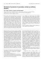
The resulting PCR products of 50, 56, 62, 68, etc. base
pairs generated a characteristic ladder with six pair
increments when separated by capilla ry electrophoresis
(ABI 3730, Applied Biosystems, Germany) (Fig 1).
Transfection
For transfection the retroviral vector S11IN was used,
which was kindly provided by Dr. Helmut Haneberd
(Dept. of Pediatric Oncology, University of Duesseldorf,
Germany). The S11IN vectors containing wild type and
mutant hTERT were constructed by subclonin g the
respective hTERT (T) cDNA sequence of the wild-type
(WT) and the mutant hTERT (DN, dominant nega tive)
from the pBABE-puro DN plasmid and the pBABE-puro
WT plasmid (kind gifts of Dr. Robert A. Weinberg,
Whitehead Institute, Cambridge, USA) using standard
protocols. Selection of S11hTDNIN and S11hTWTIN
transfected cells was carried out with geneticin (G418
sulfat e) (Invitrogen, Karlsruhe, Germany). Confirmation
of pS11 contruction insertion was proofed by PCR ana-
lysis and DNA sequencing. In addition to the
S11hTDNIN and S11hTWTIN cells were also trans-
fected with S11IN vector in order to characterise the
I
n
t
erna
l
St
an
d
ar
d
6bp-Telomer-Ladder
B. SK-N-SH-S11hTWTIN
Rox-labeled-Standard
Internal Standard
6bp-Telomer-Ladder
Internal Standard
6bp-Telomer-Ladder
Internal Standard
Internal Standard
R
ox-
l
a
b
e
l
e
d
-
St
an
d
ar
d
B. SK-N-SH-S11hTWTIN
E. CHLA-90-S11hTWTIN
Internal Standard
6bp-Telomer-Ladder
Internal Standard
Internal Standard
6bp-Telomer-Ladder
50
100 150
200
50 100 150 200
16,000
12,000
8,000
4,000
16,000
12,000
8,000
4,000
C. SK-N-SH-S11hTDNIN E. CHLA-90-S11hTDNIN
50 100 150
200
50
100 150
200
50 100
150 200
50 100
150 200
16,000
12,000
8,000
4,000
0
16,000
12,000
8,000
4,000
0
16,000
12,000
8,000
4,000
0
16,000
12,000
8,000
4,000
0
D. CHLA-90
A. SK-N-SH
Rox-labeled Standard
Rox-labeled Standard
Rox-labeled Standard
Internal Standard
Rox-labeled Standard
Rox-labeled Standard
Rox-labeled Standard
Internal Standard
6bp-Telomer-Ladder
Internal Standard
0
0
S
K
-N-S
H
S
K
-N-S
H
-S
11
hTIN
S
K
-N-S
H
-S1
1
hTWTIN
SK
-
N
-SH
-
S11-h
T
DNIN
rel. TA
0
5
10
15
20
25
C
H
LA
-90
CHLA-90-S
1
1h
TIN
C
H
LA
-9
0-S
11hTWTIN
CHLA-90
-
S11hT
D
NIN
rel. TA
0
5
10
15
20
25
Figure 1 Determination of telomerase activity. A. Telomerase activity of transfected and not-transfected CHLA-90 and SK-N-SH cells as
determined by the TRAP assay (SK-N-SH and CHLA-90: non-transfected cell lines; SK-N-SH-S11hTWTI and CHLA-90-S11hTWTI: overexpressing cell
lines; SK-NSH-S11hTDNI and CHLA-90-S11hTDNI: knockdown cell lines). B. Mean relative Telomerase activity of transfected and not-transfected
CHLA-90 and SK-N-SH cells as determined by the TRAP assay from three different passages.
Wesbuer et al. Radiation Oncology 2010, 5:66
/>Page 3 of 8
effect of vector transfection alone on proliferation, viabi-
lity, chemo- and radiosensitivity.
Statistics
GI
50
is the drug concentration that reduces the activity
of mitochondrial aldehyde dehydrogenases by 50% com-
pared to that observed in control cells incubated for
72 h without test drug. For the calculation of GI
50
sthe
following formula was used: (50% - [% viable cells
(< 50%)])/([% viable cells (> 50%)] - [% viable cells
(< 50%)]) * (drug concentration > 50% viable cells -
drug concentration < 50% viable cells) + (drug concen-
tration < 50% viable cells). Significance was determined
by using the One-Way ANOVA -Ho lm-Sidiak method,
p < 0.05 (Sigma Plot 11.0, systat.com) All experiments
were done in triplicates.
Results
Transfected cell lines
To study the effect of TA on radio- and chemo sensitiv-
ity of neuroblastomas two neuroblastoma cell lines,
CHLA-90 and SK-N-SH were stably transfected with
wild-type hTERT and a dominant negative mutant of
hTERT. Telomerase was present in the neuroblastoma
cell line SK-N-SH, while no TA was detected in CHLA-
90 cells (Fig. 1). These cells overcome telomere erosion
during cell division by an alternative lengthening of telo-
meres (ALT), which is characterized by a broad range of
telomere length within these cells (Fig. 2).
The dominant negative hTERT muta nt completely
blocked TA activity in the TA positive cell line SK-N-
SH (Fig. 1). Transfection with wild-type hTERT
increased the relativ e TA in SK-N-SH more than 10-
fold. Moreover, with increasing population doublings
the knock-down of hTERT resulted in gradual telo-
mere erosion of S11hTDNIN transfe cted SK-N-SH,
while overexpression of wild-type hTERT significantly
increased the telomere length of transfected cells (Fig.
2). SK-N-SH cells transfected with the dominant
negative hTERT mutant initially showed the same
growth characteristics compared to not transfected
cell lines. However, after more than 28 passages along
with telomere shortening cell growth slowed down.
Thecellsfinallydetachedfromthetissuecultureflask
and died. Transfection of SK-N-SH with S11hTWTIN
and S11IN, however, did not influence cell
proliferation.
Though transfection of TA-negative CHLA-90 cells
with wild-type hTERT rendered these cells TA positive
(Fig. 1) and resulted in an increase of telomere length
(Fig. 2), it had no effect on the proliferation of these cell
lines. In addition, transfection of CHLA-90 with the
dominant-negativ hTERT mu tant nor with the S11IN
vector affected cell proliferation.
Radiotherapy
Radiation reduced cell viability of the neuroblastoma cell
lines with increasing radiation dosage. The cytotoxicity
observed increased with increasing post i rradiation
interval. CHLA-90 cells were more radioresistant than
SK-N-SH cells. For the neuroblastoma cell lines an
inverse relationship between TA expression and radio-
sensitivity was observed. K nocking down TA in the TA-
expressing SK-N-SH cell line increased the radiosensi-
tivity of these cells compared to S11hTWTIN trans-
fected cells (Fig. 3). On the other hand expression of
TA in TA-negative CHLA-90 cells decreased the radio-
sensitivity (Fig. 3). Both, the radioprotective effect of
ektope TA expression as well as the radiosensitizing
effect became more prominent after longer post irradia-
tion intervals. The differences were consistently signifi-
cant for all time points.
Chemotherapy
All anticancer drugs reduced cell viability of transfected
and not-transfected cell lines in a time and dose depen-
dent manner. The effects of TA knock-down or over-
expression on chemosensitivity and -resistance were
dependent on TA, the anticancer drug, and the chemo-
sensitivity of the maternal cell line.
Transfection of wild-type and dominant negative
hTERT modulated the chemosensitivity of SK-N-SH
cells. The dominant negative t ransfected hTERT cell
lines became significantly more resistant to cisplatin,
etoposide, and doxorubicin. However, transfection with
dominant negative hTERT rendered the SK-N-SH more
sensitive against ifosfamide (Fig. 4). Modulation of drug
sensitivity/resi stance was most prominent after drug
exposure for 24 h. The differences between transfected
and not-transfected cell lines declined with increasing
duration of drug exposure (Fig.4).
Transfection of CHLA-90 only slightly modulated the
sensitivity against cisplatin, ifosfamide, doxorubicin, and
etoposide. Since there was less than two fold difference
between different transfected clones, these effects were
not considered significant
Discussion
The introduction of chemotherapy and radiotherapy
combined with tumor resection significantly improved
treatment outcome of children suffering from neuroblas-
tomas [22]. However, despite of all further efforts within
recent years the prognosis of patients with advanced
and/or disseminated disease is still poor, demonstrating
the need of new therapeutic approaches for these
patients [23-26].
During tu morigenesis the enzyme telomerase is reacti-
vated in the fast majority of these tumors promoting
tumor growth and aggressiveness [27,28]. Since
Wesbuer et al. Radiation Oncology 2010, 5:66
/>Page 4 of 8
A.
B.
7.4
5
.2
21,2
8,6
6
.1
3
.
55
4
,
2
1
,95
2
,
7
1
,55
1
,35
1
,
1
0,85
1
234 67891011125
13 14 15 16
17
19
18 20
21.2
8.6
7.4
5.0
3.55
4.2
1.95
2.7
1.55
1.35
1.1
0.85
6.1
12 345 67 8 9 101112
Figure 2 Determination of telomere length. A. Telomere length southern of transfected a nd not-transfected CHLA-90 cells. (1. DIG weight
marker; 2. DNA high: 5.5 kb; 3. DNA low: 3.2 kb; 4. CHLA-90 4.7 kb; 5. CHLA-90-IN (passage 41) 4.8 kb; 6. CHLA-90-hTDNIN (passage: 39) 4.7 kb; 7.
CHLA-90-hTWTIN (passage 42) 5.5 kb; 8. CHLA-90: 3.9 kb; 9. CHLA-90-IN (passage 40) 4.0 kb; 10. CHLA-90-hTDNIN (passage 42) 3.6 kb; 11. CHLA-
90-hTWTIN (passage 45) 4.7 kb; 12. DIG weigth marker. B. Telomere length southern of transfected and not-transfected SK-N-SH cells. (1. DIG
weight marker; 2. DNA high: 6.7 kb; 3. DNA low: 3.6 kb; 4. SK-N-SH: 4.7 kb, 5. SK-N-SH-IN (passage 20): 4.3 kb; 6. SK-N-SH-hTDNIN (passage 21): 4.3
kb; 7. SK-N-SH-hTWTIN (passage 21) 15 kb; 8. SK-N-SH: 3.8 kb; 9. SK-N-SHIN (passage 22): 4.9 kb; 10. SK-N-SH-hTDNIN (passage 23) 6.2 kb; 11. SK-
N-SH-hTWTIN (passage 23): 14.2 kb; 12. SK-N-SH: 4.3 kb; 13. SK-N-SH-IN (passage 28): 4.7 kb; 14. SK-N-SH-hTDNIN (passage 26): 4.7 kb; 15. SK-N-SH-
hTWTIN (passage 29) not evaluable; 16. SK-N-SH-IN (passage 20) 4.3 kb; 17. SK-N-SH-hTDNIN (passage 21) 4.6 kb; 18. SK-N-SH-hTWTIN (passage
21) 16.7 kb; 19. SK-N-SH-hTDNIN (passage 27) 3.2 kb; 20. DIG weight marker).
Wesbuer et al. Radiation Oncology 2010, 5:66
/>Page 5 of 8
telomerase i s almost exclu sively expressed at high levels
in most tumors it is a promising selective target for
the treatment of cancer. Hahn et al. at first demon-
strated that telomerase inhibition of telomerase expres-
sing human tumor cells effectively inhibited tumor
growth [29].
Establishing stable transfected cell lines we were able
to verify this concept for neuroblastomas, too. However,
inhibition of tumor growth as a consequence of telo-
merase inhibition only occurs after an appropriate num-
ber of cell divisio ns, when the telomeres reach a critical
length and tumor cells consequently enter a state of
senescence. Thus, telomerase inhibition alone is not a
promising approach, but it might add benefits, when
combined with chemotherapy or irradiation. We decided
to use the stable transfected cell lines to study the
effects of telomerase inhibition on chemo- and radiosen-
sitivity of neuroblastomas, since small molecules, which
inhibit TA i.e. by stabilizing the G-quadru plex structure
of telomeres, despite of high selectivity are likely to
exert off target effects, too. As standard anticancer
drugs doxorubicin, e toposide, cisplatin, and ifosfamide
were chosen, which are well established in the treatment
of neuroblastomas.
For irradiation there was an inverse relationship
between TA expression and radiosensitivity. Ektope
expression of TA which resulted in telomere elongation
in CHLA-90 cells and SK-N-SH cells rendered these
cells more r esistant against radiation. Knock-down of
TA by a dominant negative mutant in TA-positive SK-
N-SH cells induced a more radiosensitive phenotype.
These observations are in good accordance with studies,
which observed an enhanced radiosensitivity of mice
whose telomeres were shortened due to a mutant
hTERT [8,12,30,31].
Continued inhibition of TA gradually erodes telomeres
and leads to chromosome instabilities. Irradiation
induces DNA damage and it is likely that eroded and
instable chrom osomes are t argeted more easily by
irradiation.
Though the anticancer drugs tested also induce DNA
damage, this concept obviously does not apply that
strictly to the c ombination of chemotherapy and telo-
merase inhibition. TA knock down increased the sensi-
tivity to ifosfamide of SK-N-SH cells, but decreased the
sensitivity to cisplatin, doxorubicin, and etoposide.
These effects of TA-inhibition on chemosensitivity were
most prominent after an exposure for 24 h and evened
after 96 h. Knock down of TA only reduced the growth
of SK-N-SH cells after more than 28 passages. The
effects of chemotherapy were studied when the telo-
meres already shortened but before they reached their
critical length. At this time point the proliferation rate
between not-transfected, S11hTWT-, S11IN- and
S11hTDNIN-transfected cells did not differ. Thus, the
observed effects of TA-inhibition on chemo sensitivity
were not influenced by different proliferation rates. A
number of studies addressed the effect of TA inhibition
on radio- and chemose nsitivity. While radiosensitisation
by telomerase inhibition has been unambiguously
reported in l iterature, the effect s of chemotherapy com-
bined with telomerase inhibition obviously depend on
the anticancer drugs and the cell lines used. Chen et al.
treated prostate cancer cell lines antisense oligonucleo-
tides and studied the effect of the standard antiprolifera-
tive agents, paclitaxel, doxorubicin, etoposide, cisplatin,
or carboplatin at the beginning of antisense treatment
and after erosion of telomeres. They found no effects of
TA inhibition on chemosensitivity at the beginning of
antisense treatment. When telomeres were shortened
the cells were more sensitive to cisplatin and carboplatin
but not to paclitaxel, doxorubicin, and etoposide [32].
A.
B.
SK-N-SH - 20 Gy
24h 48h 72h 96h
cell viability [%]
compared to untreated controls
0
20
40
60
80
100
120
CHLA-90 - 20 Gy
24h 48h 72h 96h
cell viability [%]
compared to untreated controls
0
20
40
60
80
100
120
Figure 3 Cytotoxicity of irradiation on S11hTDNIN (Black line),
S11hTWTIN (Grey line), and S11hTIN (Dark grey line)
transfected CHLA-90 (A.) and SK-N-SH (B.) 24 h, 48 h, 72 h, and
96 h post irradiation.
Wesbuer et al. Radiation Oncology 2010, 5:66
/>Page 6 of 8
However, long telomeres and high telomerase activity
are distinct features of highly proliferating cells (e.g.
germ cells, stem cells) and are reported to be essential
vitality factors of cancer stem cells [33-35]. These cells
are defined as a small subpopulation of cancer cells,
which have t he ability of self-renewing and to produce
heterogeneous lineages of cancer cells that comprise the
tumor [18]. Should it be proved to be true that these
cell s are more resistant towards therapeutic regimens, it
follows that they can limit the therapeutic outcome and
impair long term curability. However, the stem cell mar-
ker telomerase i nfluences radiation response an d che-
moresistance and therefore, could be one potential
factor influencing cancer stem cell survival under ther-
apy. The here described construct with telomerase
knock-down in combination with other stem cell mar-
kers like CD133, CD44/CD24, ALDH-1 and SP may be
useable to verify this in further experiments.
Conclusions
In summary, our results support the concept of telomer -
ase inhibition a s an antiproliferative treatment approach
for neuroblastomas. Regarding irradiation our data
further suggest t hat telomerase inhibition improves
radiation response of neuroblastomas. With respect to
the varying effects reported for telomerase inhibition
combined with chemotherapy our data complete this pic-
ture of drug- and cell line-dependent additive/synergistic
or antagonistic effects of telomerase inhibition combined
with chemotherapy and suggests p ositive effects of com-
binations with certain anticancer drugs. Further experi-
ments should clarify the role of telomerase acticity on the
long term curability of radio- and chemotherapy by tar-
geting cancer stem cells which are known to have long
telomeres and high telomerase activity.
Conflicts of interests
The authors declare that they participated in the here
listed contributions made to the study and that they
have seen and approved the final version. They declare
no conflict of interest or financial relationship influen-
cing the conclusions of the work.
Acknowledgements
We would like to thank Christopher Poremba for providing the cell lines
used. We greatfully acknowledge the excellent technical assistance of
Annette van Dülmen. This work was supported by a grant of the Josef-
Freitag-Stiftung, Paderborn, Germany
Author details
1
Department of Radiotherapy -Radiooncology-, University Hospital Münster,
Albert-Schweitzer-Straße 33, D-48149 Münster.
2
Department of Paediatric
SK-N-SH - Etoposide - 10 µmol/L
24h 48h 72h 96h
cell viability [%]
compared to untreated controls
0
20
40
60
80
100
120
140
*
*
*
*
SK-N-SH - Cisplatin - 10 µmol/L
24h 48h 72h 96h
cell viability [%]
compared to untreated controls
0
20
40
60
80
100
120
140
*
*
*
*
SK-N-SH - Doxorubicin - 0.5 µmol/L
24h 48h 72h 96h
cell viability [%]
compared to untreated controls
0
20
40
60
80
100
120
140
SK-N-SH - Ifosfamide - 10 µmol/L
24h 48h 72h 96h
cell viability [%]
compared to untreated controls
0
20
40
60
80
100
120
140
*
*
*
*
A.
B.
.D.C
Figure 4 Cytotoxicity of etoposide (A.), cisplatin (B.), ifosfamide (C.), and doxorubicin (D.) on S11hTDNIN (Black line), S11hTWTIN (Grey
line), and S11hTIN (Dark grey line) transfected SK-N-SH cells after 24 h, 48, 72 h, and 96 h.
Wesbuer et al. Radiation Oncology 2010, 5:66
/>Page 7 of 8
Haematology and Oncology, University Hospital, Münster, Germany.
3
Institute of Pathology, Heinrich-Heine University Düsseldorf, Germany.
Authors’ contributions
SW and CLK have contributed to the same extent to the manuscript and
carried out most of the experiments shown here. IDS and TB did parts of
the statistical analysis and helped in discussion of data. KLSCH and YB
carried out generation of the transformed cell lines. NW participated
substancially in the design of this study and BG worked out the study
design and carried out the telomer-length experiments. All authors read and
approved the final manuscript.
Received: 12 May 2010 Accepted: 19 July 2010 Published: 19 July 2010
References
1. Kim Nw, Piatyszek Ma, Prowse Kr, Harley Cb, West Md, Ho Pl, Coviello Gm,
Wright We, Weinrich Sl, Shay Jw: Specific association of human
telomerase activity with immortal cells and cancer. Science 1994,
266:2011-5.
2. Shay Jw, Keith Wn: Targeting telomerase for cancer therapeutics. Br J
Cancer 2008, 98:677-83.
3. Arifin M, Tanimoto K, Putra Ac, Hiyama E, Nishiyama M, Hiyama K:
Carcinogenesis and cellular immortalization without persistent
inactivation of p16/Rb pathway in lung cancer. Int J Oncol 2010,
36:1217-27.
4. Dhaene K, Van Marck E, Parwaresch R: Telomeres, telomerase and cancer:
an up-date. Virchows Arch 2000, 437:1-16.
5. Meyerson M: Role of telomerase in normal and cancer cells. J Clin Oncol
2000, 18:2626-34.
6. Shay Jw, Bacchetti S: A survey of telomerase activity in human cancer.
Eur J Cancer 1997, 33:787-91.
7. Marian CO, Cho SK, Mcellin BM, Maher EA, Hatanpaa KJ, Madden CJ,
Mickey BE, Wright WE, Shay JW, Bachoo RM: The telomerase antagonist,
imetelstat, efficiently targets glioblastoma tumor-initiating cells leading
to decreased proliferation and tumor growth. Clin Cancer Res 2010,
16:154-63.
8. Castella M, Puerto S, Creus A, Marcos R, Surralles J: Telomere length
modulates human radiation sensitivity in vitro. Toxicol Lett 2007,
172:29-36.
9. Barwell J, Pangon L, Georgiou A, Docherty Z, Kesterton I, Ball J,
Camplejohn R, Berg J, Aviv A, Gardner J, Kato Bs, Carter N, Paximadas D,
Spector Td, Hodgson S: Is telomere length in peripheral blood
lymphocytes correlated with cancer susceptibility or radiosensitivity? Br J
Cancer 2007, 97:1696-700.
10. Genesca A, Martin M, Latre L, Soler D, Pampalona J, Tusell L: Telomere
dysfunction: a new player in radiation sensitivity. Bioessays 2006,
28:1172-80.
11. Goytisolo Fa, Samper E, Martin-Caballero J, Finnon P, Herrera E, Flores Jm,
Bouffler Sd, Blasco Ma: Short telomeres result in organismal
hypersensitivity to ionizing radiation in mammals. J Exp Med 2000,
192:1625-36.
12. Wong Kk, Chang S, Weiler Sr, Ganesan S, Chaudhuri J, Zhu C, Artandi Se,
Rudolph Kl, Gottlieb Gj, Chin L, Alt Fw, Depinho Ra: Telomere dysfunction
impairs DNA repair and enhances sensitivity to ionizing radiation. Nat
Genet 2000, 26:85-8.
13. Lanvers-Kaminsky C, Winter B, Koling S, Frodermann B, Braun Y, Schaefer Kl,
Diallo R, Koenemann S, Wai D, Willich N, Poremba C, Schuck A:
Doxorubicin modulates telomerase activity in Ewing’s sarcoma in vitro
and in vivo. Oncol Rep 2005, 14:751-8.
14. Poremba C, Heine B, Diallo R, Heinecke A, Wai D, Schaefer Kl, Braun Y,
Schuck A, Lanvers C, Bankfalvi A, Kneif S, Torhorst J, Zuber M, Kochli Or,
Mross F, Dieterich H, Sauter G, Stein H, Fogt F, Boecker W: Telomerase as a
prognostic marker in breast cancer: high-throughput tissue microarray
analysis of hTERT and hTR. J Pathol 2002, 198:181-9.
15. Schuck A, Poremba C, Lanvers C, Konemann S, Schleifer T, Wai D, Horn K,
Hesselmann S, Braun Y, Frodermann B, Schafer Kl, Diallo Ri, Rube Ce,
Rube C, Dockhorn-Dworniczak B, Willich N: Radiation-induced changes of
telomerase activity in a human Ewing xenograft tumor. Strahlenther
Onkol 2002, 178:701-8.
16. Neuhof D, Auberger F, Ruess A, Wenz F, Weber Kj: Abrogation of
radiation-inducible telomerase upregulation in HPV16 E6 transfectants
of human lymphoblasts. Strahlenther Onkol 2004, 180:52-6.
17. Saretzki G: Telomerase inhibition as cancer therapy. Cancer Lett 2003,
194:209-19.
18. Baumann M, Krause M, Hill R: Exploring the role of cancer stem cells in
radioresistance. Nat Rev Cancer 2008, 8(7):545-54.
19. Simon T, Hero B, Bongartz R, Schmidt M, Muller Rp, Berthold F: Intensified
external-beam radiation therapy improves the outcome of stage 4
neuroblastoma in children > 1 year with residual local disease.
Strahlenther Onkol 2006, 182:389-94.
20. Lanvers-Kaminsky C, Bremer A, Dirksen U, Jurgens H, Boos J: Cytotoxicity of
treosulfan and busulfan on pediatric tumor cell lines. Anticancer Drugs
2006, 17:657-62.
21. Sambrook J, Fritsch Ef, Maniatis T: Molecular Cloning: A Laboratory
Manual 2nd edn. Cold Spring Harbor Laboratory Press, New York 1989.
22. Eich Ht, Muller Rp, Micke O, Kocher M, Berthold F, Hero B:
Esthesioneuroblastoma in childhood and adolescence. Better prognosis
with multimodal treatment? Strahlenther Onkol 2005, 181:378-84.
23. Hero B, Simon T, Spitz R, Ernestus K, Gnekow Ak, Scheel-Walter Hg,
Schwabe D, Schilling Fh, Benz-Bohm G, Berthold F: Localized infant
neuroblastomas often show spontaneous regression: results of the
prospective trials NB95-S and NB97. J Clin Oncol 2008, 26:1504-10.
24. Simon T, Hero B, Benz-Bohm G, Von Schweinitz D, Berthold F: Review of
image defined risk factors in localized neuroblastoma patients: Results
of the GPOH NB97 trial. Pediatr Blood Cancer 2008, 50:965-9.
25. Katzenstein HM, Kent PM, London WB, Cohn SL: Treatment and outcome
of 83 children with intraspinal neuroblastoma: the Pediatric Oncology
Group experience. J Clin Oncol 2001, 19:1047-55.
26. Stram DO, Matthay KK, O’leary M, Reynolds CP, Haase GM, Atkinson JB,
Brodeur GM, Seeger RC: Consolidation chemoradiotherapy and
autologous bone marrow transplantation versus continued
chemotherapy for metastatic neuroblastoma: a report of two concurrent
Children’s Cancer Group studies. J Clin Oncol 1996, 14:2417-26.
27. Poremba C, Scheel C, Hero B, Christiansen H, Schaefer Kl, Nakayama J,
Berthold F, Juergens H, Boecker W, Dockhorn-Dworniczak B: Telomerase
activity and telomerase subunits gene expression patterns in
neuroblastoma: a molecular and immunohistochemical study
establishing prognostic tools for fresh-frozen and paraffin-embedded
tissues. J Clin Oncol 2000, 18:2582-92.
28. Poremba C, Willenbring H, Hero B, Christiansen H, Schafer Kl,
Brinkschmidt C, Jurgens H, Bocker W, Dockhorn-Dworniczak B: Telomerase
activity distinguishes between neuroblastomas with good and poor
prognosis. Ann Oncol 1999, 10:715-21.
29. Hahn WC, Stewart SA, Brooks MW, York SG, Eaton E, Kurachi A,
Beijersbergen RL, Knoll JH, Meyerson M, Weinberg RA: Inhibition of
telomerase limits the growth of human cancer cells. Nat Med 1999,
5:1164-70.
30. Agarwal M, Pandita S, Hunt CR, Gupta A, Yue X, Khan S, Pandita RK, Pratt D,
Shay JW, Taylor JS, Pandita TK: Inhibition of telomerase activity enhances
hyperthermia-mediated radiosensitization. Cancer Res 2008, 68:3370-8.
31. Ji XM, Xie CH, Fang MH, Zhou FX, Zhang WJ, Zhang MS, Zhou YF: Efficient
inhibition of human telomerase activity by antisense oligonucleotides
sensitizes cancer cells to radiotherapy. Acta Pharmacol Sin 2006,
27:1185-91.
32. Chen Z, Koeneman KS, Corey DR: Consequences of telomerase inhibition
and combination treatments for the proliferation of cancer cells. Cancer
Res 2003, 63:5917-25.
33. Armanios M, Greider CW: Telomerase and cancer stem cells. Cold Spring
Harb Symp Quant Biol 2005, 70:205-8.
34. Phatak P, Burger AM: Telomerase and its potential for therapeutic
intervention. Br J Pharmacol 2007, 152:1003-11.
35. Harley CB: Telomerase and cancer therapeutics. Nat Rev Cancer 2008,
8:167-79.
doi:10.1186/1748-717X-5-66
Cite this article as: Wesbuer et al.: Association of telomerase activity
with radio- and chemosensitivity of neuroblastomas. Radiation Oncology
2010 5:66.
Wesbuer et al. Radiation Oncology 2010, 5:66
/>Page 8 of 8